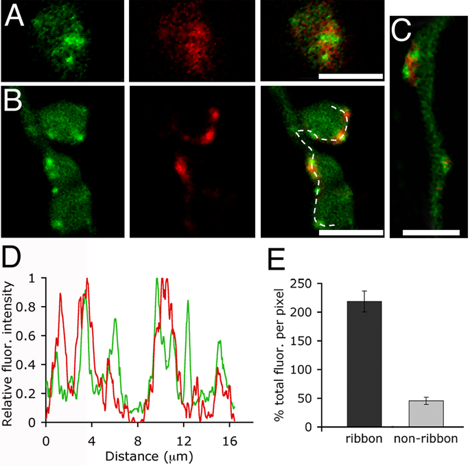
Figure 3.
Sites of endocytosis correlate with the position of synaptic ribbons in the axon and terminals. A, Confocal images of a live bipolar cell loaded with FM4-64 (red) and RIBEYE-binding peptide (green). To load recycling vesicles, cells were bathed in a high K+ solution containing 20 µM FM4-64 for 90 s and washed with Advasep-7. The last panel is an overlay of the red and green channels. Scale bar represents 2.5 µm. B, Confocal images of a different terminal loaded with RIBEYE-binding peptide (green) via a patch pipette and depolarized from a holding potential of −60 mV to 0 mV for 250 ms in the presence of FM4-64. Dye was removed after ~12 seconds, and the cell was washed with Advasep-7 to expose the location of trapped dye (middle panel). Far right panel is the overlay. Scale bar represents 5.0 µm. C, FM4-64 labeling (red) also concentrates around axonal ribbons (green). Scale bar represents 2.5 µm. D, Fluorescence intensity profile of synaptic ribbons (green) and FM4-64 (red) along the dashed line shown in B. E, Graph showing the percent total FM4-64 fluorescence per pixel at ribbon and non-ribbon locations within the axon and terminals (p< 0.001, n=10 cells). See text for a description of the measurement procedure.

