Abstract
The licensed smallpox vaccine, comprised of infectious vaccinia, is no longer popular as it is associated with a variety of adverse events. Safer vaccines have been explored such as further attenuated viruses and component designs. However, these alternatives typically provide compromised breadth and strength of protection. We conducted a genome-level screening of cowpox, the ancestral poxvirus, in the broadly immune-presenting C57BL/6 mouse as an approach to discovering novel components with protective capacities. Cowpox coding sequences were synthetically built and directly assayed by genetic immunization for open-reading-frames that protect against lethal pulmonary infection. Membrane and non-membrane antigens were identified that partially protect C57BL/6 mice against cowpox and vaccinia challenges without adjuvant or regimen optimization, whereas the 4-pox vaccine did not. New vaccines might be developed from productive combinations of these new and existing antigens to confer potent, broadly-efficacious protection and be contraindicated for none.
Keywords: smallpox, cowpox, antigen discovery, gene vaccine, DNA vaccine, expression library immunization, genomics
INTRODUCTION
Despite having a vaccine in hand that eliminated natural smallpox infections, new vaccines are needed to protect against the threat of deliberately released variola in addition to other endemic and emerging poxviruses. Poxviridae contains viruses with linear genomes of double-stranded DNA that replicate in the host cytoplasm and can encode more than 200 proteins, making them one of the largest among all viruses (Henderson, 1999). The orthopoxviruses produce several structural forms, including two infectious particles that have distinct membrane surfaces and roles in the viral life cycle. The intracellular mature virion (IMV) is important for host to host transmission (Lustig et al., 2005; Vanderplasschen et al., 1998), while the extracellular enveloped virion (EEV) contributes to efficient dissemination within a host (Lustig et al., 2005; Vanderplasschen et al., 1998). The orthopox genus contains variola, cowpox, vaccinia, ectromelia (mousepox), monkeypox, and several other closely related viral species that show extensive serological cross-reactivity and vaccine cross-protection (Edghill-Smith et al., 2005; Heraud et al., 2006; Parrino and Graham, 2006). However, variola (VARV) survivors are protected from smallpox disease for life whereas people immunized with live VACV display reduced immunity within 10 to 20 years (Jacobs, 2002; Viner and Isaacs, 2005). Cowpox (CPV) appears to be the ancestral poxvirus (Dasgupta et al., 2007; Shchelkunov et al., 1998), maintained for thousands of years in its natural rodent host, though cattle infections are also common (Wang et al., 2008) and human infections are considered an emerging hazard (Vorou, Papavassiliou, and Pierroutsakos, 2008). VACV has no known natural host nor clear origin; it has been suggested to be a divergent CPV or VARV species that emerged during early variolation practices (Buller and Palumbo, 1991).
Live CPV was used by Jenner in his original demonstration of protective immunity against smallpox. CPV was gradually replaced with VACV because of this newer virus’ lower virulence (Buller and Palumbo, 1991; 2002), although both viruses can cause disease in humans (Moore, Seward, and Lane, 2006). A process for large-scale production of lyophilized VACV from infected calf lymph was developed in the 1940s (Moss, 1991). This product ironically made research toward a better smallpox vaccine superfluous for decades. However, our current society expects greater vaccine product performance, uniformity, and safety. The US DryVax product was not produced under good manufacturing practice (GMP) protocols, and its efficacy varied among batches (Slifka, 2005). Lifethreatening complications (Henderson, 1999) and cardiac related adverse events (Karron et al.) remain concerns. Estimates indicate that half of the US population would be excluded from receiving a vaccination due to histories of eczema or dermatitis (Wiser, Balicer, and Cohen, 2007). It remains contraindicated for infants, elderly, pregnant women, and our increasing immunocompromised sector (LeDuc and Becher, 1999).
To address these difficulties, newer generation vaccines have been developed from VACV grown in cell culture (Weltzin et al., 2003). This improved batch consistency but showed reactogenicity similar to that of the original product (Ward et al., 2005). Attenuated VACVs have been widely investigated, such as the LC16m8 strain, Modified Vaccinia Ankara (MVA), and a number of other more recent ones (Vollmar et al., 2006). The potency and safety of these further modified live-virus inocula are under continued study.
Poxviruses are notable for their abilities to interfere with or circumvent many critical processes of the host innate and adaptive immune responses upon infection (Dasgupta et al., 2007; Dunlop et al., 2003; Thornburg et al., 2007). Since these properties are counterproductive for vaccine inocula, component designs may hold advantage. Unfortunately optimal subunit selection is stymied by our incomplete knowledge of the immune mechanisms needed to protect against these complex viruses and the diversity of systems used to model human variola disease. Researchers are currently using the VACV, ectromelia (ECTV), and CPV viruses in mice, in addition to the MPV in primates to evaluate vaccine designs. Immune studies have indicated that both antibody and cellular responses are involved (Cornberg et al., 2007), but relatively few antigens have been tested. USAMRIID scientists assembled four VACV genes (A27L, A33R, B5R, and L1R), encoding two major IMV and two major EEV surface proteins, and showed that biolistic co-immunization protects BALB/c mice against an intraparenterally (i.p.) delivered VACV challenge (Hooper, Custer, and Thompson, 2003) or CPV challenge (Pickup, 2007). Primates are also protected against an intravenous MPV challenge (Hooper et al., 2004). This tetravalent inoculum has been referred to as “4-pox”. Subsets of these four antigens delivered as gene, replicon or protein, have shown various levels of protection against VACV challenges in mice (Golden and Hooper, 2008; Hooper et al., 2000; Pulford et al., 2004; Sakhatskyy et al., 2008; Thornburg et al., 2007; Xiao et al., 2007). Other mouse protection studies showed immunization with H3L or a codon-optimized D8L provides some protection against VACV challenge (Davies et al., 2005; Sakhatskyy et al., 2006), and an antibody against A28L is passively protective (Nelson et al., 2008). Using the ECTV murine model, a peptide from B8R and protein B19R VACV homologues were shown to confer partial protection (Tscharke et al., 2005; Xu et al., 2008). Finally, VARV antigens A30, B7, and F8 have shown to be protective against VACV challenge in mice (Sakhatskyy et al., 2008). While it is not clear whether or not these particular surface proteins or their VARV homologues would be protective against exposure to VARV, these studies demonstrate that isolated viral components can protect against orthopoxvirus infections. They also indicate that biolistic genetic immunization is at least one effective modality of vaccine delivery. Unfortunately, none of the inocula were as effective as live VACV and all required relatively large doses and multiple immunizations. Additional candidates, with perhaps unique immune capacities, may be needed to successfully develop a modern vaccine.
We report here a comprehensive screen of the cowpox virus for protective components encoded by its genome, using expression library immunization (ELI) (Barry et al., 2004) and a pulmonary challenge assay in C57BL/6 mice. In this approach, the full protein coding capacity of a pathogen is introduced into host animals as matrix-organized pools of genes or subgenes by genetic immunization (Tang, DeVit, and Johnston, 1992). Animals are exposed to pathogen and disease is monitored as a readout of inoculum utility. Optimally, all open-reading frames (ORFs) should be expressed at similar levels to avoid bias in the screen. We chose to synthetically build the ORFs as codon-optimized versions. This enabled us to uniformly increase expression, and normalize antigen dose. Even though nearly all vaccine studies to date have been conducted with the BALB/c mouse, we selected C57BL/6 mice as the host strain for our vaccine assay because the BALB/c does not produce a broad profile of cellular immune responses against viral infection (Dieli et al., 1988; Janeway et al., 2005) whereas the C57BL/6 does (Moutaftsi et al., 2006); furthermore, any widely useful candidate would need to be effective in more than one genetic background. Research has indicated that delivery routes can influence which components of 4-pox are needed for protection (Kaufman et al., 2008). Since we are targeting candidates for protection against aerosol exposure to virus, a pulmonary challenge delivery route was chosen for our antigen screen. In particular, viral infections were administered intratracheally (i.t.), as this is most reliable method for delivering viral particles to the lower respiratory tract (MacNeill, Moldawer, and Moyer, 2009).
Nine new protective antigen candidates are presented, including six that are either non-membrane or hypothetical proteins, and unlikely to have been rationally selected. This contrasts with previously tested set of antigens, which have all been surface proteins. We propose that these new functionally-identified antigens, in combination with existing ones, constitute a uniquely diverse panel of molecular candidates from which a broadly efficacious subunit vaccine against smallpox and other emergent poxvirus diseases might be developed.
MATERIALS AND METHODS
Gene recoding, oligonucleotide design and oligo synthesis
All 233 protein coding CPV genes annotated in GenBank were extracted from the genomic database. The coding sequences were redesigned for optimal mammalian translation based on the codon usage of a set of highly expressed mammalian genes from EMBOSS. Using codon adaptation index (CAI) criteria, genes were converted to sequences with CAI values of greater than 0.8. The predicted new genes were electronically organized into 789 open-reading-frames (ORFs) of approximately 500bp in length. The 81 CPV genes of less than 500bp were represented as full length fragments. The 152 longer genes were split into smaller fragments that overlap by at least 50bp. ORF names for subgenes were assigned using the corresponding gene name followed by progressive lettering. These ORF sequences were used to predict two sets of oligonucleotides (oligos) for synthetic gene assembly: gene-building oligos (GBOs) and gene amplification primers (GAPs). GBO sequences were obtained by partitioning both DNA strands of each ORF into sets of 60nt fragments, which overlapped by 30nt. GAPs were designed as Tm matched primer pairs for each ORF. The GAPs consisted of two regions: common and gene-specific. All forward GAPs started with TATAGGCGGAAGCGGATTG and all reverse with GTGGGAGGGAGGTTAGGT followed by gene-specific sequences. All GAP and GBO oligonucleotides were synthesized in-house on a MerMade-192 oligonucleotide synthesizer (BioAutomation, Dallas TX) using standard phosphoramidite chemistry and used after desalting without any additional purification. All oligonucleotides were normalized by dilution to 50 µM with 10 mM Tris-HCl, pH 7.5 and stored at −80°C.
Synthetic gene assembling and evaluation
GBOs were diluted to 5µM and equal volumes (5µl) of each oligo were mixed together as 233 separate pools, each comprised of the oligos for assembly a single ORF. Individual ORFs were assembled from these pools in a three steps: i) oligo assembling, ii) ORF amplification and iii) attachment of dU-rich region. Assembling reactions were performed by mixing 5µl of individual GBO pools (8 to 35 oligos/pool) with 1µl of 10mM dNTPs, 5µl of 10× Taq PCR with MgCl2 buffer (Promega), 0.5u Taq polymerase (Promega) in total volume of 50µl. The mixture was incubated at 95°C for 1 min, followed by 35 cycles of incubations at 94°C for 1 min, 50°C 1 min, 72°C 2 min and final extension at 72°C for 5 min. For ORF amplification 1µl of an assembly reaction was mixed with 1ul of 1uM forward and reverse GAPs, 0.2µl of 10mM dNTPs, 1µl of 10× Taq PCR with MgCl2 buffer (Promega), 0.1u Taq polymerase (Promega) in total volume of 10µl. The mixture was incubated at 95°C for 1 min, followed by 5 cycles of incubations at 94°C for 1 min, 50°C 1 min, 72°C 2 min, 20 cycles of 94°C for 1 min, 64°C 1 min, 72°C 2 min, and final extension at 72°C for 5 min. Next the PCR generated products were extended with uracil containing sequences by adding 90ul of a master mix composed of 1×Taq PCR with MgCl2 buffer, 1mM dNTPs, 250nM universal dU forward (5’AGUAGUAGUAGUAGUGGTATAGGCGGAAGCGGATTG3’) and reverse (5’AUGAUGAUGAUGAUGAUGTGGGAGGGAGGTTAGGT3’) primers, and 5u of Taq polymerase and performing 20 cycles of incubation at 94°C for 1 min, 55°C 1 min, 72°C 2 min and final extension at 72°C for 5 min. The gene products were evaluated by agarose gel electrophoresis and quantified by PicoGreen assay according to the manufacturer’s protocol (Invitrogen). A random set of PCR products was sequenced directly without cloning to assess fidelity of assembly. All base calls corresponded to the designed sequence, which demonstrated that as a population the molecules displayed the intended ORF. This indicated predominantly proper gene assembling and the absence of major rearrangements, deletions, insertions or duplications. To evaluate uniformity of the generated products, types and frequencies of the aberrations several independently assembled genes were sequenced after cloning into pCR3.1 cloning vector. Approximately half of the analyzed clones carried perfect sequences. The remaining half predominantly carried single nucleotide deletions and insertions which would encode partial products.
Design and arraying of ORFs into pools
The complete CPV library of 789 new ORFs were randomly arrayed into 25 pools of 30–32 ORFs each, such that each ORF was resident in only one pool. The same ORFs were re-arrayed into 25 new pools two more times, to obtain three complete library sets, designated X, Y, and Z, and 75 pools. After all 789 ORFs were generated they were quantified using picogreen reagent (Invitrogen) following the manufacturer protocol and normalized to 1µM. Normalized ORFs were robotically pooled into the 75 groups, and aliquots were used for constructing plasmid sub-libraries. Remaining ORF sample was stored at −80°C.
Cloning PCR generated products
Two mammalian expression vectors pCMViLS and pCMViUB (Sykes and Johnston, 1999) were to provide expression of each ORF with both an N terminal secretory leader (LS) and as ubiquitin subunit (UB) fusion, respectively. The two vectors were prepared for by ligation of single-stranded dU complimenting adapters onto the restricted BglII and HindIII recognition sites. In particular, each plasmid (10µg) was digested with the corresponding enzymes, dephosphorylated, gel purified. Next, 5’ phosphorylated BglII (GATCTGAGTAGTAGTAGTAGT) and HindIII (AGCTTAGATGATGATGATGATGAT) adapter-oligos were ligated to the corresponding DNA ends in 50ul of 1×T4 ligase buffer with 50u of T4 ligase at 15oC overnight using 75 fold molar excess of the adapters. After the reaction unligated adapters were removed by gel purification. The resulting vector was precipitated with ethanol and resuspended in TE at 1µg/ul.
Prior to incubating with the prepared vectors, 1 µg of dU flanked PCR products was incubated in 50ul of 1×UDG Buffer (NEB) with 10u of uracil-DNA glycosylase (UDG). After incubation for 45min at 37°C DNA was precipitated with ethanol, resuspended in 5ul of water, and mixed with the prepared vectors at 1:3 plasmid to insert molar ratio in 10 µl of water. Insert was annealed to the vector by sequential incubation of the mixture at 65°C, 37°C, and room temperature for 10 min and then directly used for transformation of chemically competent E. coli (DH10b) cells (Invitrogen).
Expression immunization library construction and DNA preparation
Small aliquots of the primary transformants from the dU annealing reaction were plated onto LB agar, grown overnight, and used to determine complexity of the 75 pool libraries. The remaining cells were inoculated into 2ml LB/Amp culture and grown at constant agitation at 37°C for 4hrs. Then the entire culture was used for inoculation of 100ml LB/Amp, which was grown at 37°C until mid log phase (~1OD660). Then cells were harvested by centrifugation and used for plasmid preparation using an endotoxin free plasmid preparation kit following the recommended protocol (Qiagen, Hercules, CA). Sub-library transformations generating less than 104 original transformants were remade. High diversity (>104) libraries were used for plasmid DNA isolation. At least 50µg of plasmid DNA was prepared from each sub-library. The corresponding pCMViLS and pCMViUB sub-libraries were mixed together at 1:1 ration and divided into four 25µg aliquots. The aliquots and unused plasmid DNA were catalogued and stored at −80°C.
Preparation of gene gun bullets
Gold spherical microparticles (1–2µm, RDAU11K-20, Ferro Electronic Material Systems, South Plainfield, NJ) were washed twice with water and ethanol, and stored frozen in low retention tubes as 50mg/ml water slurry. To prepare 100 bullets 1ml of gold slurry was mixed with 100µg of DNA and total volume adjusted to 2ml with water. Then 2ml of cold 2.5M CaCl2 and 100ul of 1M spermidine were quickly added, the mix was briefly vortexed for few seconds and left on ice for 10–15min. Then gold was spun down by gentle centrifugation (1 min at 200 rcf), supernatant aspirated, and pellet resuspended in ~5ml of 100% ethanol. After two more washes with ethanol the gold pellet was resuspended in 5.5ml of absolute ethanol with 0.005% polyvinylpyrrolidone and transferred into Teflon tubing (0.125”OD, 0.93”ID, Bio-Rad, Hercules, CA ) and left on an even surface for gold to settle. After gold settled, ethanol was gently drained from the tubing and gold dried by gently flowing dry nitrogen gas through the tubing. Individual gene gun cartridges loaded with 1µg DNA on 0.5mg of gold microparticles were made by cutting the tubing into 0.5” sections. The bullets were packed over Drierite and used within a week.
Large-scale immunizations for screen
Bullets coated with the CPV pool DNAs described above were used for biolistic vaccination of six- to eight-week old female C57BL/6 mice (Harlan Sprague Dawley, Indianapolis, IN). Bullets were shot into dorsal ear pinna with Helios Gene Gun (BioRad) at a helium pressure of 400psi. Each mouse received 2 shots, one into each ear per immunization, for a total dose of 2µg. Boosts were administered at weeks 4 and 8 post-prime with the same dose and delivery protocol. During immunization animals were housed 5 mice per case (2 cases per immunization group) in a pathogen-free environment at the University of New Mexico. Mice were given food and water ad libitum, and all procedures were conducted in accordance with federal guidelines for animal experimentation.
Viral Propagation
CPXV-Brighton Red and VACV Western Reserve were grown in Vero E6 cells and purified as described (Joklik, 1962).
Challenge-protection assays
We have previously developed the murine-CPV model of smallpox disease in C57BL/6 mice and the i.t. route for pulmonary delivery of virus, (MacNeill, Moldawer, and Moyer, 2009). Control experiments were conducted on naïve and vaccinated C57BL/6 mice. The cowpox and VACV were clonally purified and quality controlled to ensure virulence. These were used as live inocula to intraperitoneally vaccinate mice to establish a positive control for vaccination (5 × 10² pfu/ml, 50µl per mouse). Challenge material stocks were prepared and lethal dose titrations were conducted to establish the minimum dose necessary for lethality. This was designed to maximize the sensitivity of the screen. To assay protection, animals were challenge with 1 LD100 (5×10⁴ pfu/mouse) by i.t. route at week 14. Mouse survival was monitored and recorded twice daily for 14 days. Data were analyzed as described in results.
Production of clonal genetic immunization constructs for the validation studies
ORFs selected by the protection assay screen were re-synthesized as described above and individually dU cloned into into pCMViLS and pCMViUB. Randomly selected clones were sequenced to identify those carrying the intended ORF sequence. Perfect clones were used for large scale DNA production and purification using endotoxin free plasmid preparation kit (Qiagen).
Generation of prokaryotic expression constructs and protein production
For the large-scale protein production sequence-confirmed ORFs described above were sub-cloned into one of the commonly used prokaryotic expression vectors: pGEX52 (Amersham Biosciences, Piscataway, NJ); pEXP5-NT (Invitrogen, Carlsbad, CA); or pBAD/Thio (Invitrogen). The constructs were used for transformation of the appropriate host strains: BL21(λ)DE3 for those cloned into pEXP5-NT and pGEX52; and LMG194 for those in pBAD/Thio. Cells were grown and induced according to the recommended protocols. Cells were harvested 3–4 hrs after induction by centrifugation and the resulting cell pellet lysed by resuspension in phosphate-buffered saline (PBS) containing 1% Triton X-100, 1 mM phenylmethylsulfonylfluoride (PMSF), and protease inhibitor cocktail (Complete, Roche, Indianapolis, IN). Cell walls were permeabilized with 10 mg of lysozyme and subjected to 3 freeze/thaw cycles between −80°C and room temperature. The viscous lysate was cleared in a 1h-incubation at 4°C with 10 µg/mL of DNase I and 20 mM MgCl2. The lysate was centrifuged at 27,000 × g for 10 minutes at 4°C, and the supernatant containing the soluble material was transferred to a fresh tube. The insoluble material, remaining in the pellet of the cleared lysate, was washed 4 times in PBS containing 1% Triton X-100 and 0.5 M guanidine followed by 3 washes with PBS. Cells were collected between washes by centrifugation at 3,000 × g for 5 min at room temperature. After the final PBS wash, the inclusion bodies were resuspended in PBS, flash-frozen in liquid nitrogen, and stored at −80°C until ready for use. To solublize the inclusion bodies, the pellets were resuspended in PBS containing 8 M urea and 10% glycerol. Insoluble material was removed by centrifugation at 14,000 × g for 5 min at room temperature, and the soluble protein was collected in the supernatant and dialyzed against PBS.
Immunization regimen for immune analyses
For the immune studies six- to eight-week old female C57BL/6 mice (Harlan Sprague Dawley, Indianapolis, IN) were housed in a pathogen-free environment at the Biodesign Institute at Arizona State University. Mice were given food and water ad libitum, and all procedures were conducted in accordance with federal guidelines for animal experimentation.
The ORF clones in pCMViUB and pCMViLS were mixed at a 1:1 ratio, precipitated onto bullets, and delivered biolistically into mice as described above. Each mouse received 2µg of DNA at weeks 0, 1, and 5. Mice received a final boost of DNA at week 22 (samples CPV100, CPV225, CPV106, and CPV204) or a boost of protein at week 23 (remaining samples). Mice immunized with protein were injected with 25 µg of antigen emulsified 1:1 with TiterMax adjuvant (TiterMax, Norcross, GA) delivered in three 50 µL subcutaneous injections. Mice were sacrificed 11 days later, and both sera and splenocytes were harvested.
ELISA assays
Antigen-specific antibody titers were measured in an indirect ELISA using selected peptides or peptide pools since recombinant proteins produced in E. coli were insoluble. Immunlon 2-HB 96-well plates (Thermo Fisher Scientitic, Waltham, MA) were coated overnight at 4°C with 2µg /ml of peptide or peptide pool diluted in PBS. Wells were blocked with 2% (w/v) bovine serum albumin (BSA) in PBS. Sera, diluted two-fold serially in PBS containing 0.05% (w/v) BSA and 0.1% polyoxyethylene sorbitol monolaurate (Tween 20) starting at 1/100, were added to blocked wells and incubated for 1 hour at 37°C. Bound antibody was detected by sequential 1-hour incubations at 37°C with horseradish peroxidase (HRP)-conjugated goat anti-mouse IgG/IgM-specific polyclonal antibody and 2,2’ azino-bis (3-ethylbenzylthiazoline-6-sulphonic acid, ABTS) substrate (both from Kirkegaard & Perry Laboratories, KPL, Gaithersburg, MD). Conversion of the substrate was quantified at 405 nm using an ELISA plate reader (Molecular Devices, Sunnyvale, CA). Reactivity of sera from mice immunized with constructs expressing candidates CPV 225a, CPV 110a, CPV 100a, a fragment of CPV 097b, CPV 066, and CPV 030a were evaluated against synthetic peptides derived from regions of the corresponding proteins in ELISA assays. All sera were tested in two-fold dilution series. Endpoint titers were determined as the reciprocal dilution at which the mean absorbance value of the test serum was at least twice that of the mean absorbance value of the background (diluent alone). Sequences of specific peptides selected for use in ELISAs are as follows. CPV225a_pep3 B12: DVPYEHINGKCNGTDYNSNN, CPV110a_pep1: DEQIYAFCDANKDDIRCKCI, CPV110a_pep2: NKDDIRCKCIHPDKSIVRIG, CPV110a_pep3: HPDKSIVRIGIDTRLPYYCW, CPV110a_pep4: IDTRLPYYCWYEPCKRSDAL, CPV110a_pep5: YEPCKRSDALLPASLKKNIS, CPV110a_pep6: LPASLKKNISRCNVSDCTIS, CPV110a_pep7: RCNVSDCTISLGNVSITDSK, CPV110a_pep8: LGNVSITDSKLDVNNVCDSK, CPV110a_pep9: LGNVSITDSKLDVNNVCDSK, CPV110a_pep10: RVATENIAVRYLNQEIRYPI, CPV110a_pep11: YLNQEIRYPIIDIKWLPIGL, CPV110a_pep12: IDIKWLPIGLLALAILILAF, CPV110a_pep13: LALAILILAFF ,CPV100a_pep16 H6: SDRTAEGQQSLINLYNKMQT, CPV097b_pep14 F8: SGKEPISDYSAEVERLMELP, CPV097b_pep16 F10: VKTDIVNTTYDFLARKGIDT, CPV066a_pep1: TINEKNLEFDTWKDVIHNDE, CPV030a_pep7 C3: AVRYYDGDIYELAKEINAMS, CPV030a_pep15 C11: LSHLKVALYRRIQRRYPIDD, CPV030a_pep16 C12: RIQRRYPIDDDVDR. Background reactivity was too high on the remaining peptides to evaluate sera of mice immunized with CPV171a and CPV120f, by ELISA. For these immune sera, only immunoblot analyses were conducted.
Immunoblot analyses
Antibody responses to the vaccine candidates were evaluated by immunoblot analysis. CPV 171a and CPV 172 were expressed with an N-terminal thioredoxin fusion. CPV 165 was generated as two glutathione-s-transferase (GST) tagged fragments spanning the full length protein. Insoluble inclusion bodies of CPV 030a, CPV 066a, CPV 110a, and CPV 171a were used. Recombinant proteins were solubilized in urea, fractionated in SDS-PAGE, and transferred to nitrocellulose membranes (BioRad, Hercules, CA). Blots were blocked with casein in PBS (Pierce, Rockford, IL) and probed with a 1:250 dilution of mouse anti-sera followed by HRP-conjugated, goat anti-mouse IgG/IgM antibody (KPL). Reactive bands were detected with 3,3’,5,5’-tetramethylbenzidine (TMB) substrate (Pierce).
IFN-γ ELISPOT assays
CD4+ and CD8+ T cell responses to the CPV vaccine candidates were evaluated by quantifying the number of splenocytes of vaccinated mice secreting IFN- γ in response to antigen-specific stimulation. ImmunoSpot plates (Millipore, Billerica, MA) were coated with rat anti-mouse IFN-γ monoclonal antibody (BD Pharmingen, San Diego, CA) and blocked with RPMI-1640 medium containing L-glutamine and supplemented with 10% (v/v) FBS (Gemini Bio-Products, West Sacramento, CA) and penicillin/streptomycin solution (Cambrex, East Rutherford, NJ). Splenocyte suspensions isolated from vaccinated mice were seeded in triplicate or quadruplicate wells of ImmunoSpot plates at densities from 2.5 × 10⁵ cells/well to 1 × 10⁶ cells/well. Cells were stimulated with full-length protein or peptide pools consisting of 20-mers overlapping in sequence by 10 amino acids (Alta Bioscience, Edgbaston, Birmingham, UK). All peptide sequences are available upon request. Proteins were assayed at 20µg/mL while the peptide pools were assayed at 10µg/mL. Control wells contained splenocytes incubated with medium alone (no antigen). After 18 – 22 hours, captured IFN-γ was detected by the sequential addition of biotin-labeled rat anti-mouse IFN- γ monoclonal antibody (BD Pharmingen) and horseradish peroxidase-labeled avidin D (Vector Labs, Burlingame, CA). Spots produced by the conversion of 3-amino-9-ethylcarbazole substrate (AEC, BD Pharmingen) were quantified with an ImmunSpot Analyzer (Cellular Technology Ltd., Cleveland, OH). Data are presented as the number of antigen-specific Spot Forming Units (SFU) per million splenocytes. SFU counts were adjusted for background by subtracting the number of spots in wells containing splenocytes in medium alone.
RESULTS
Premise for new antigen discovery
The most characterized VACV genes identified as orthopoxvirus vaccine candidates are the constituents of the 4-pox gene inoculum. A desirable orthopox vaccine would be cross-protective against more than one these related viral species; indeed VACV 4-pox has been shown to cross-protect against CPV infections in BALB/c mice, and MPV in primates. We reasoned that a successful vaccine will also need to protect a diverse host species population. To address this question, we tested the performance of the 4-pox inoculum in C57BL/6 mice relative to its effectiveness in the well-studied BALB/c strain.
Evaluation of the 4-pox gene constructs as a vaccine candidate
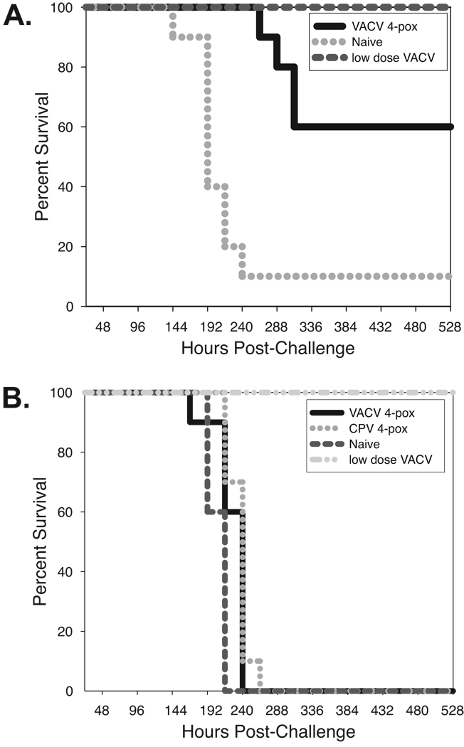
The VACV genes encoding A27L, A33R, B5R, and L1R, and also the corresponding CPV homologues were extracted from GenBank and redesigned as codon-optimized versions using our GeneBuilder program. These were built as described in Material and Methods and cloned into plasmid vector pCMViLS, which expresses inserted sequences with the secretory leader peptide (LS) from human alpha-1 antitrypsin. The four plasmids were co-administered biolistically into both sides of both ears (4µg total DNA per mouse) of groups of ten BALB/c and C57BL/6 mice. Positive control groups were administered a VACV or CPV live vaccine and negative controls were naïve age-matched. Boosts were administered at week 2 post-prime and animals were homologously challenged i.t. with a lethal dose of VACV or CPV at week 6. The survival results plotted in Figure 1A show that the live vaccines were fully protective and the 4-pox inocula partially protected BALB/c mice from VACV or CPV exposure. These results are consistent with those published by others (Hooper, Custer, and Thompson, 2003). In the C57BL/6 model, the live vaccines were also fully protective; however, the same 4-pox inocula did not protect these mice (Fig. 1B).
Fig. 1. Comparison of protection conferred by 4-pox gene vaccine in two strains of mice.
Animals (10 mice per group) were biolistically immunized with a pool of VACV A27L, A33R, B5R, and L1R or the CPV homologues of these genes and boosted 2 weeks later. A lethal dose of virus (VACV strain Western Reserve, or CPV Brighten Red) was administered 6 weeks post prime by i.t. route, and survival was monitored for 21 days. Kaplan-Meir plots were drawn and relative to live virus vaccinated and naïve mice.
A. The BALB/c mouse strain was used as host.
B. The C57BL/6 mouse strain was used as host.
Experimental design and pilot studies
The ELI screen was designed so as to assay the vaccine potential of each CPV gene or subgene three times, within different antigenic contexts. This was done by applying three different strategies for distributing the library of ORFs into 25 sub-library pools. Every ORF resided once in each library set (named X, Y, and Z) and thereby was a component of three of the total collection of 75 uniquely comprised pools. This overlapping distribution strategy can be envisioned as a three-dimensional array of ORFs within a cube. Each ORF’s position can be defined using an X, Y, and Z planar coordinate and therefore matrix-based analyses can pinpoint individual ORFs that are in common within any X, Y, and Z pool. These organized overlaps enable a single animal experiment to be used to infer which ORFs are responsible for animal protection, and provide each ORF with a calculated rank relative to its apparent ability to induce protective immunity. Several pilot studies were conducted to demonstrate the feasibility of performing this multiplexed protection experiment.
Validating immunogenicity of synthetic gene-pools
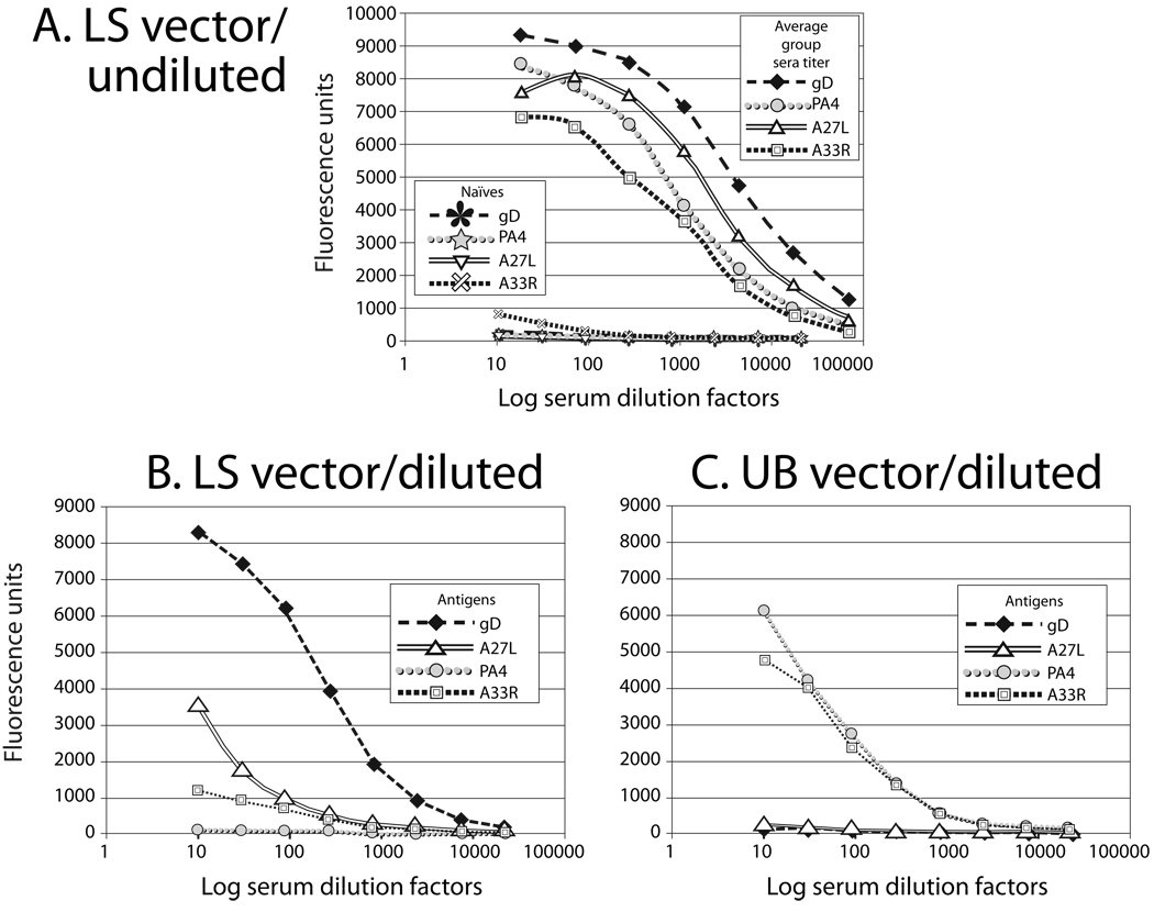
Before constructing the library, we built a set of test genes encoding well-known antigens. This would enable us to confirm that pools of synthetic gene expression constructs work in vivo to stimulate specific immune responses. In addition to using the CPV homologues of vaccinia virus genes A27L, A33R, B5R, and L1R, we included the HSV-1 glycoprotein D (gD) gene, the Yersinia (Y.) pestis V antigen, and domain 4 of the anthrax protective antigen (PA4) (Flick-Smith et al., 2002b). The GenBank sequences were extracted, recoded, and built cloned into the plasmid vector pCMViLS. The same cloning strategy was used as planned for library construction. The seven plasmids were co-administered biolistically (1µg total DNA per shot) into both ears of a group of ten C57BL/6 mice. Boosts were administered at week 4 post-immunization and blood was drawn two weeks after the boost from both immunized and naïve age-matched control animals. Pooled sera were tested for specific reactivity against each of the antigens by ELISA. The reciprocal dilution curves in Figure 2A show antigen-specific antibody responses were generated by four of the seven constructs in the immunization inoculum. The immune sera reacted with gD, PA4, A27L, and A33R but not V antigen, B5R, or L1R. As gene vaccines, B5R, L1R, and V antigen have been found to be weaker B cell immunogens relative to the other antigens in the group (Flick-Smith et al., 2002a), therefore the undetectable responses may be attributed to the seven-fold dose reduction relative to a typical full shot dose of 1µg of a single gene. Alternatively, these lower level responses were suppressed in the context of the other antigens. These results demonstrated that i) the construct design strategy is sufficient for genetic immunization, and ii) within a pool of seven genes encoding antigens known to hold a range of immunogenicity levels, the antibody response to the four stronger immunogens concomitantly stimulated immune responses.
Fig. 2. Evaluating immunogenicity of gene pools.
A. In vivo activity of test-antigen expressing constructs. A mammalian expression vector (pCMViLS) carrying genes encoding antigens from cowpox (homologues of VACV A27L, A33R, B5R, and L1R), HSV-1 (gD), Y. pestis (V antigen) and anthrax (PA4) were biolistically co-delivered into the ears of a group of 10 C57BL/6 mice (2 × 1µg total DNA dose) at weeks 0 and 4. Two weeks after the boost, sera from the immunized group and a naïve control group were collected. Reactivities of pooled sera against each specific antigen were assayed by ELISA. Log serum dilution factors are plotted against fluorescence-units. For figure clarity, data on B5R, L1R, and V antigen were not plotted since they were not significantly different from naïve sera.
B. Immune responses stimulated by the leader sequence fusion constructs within a complex pool of gene-expressing constructs. A pool of 250 ORFs from the genome of an irrelevant pathogen was spiked with the LS antigen expression constructs. A group of 10 mice were biolistically immunized (2 × 1µg shots) at weeks 0 and 4 with this complex pool and ORFs. Sera were drawn 2 weeks after the last boost, pooled and assayed against each of the four antigens by ELISA. Log serum dilution factors are plotted against fluorescence-unit readouts.
C. Specific immune responses stimulated by ubiquitin fusion constructs within a complex pool of gene constructs. A pool of 250 ORFs from the genome of an irrelevant pathogen was spiked with the set of UB expression constructs. Mice (10 per group) were gene-gun immunized with the mixed inocula (2 × 1µg shots) at weeks 0 and 4. Pooled sera were analyzed and plotted as described in Figure 1B.
To facilitate broad antigenicity, the seven test genes were subcloned into a second mammalian vector, pCMViUB, which expresses inserted coding sequences with an N-terminally fused ubiquitin (UB) subunit. Both the LS and UB fusion constructs were used to assess humoral immune responses to antigens delivered genetically within a more complex pool of other ORFs. Each set of seven constructs was diluted into a mixture 250 other constructs expressing ORFs from Bacillus anthracis. These spiked LS and UB construct pools were delivered by gene-gun into groups of 10 C57BL/6 mice; the control group was unimmunized. A booster immunization was given at week 4; pooled sera were analyzed two weeks after the boost for specific antigen reactivity by ELISA in Figure 2B and 2C. The positive titers found in the pool-immunized mice demonstrate the feasibility of stimulating antibody responses by immunizing with highly complex pools of genes. Notably, neither expression vector elicited significant titers to all of the antigens; however, taken together the LS and UB vectors stimulated responses to four out of the same four antigens found to be immunogenic without dilution (Fig. 2A). The optimal vector was antigen dependent. For example, the LS fusion construct generated the highest titers anti-gD titers while the UB constructs showed higher PA4 and A33R titers than the LS constructs expressing the same antigens.
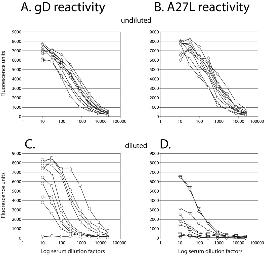
The individual sera titers elicited by mice immunized with the gD and A27L expressing LS constructs were also determined so as to facilitate a comparison of the diluted and undiluted genetic inocula shown in Figure 2A and 2B. Importantly, the results plotted in Figure 3 corroborate the pooled sera data in that differences in response levels are not linearly proportional to differences in the inoculating dose. (Compare Fig. 2A versus Fig. 2B; Fig. 3A versus Fig. 3C; Fig. 3B versus Fig. 3D.) Namely, a 250-fold reduction in the dose of gD and A27 expressing plasmids led to only an approximate 10-fold reduction in the elicited titers. Responder frequencies were also assessed. Without dilution all animals responded (10 of 10 for both gD and A27L), and even with a 250-fold dose dilution responder frequencies were high (9 of 10 for gD; 6 of 10 for A27L).
Fig. 3. Individual sera analysis of pools of antigen-expressing test constructs.
To discern responder frequencies, the sera from the mice described in Figure 2A and 2B were individually tested by fluorescent ELISA. Reactivities are shown against HSV1 glycoprotein (g)D in sera from mice immunized with the LS test constructs undiluted (A) or in mice immunized with the test constructs diluted into a pool of 250 irrelevant ORF expression constructs (B). Similarly, VACV A27L reactivity was measured in mice immunized with the LS test constructs undiluted (C) or diluted into the same pool of 250 irrelevant expression constructs (D).
Methods for high throughput plasmid construction
Gene building techniques based on polymerase chain reactions (PCR) generate mixtures of molecular species that perfectly or imperfectly correspond to an intended gene sequence. Standard cloning procedures enable a single molecule to be isolated and amplified. After generating transformants, researchers typically purify plasmid DNAs and sequence a collection of these individual molecules to identify a bona fide sequence. For the large program presented here, these cloning and sequence-searching procedures would have rendered library production timeframe and costs untenable. As an alternative, we explored using batched recombinant-DNAs. Standard bacterial cloning involves amplification of E. coli transformants carrying a single plasmid species. By contrast if bacteria transformed with a mixed population of plasmids are co-cultured, then growth biases occur as the transformants are unequally fit. Hence, the amplified plasmid population may not reflect the original molecular diversity. Our method would be possible if i) collectively the gene building reaction products included all the desired nucleotide sequence space (and thereby the encoded epitope space) and ii) the molecular diversity of any mixed batch of bacteria could be maintained. In particular we supposed generating 150 mini-libraries, each comprised of ~30 different synthetic ORF products, non-clonally engineered into the LS and UB vectors.
To investigate how ORF-species diversity would drift during bacterial amplification, we conducted the following experiment. Sixteen different PCR-generated ORFs with known sequences were built with deoxyuracil (dU) ends and treated with uracil DNA-glycosylase (UDG) to expose 3’ overhangs. These were annealed into the LS plasmid via a complementary overhang sequence. This method of inserting DNA into a plasmid is more amenable to a high throughput protocol since restriction enzymes are not used. ORF sequences do not need to be individually analyzed to identify and accommodate internal restriction sites and no ligation reaction steps are needed. Linkage products were used to transform E. coli and cells were plated to determine the transformation efficiency by counting colonies the following day. To assess any molecular population drift, the same experiment was repeated however the transformants were not directly plated. Instead, dilutions that approximated from 106 to 102 transformants were used to inoculate an overnight culture (2ml). Aliquots of these stationary cultures were plated and colonies grown. Plasmids were purified from selected colonies and sequenced to determine the identity of the ORF species. These results are summarized in Table 1. The distribution of the 16 ORF sequences in the original transformants is shown alongside the distribution of sequences found in amplified cells. Inoculation of a culture with as few as 103 transformants was sufficient to maintain most of the population’s diversity with similar distribution. With only half as many colonies screened, all but two species in the direct plating were found in the amplified cells, and the two lost had shown low representation even in the original population. This contrasts with the lost diversity of the culture started with 102 transformants. A bottlenecking down to largely one ORF species occurred, with either zero or a single representative of all others. Based on these data, we inferred that the diversity of a mixed plasmid sample could be sufficiently maintained by inoculating growth cultures with 103 or more original bacterial transformants.
Table 1.
Validating CPV library amplification scheme
| ORF | Direct plating |
% of total |
~106 inoc. |
% of total |
~105 inoc. |
% of total |
~104 inoc. |
% of total |
~103 inoc. |
% of total |
~102 inoc. |
% of total |
|---|---|---|---|---|---|---|---|---|---|---|---|---|
| Sequence #1 | 1 | 1.3 | 1 | 3.3 | 3 | 9.7 | 5 | 15.2 | 2 | 4.9 | 0 | |
| Sequence #2 | 2 | 2.5 | 0 | 0 | 4 | 13.0 | 7 | 21.2 | 2 | 4.9 | 8 | 14.8 |
| Sequence #3 | 1 | 1.3 | 1 | 3.3 | 1 | 3.2 | 2 | 6.1 | 1 | 2.4 | 43 | 79.6 |
| Sequence #4 | 13 | 16.3 | 4 | 13.3 | 0 | 0 | 1 | 3.0 | 2 | 7.3 | 0 | |
| Sequence #5 | 5 | 6.3 | 4 | 13.3 | 0 | 0 | 0 | 0 | 1 | 2.4 | 1 | 1.9 |
| Sequence #6 | 4 | 5 | 1 | 3.3 | 5 | 16.1 | 2 | 6.1 | 3 | 7.3 | 0 | |
| Sequence #7 | 12 | 15 | 6 | 20 | 1 | 3.2 | 4 | 12.1 | 5 | 12.2 | 0 | |
| Sequence #8 | 13 | 16.3 | 3 | 10 | 3 | 9.7 | 1 | 3.0 | 9 | 22.0 | 0 | |
| Sequence #9 | 5 | 6.3 | 2 | 6.7 | 0 | 0 | 0 | 0 | 5 | 12.0 | 0 | |
| Sequence #10 | 4 | 5 | 2 | 6.7 | 5 | 16.1 | 4 | 12.1 | 4 | 7.6 | 1 | 1.9 |
| Sequence #11 | 2 | 2.5 | 1 | 3.3 | 0 | 0 | 0 | 0 | 0 | 0 | 0 | |
| Sequence #12 | 11 | 13.8 | 2 | 6.7 | 1 | 3.2 | 1 | 3.0 | 2 | 4.9 | 0 | |
| Sequence #13 | 2 | 2.5 | 0 | 0 | 0 | 0 | 0 | 0 | 1 | 2.4 | 0 | |
| Sequence #14 | 1 | 1.3 | 0 | 0 | 2 | 6.5 | 3 | 9.1 | 0 | 0 | 0 | |
| Sequence #15 | 1 | 1.3 | 3 | 10 | 1 | 3.2 | 2 | 6.1 | 3 | 7.3 | 1 | 1.9 |
| Sequence #16 | 3 | 3.8 | 0 | 0 | 5 | 16.1 | 1 | 3.0 | 1 | 2.4 | 0 | |
|
Total clones sequenced |
80 | 30 | 31 | 33 | 41 | 54 |
Construction and pooling of the synthetic cowpox expression library
The coding sequences of the CPV Brighton Red strain were recoded and organized into 789 overlapping ORFs of ~500bp in length as described in the Materials and Methods. ORF size was guided by our previous studies demonstrating that fragments of ~200 amino acids elicit a broader immune profile than do either larger or smaller ones (Sykes and Johnston, 1999). Each ORF was assembled, and then product yield was quantitated by fluorimetry and verified for length and quality by agarose gel analysis. Quantities were normalized and the 75 unique pools of ~30 ORFs were formed. The dU flanked mixtures of ORFs were annealed into both pCMViLS and pCMViUB vectors and used to transform E. coli. An aliquot of the each of the 150 transformation mixes was plated to determined transformation frequency. If at least 104 original transformants were counted then the remaining culture (2ml) was used to inoculate a growth culture (100ml). This corresponds to ten-fold more inoculating bacteria than we demonstrated were required to maintain diversity. Otherwise, the transformation was repeated. Cells were harvested at mid-log phase for plasmid purification. All 150 prepared plasmid preparations were quantitated and assessed by restriction digest to confirm DNA quality and to visualize range of inserts.
Screening ORF pools as vaccines in challenge-protection assays
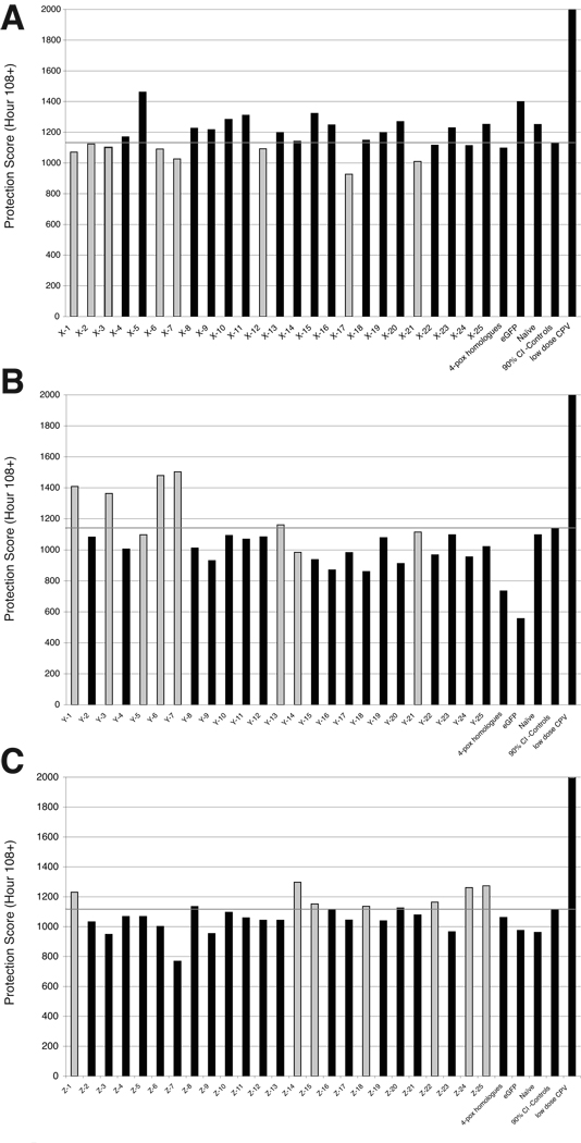
To prepare the inocula, the LS and UB plasmid preparations carrying of the same ORF pools were mixed in equal proportion. These 75 DNA samples were precipitated onto gold microprojectiles and biolistically delivered into C57BL/6 mice. Two negative control groups were immunized with a plasmid expressing green fluorescent protein (GFP) or naive. A positive control group was immunized with of a sublethal dose of live CPV. Another group was genetically immunized with the CPV homologues of the VACV 4-pox vaccine constituents A27L, A33R, B5R, and L1R. Animals were boosted twice, at weeks 3 and 8. At week 14 post-prime, the animals were i.t. challenged with one lethal dose (1LD100) of CPV. This dose was defined as the lowest dose of virus required to kill 100% of the control animals in a titration experiment conducted 2 weeks earlier with the same viral stock. Challenges were cascaded as three sub-experiments X, Y, and Z, with controls in each set. Animal survival was checked at approximately 12-hour intervals and data recorded twice daily due to the rapid course of the disease. The hours survived by each mouse within all 75 experimental and control groups were analyzed by two methods. First a single value was also assigned to each group as its protection score by calculating an average survival time. This was obtained by multiplying the time-value (in hours post challenge) at an observation by the number of mice alive at that time-value. This total survival time-value was divided by the starting number of mice to derive an average group survival time, which would be the group’s protection score. The data from the two negative control groups were combined into a single control group for data analyses. These data are plotted for all groups within the X (Fig. 4A), Y (Fig. 4B), and Z (Fig. 4C) libraries. A line is drawn at the lower (X plot) or upper (Y and Z plots) values of the 90% confidence interval. The protection scores were used to rank order each group within the three libraries; both score and rank are displayed in Table 2. As a second analysis, a Mantel-Haenszel survival curves (Prism 4.0, Graphpad Software, San Diego, CA) were determined in order to calculate p values and identify groups with significantly extended times to death. The Y and Z groups shown in bold were significantly protected compared to the negative controls (p <0.05) and those in bold italics had a trend towards protection (p < 0.10). There were no significantly protective groups from the X library; therefore, we could not select any positives. In general mice in the X library lived longer than mice in the Y and Z libraries, suggesting a weaker challenge was administered to the X set. Nonetheless, we could statistically eliminate eight of the groups as negative. Namely, the groups shown in bold (p < 0.05) or bold italics (p < 0.10) succumbed more quickly than the negative controls. Since the remaining 17 could not be eliminated from the screen, all of these groups were included in our next round of screening, and are shaded in Table 2. By contrast, several groups in the Y and Z sets displayed statistically improved survival and all but one of these rank-sorted as the top seven protective groups by scoring. We selected the top seven groups of the Y and Z libraries and one rank-outlying but statistically-protective group Y-14 to carry forward. These groups are shaded in Table 2. In the Z library another three groups scored within the 90% confidence interval; however we did not carry these forward because they were not significantly different from the controls by survival curve analysis. Since the ORFs comprising the selected X, Y, and Z pools are known, a matrix, overlap analysis was performed. All ORFs commonly found within the three lists of ORFs comprising the selected X, Y, and Z pools were identified. This can be envisioned as a triangulation of positive X, Y, and Z coordinates and indicated 48 ORFs as possibly responsible for conferring the observed level of protection. This output is a function of the number of selected groups and the percent of loci assigned ORFs within the virtual cube that was used for arraying. If we assume each pool contains no more or less than one protective ORF, the anticipated number of protective ORFs among the 48 intersections should approximate the smallest number of groups selected from any one library.
Fig. 4. ELI screen protection scores.
Expression libraries built from these ORF pools were evaluated in a murine pulmonary challenge-protection assay. Each set of 25 groups represented the complete library and was designated X1–X25 (A), Y1–Y25 (B), or Z1–Z25 (C). C57BL/6 mice (10 per group) were challenged i.t. as described in Materials and Methods and monitored twice daily for 21 days. A protection score for each of the 75 groups was determined and plotted against controls. A27L/A33R/L1R/B5R is a mixed inoculum comprised of the cowpox homologues of the 4-component vaccinia vaccine candidate (4-pox). The eGFP plasmid expresses a codon-optimized green-fluorescence gene used as an irrelevant construct. Naïve and low-dose cowpox immunized groups are negative and positive controls, respectively. Groups significantly different from the negative control group are shown as gray bars. Note that for sets Y and Z, groups with significant protection are shown in gray. By contrast for set X, groups displaying enhanced disease are shown in gray. The 95% confidence interval relative to the irrelevant vaccine controls is shown. Nonparametric tests using the Mantel-Haenszel Logrank test showed identical or nearly so values. Risks significantly greater than the average odds-ratio were considered as candidates. All survival data were considered both parametrically and nonparametrically in order to prevent bias due to non-normal data distribution. Due to space consideration, survival plots are not shown.
Table 2.
| Group | Score | XRank | Group | Score | YRank | Group | Score | ZRank |
|---|---|---|---|---|---|---|---|---|
| X-5 | 1460 | 1 | Y-7 | 1504 | 1 | Z-14 | 1297 | 1 |
| X-15 | 1322 | 2 | Y-6 | 1478 | 2 | Z-25 | 1273 | 2 |
| X-11 | 1310 | 3 | Y-1 | 1409 | 3 | Z-24 | 1260 | 3 |
| X-10 | 1284 | 4 | Y-3 | 1364 | 4 | Z-1 | 1230 | 4 |
| X-20 | 1268 | 5 | Y-13 | 1160 | 5 | Z-22 | 1164 | 5 |
| X-25 | 1252 | 6 | Y-21 | 1114 | 6 | Z-15 | 1152 | 6 |
| X-16 | 1247 | 7 | Y-5 | 1097 | 7 | Z-18 | 1135 | 7 |
| X-23 | 1229 | 8 | Y-23 | 1097 | 8 | Z-8 | 1134 | 8 |
| X-8 | 1225 | 9 | Y-10 | 1092 | 9 | Z-20 | 1126 | 9 |
| X-9 | 1217 | 10 | Y-12 | 1083 | 10 | Z-16 | 1115 | 10 |
| X-19 | 1198 | 11 | Y-2 | 1081 | 11 | Z-10 | 1096 | 11 |
| X-13 | 1196 | 12 | Y-19 | 1077 | 12 | Z-21 | 1079 | 12 |
| X-4 | 1170 | 13 | Y-11 | 1069 | 13 | Z-4 | 1068 | 13 |
| X-18 | 1148 | 14 | Y-25 | 1021 | 14 | Z-5 | 1068 | 14 |
| X-14 | 1141 | 15 | Y-8 | 1012 | 15 | Z-11 | 1059 | 15 |
| X-2 | 1123 | 16 | Y-4 | 1004 | 16 | Z-17 | 1044 | 16 |
| X-22 | 1116 | 17 | Y-14 | 984 | 17 | Z-13 | 1043 | 17 |
| X-24 | 1113 | 18 | Y-17 | 983 | 18 | Z-12 | 1042 | 18 |
| X-3 | 1100 | 19 | Y-22 | 968 | 19 | Z-19 | 1039 | 19 |
| X-12 | 1091 | 20 | Y-24 | 954 | 20 | Z-2 | 1032 | 20 |
| X-6 | 1091 | 21 | Y-15 | 937 | 21 | Z-6 | 1001 | 21 |
| X-1 | 1070 | 22 | Y-9 | 931 | 22 | Z-23 | 966 | 22 |
| X-7 | 1026 | 23 | Y-20 | 911 | 23 | Z-9 | 953 | 23 |
| X-21 | 1009 | 24 | Y-16 | 870 | 24 | Z-3 | 948 | 24 |
| X-17 | 926 | 25 | Y-18 | 859 | 25 | Z-7 | 769 | 25 |
Footnotes:
Shaded groups were carried into analysis for antigen identification
Groups in bold were statistically different from controls at p <0.05
Groups in bold italics showed a statistical trend as different from controls at p < 0.10
Groups in X library were statistically analyzed to remove those groups showing enhanced disease relative to negative controls. By contrast, the Y and Z libraries were statistically analyzed to identify those groups showing extended survival relative to the negative controls.
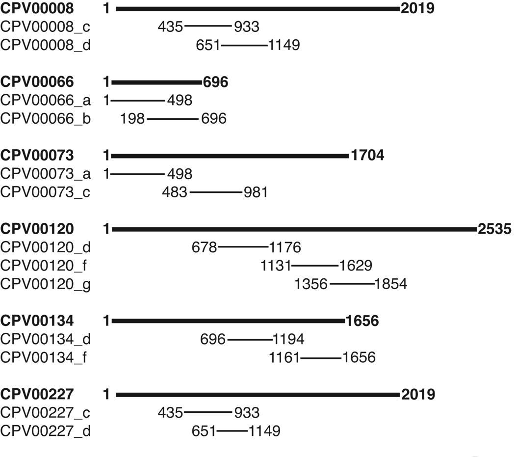
None of the CPV homologues (CPV 165, CPV 172, CPV 204 and CPV 106) of the VACV 4-pox inoculum (A27L, A33R, B5R, and L1R) were identified by our analysis of the survival data. Consistent with this result, the CPV 4-pox homologues did not show protection in the model even when delivered directly as an undiluted vaccine (Fig. 1 and Fig. 4). Among the 48 inferred candidates, five do not have variola virus homologues, and one of these is not found in variola or vaccinia. These ORFs may warrant investigation as CPV or MPV candidates; however, we chose to concentrate our most immediate efforts on the ORFs most likely to comprise a smallpox vaccine. Among the remaining ORFs, several were found to be different portions of the same gene, such that 13 of the ORFs correspond to only 5 genes. Furthermore, these ORFs have overlapping sequences. We suggest this indicates that the protection screen sorted the coding sequences non-randomly. Fig. 5 displays a mapping of these ORFs with respect to their derivative genes. Analysis also established that CPV008 and CPV227 are two independent loci carrying identical genes. The ELI screen identified both of the gene duplicates, and was able to identify the same two overlapping ORFs within them. This redundancy indicates that a complete screening of the library was accomplished. With the final removal of one of these chromosomal duplicates we arrived at a shortlist of 41 ORFs, and 36 of these were selected to test as individually delivered vaccine candidates in the CPV-C57BL/ 6 protection assay.
Fig. 5. Multiple ELI-selected ORFs map to the same genes.
The genomic locations of the 36 individual CPV ORFs selected by matrix analysis of the protection assay results were determined. Sets of ORFs were identified that correspond to distinct but overlapping regions of the same gene. These genes are shown, represented as bold bars; the ORFs are drawn below the corresponding gene with its nucleotide endpoints indicated.
Identification of cowpox ORFs that individually confer protection against a lethal pulmonary challenge
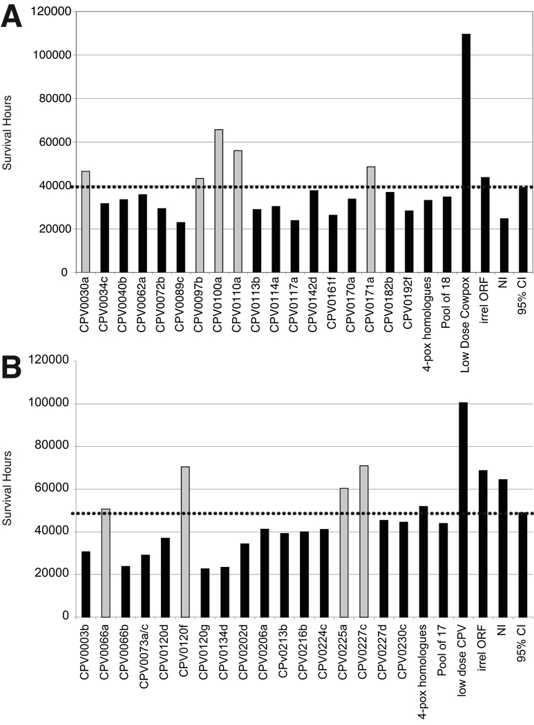
The 36 CPV gene fragments identified in the ELI screen were synthetically regenerated and inserted into the LS and UB plasmids. These ORF-expressing constructs were now clonally purified by standard bacterial transformation and colony growth. Both UB and LS expression constructs for each ORF were isolated and sequenced to identify bona fide DNAs. UB and LS constructs carrying the same ORFs were mixed at equal molar ratios. CPV073 fragments a and b were combined since they are derived from the same gene and shared only minimal overlap (15 nucleotides) such that combining them would not skew epitope doses. The DNAs were precipitated onto gold microprojectiles, as before, and biolistically delivered into groups of 10 C57BL/6 mice with a Helios gene gun (BioRad, Inc). Control groups were immunized with a low dose infection of live virus (positive), an irrelevant gene vaccine from B. anthracis, (negative), or unimmunized (negative). Animals were immunized with 2 × 1µg shots at weeks 0, 3 and 8. The test groups were randomly split into two phases for the challenges. At week 13 or 14 post-prime, mice were challenged by i.t. route with a lethal dose of CPV, and then monitored at 12 h intervals for 14 days. Deaths were recorded and survival hours tabulated. Average survival hours for each group were calculated and compared to that of all test samples. The protection scores are graphed in Fig. 6A and B, and the 95% confidence interval is delineated with a dotted bar. As before, the CPV homologues of the VACV 4-pox inoculum did not confer significant protection in this murine-cowpox model of pulmonary disease; however, the live virus vaccine conferred strong protection. The ORFs tested in each phase were also combined and delivered into mice as a pooled inoculum. Neither of these collections of protective and non-protective ORFs conferred significant protection. By contrast, nine of the individually delivered ORFs did confer C57BL/6 significant protection against a lethal pulmonary CPV infection.
Fig. 6. ORFs selected in the ELI screen were individually tested as vaccines in the challenge-protection assays.
Analyses of the protection scores identified a shortlist of ORFs to further test as single-gene inocula. Groups of C57BL/6 mice (10 per group) were immunized with one plasmid, without adjuvant, and then i.t. challenged with a previously titrated lethal dose of CPV. Differences in cumulative survival hours were compared using the difference in means between control samples and test samples. Confidence intervals of the resulting difference in means (control minus test) that did not include 0 were considered significant and are colored grey. The dotted bar displays the upper limit of the 95% confidence interval for the test groups.
Analysis of vaccine candidates
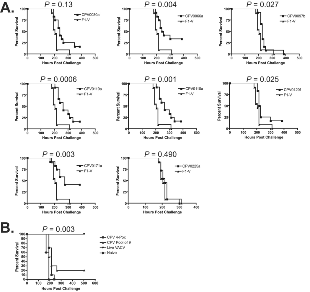
In Table 3, the identities of all nine cowpox genes selected as candidates are presented alongside the corresponding VACV and VARV genes. Known or putative functions are indicated, based on homologies to previously characterized proteins; two ORFs encode hypothetical proteins. To confirm these protection results, a new similarly designed experiment was initiated. CPV227c was left out of this round because it is a putative host range factor (Antoine et al., 1998), and therefore may not be broadly useful against the poxvirus family. Homologues, however, may be useful as an orthopox-specific antigen. The remaining eight plasmid constructs expressing the ORFs displaying protection in the first experiment were again precipitated onto gold and prepared for biolistic immunization of C57BL/6 mice (12 per group). Controls were similar to the previous experiments except the irrelevant ORF was a gene encoding a protective chimeric antigen from an irrelevant pathogen (Y. pestis F1-V). A similar immunization regimen was carried out and at week 13 post prime the mice were i.t. challenged with 1LD100 of CPV. The survival results of the test groups were analyzed against survival of mice immunized with the irrelevant gene vaccine, F1-V, and plotted in survival graphs (Fig. 7A). Using the Mantel-Haenszel survival curve analysis, p-values were calculated and shown above each plot. All but two of the candidates (CPV 030 and CPV 225) showed reproducibly significant protection (p values of ≤ 0.027).
Table 3.
Bioinformatic analysis of confirmed protective CPV candidates.
| CPV ORF | VACCINIA HOMOLOGUE |
VARIOLA HOMOLOGUE |
FUNCTION OR HOMOLOGY |
SURVIVAL1 (P-VALUE) |
|---|---|---|---|---|
| CPV0030a | C6L | D9L | hypothetical protein | 0.013 |
| CPV0066a | F16L | C20L | hypothetical protein | 0.004 |
| CPV0097b | G7L | H7L | core protein | 0.027 |
| CPV0100a | G9R | H9R | myristylation site, trans-membrane protein involved in entry/fusion |
0.0006 |
| CPV0110a | J5L | L5L | myristylation site, trans-membrane protein of unknown function |
0.001 |
| CPV0120f | D1R | F1R | mRNA capping enzyme; termination and initiation factor |
0.025 |
| CPV0171a | A32L | A35L | GTP-ATPase, DNA packaging, virion assembly |
0.003 |
| CPV0225a | C3L | G4R | Tumor necrosis factor receptor (TNFR) domain, binds secreted complement C3b/C4b |
0.490 |
| CPV0227c | C9L | B19R | ankyrin-like repeats, host range and tissue tropism, pathogenesis |
n.d.2 |
Footnotes:
p-values were calculated by a Mantel-Haenszel log-rank survival curve analysis
In the first challenge-protection assay, CPV0227c conferred protection above the 95% confidence interval, but it was not included in the confirmatory study.
Fig. 7. Confirmation of protective capacities of new C57BL/6 vaccine candidates.
A. Survival of mice immunized with individual CPV gene vaccine candidates and challenged in an independently conducted protection experiment. Mantel- Haenszel survival plots (Prism, GraphPad Inc, LaJolla, CA) display differential survival rates of mice immunized with one of eight CPV vaccine candidates relative to mice immunized with a gene expressing an irrelevant vaccine antigen, F1-V. The calculated p values for each candidate are provided above the corresponding survival curve. Ten mice per group were used; an a posteriori power analysis indicates 6 mice per group were needed to observe differences in cumulative survival of less than 10% at 95% confidence and 80% power.
B. Evaluation of a pool of the top nine ORF candidates in the vaccine assay. Mice (15 per group) were immunized with the ORFs identified in the ELI screen alongside positive and negative controls: live vaccine and naïve mice, respectively. Survival results following a lethal CPV challenge are displayed as Mantel- Haenszel plots. Data was analyzed as above.
In another experiment, constructs expressing the nine ORFs selected from the ELI screen were pooled and biolistically delivered into C57BL/6 mice using an abbreviated regimen relative to the prior experiment. For comparison, a group was immunized with the CPV homologues of 4-pox. A lethal i.t. challenge was administered six weeks post prime, and survival was monitored. These results are plotted in Figure 7B along with positive (live VACV) and negative (naive) control groups, and show that the pool of 9 CPV ORFs was partially protective in the C57BL/6 strain (p = 0.003), whereas co-delivery of homologous CPV 4-pox genes was not.
Host immune responses to candidate vaccine antigens
A preliminary investigation of the immunogenicity of the nine ORFs as gene vaccines was conducted. Since no pre-screen for immunogenicity was performed, the level or type of B and T cell immune responses elicited by these ORFs would be unknown. Mice were individually gene gun immunized with the nine ORFs and then boosted with either DNA or protein as described in Material and Methods. Blood and spleens were harvested ten days later. Antibody responses to the antigens were assessed in ELISAs or immunoblots by measuring reactivity of sera to peptides or protein antigen, respectively. These antibody results, summarized in Table 4, indicate that all of the tested candidates stimulated production of antigen-specific antibodies, as measured by one or both of the assays. For comparison of sera reactivities to know orthopox immunogens, the CPV homologues of the VACV antigens CPV 165 (VACV A27L) and CPV172 (VACV A33R) were also expressed in E. coli and used to evaluate sera from mice immunized with the corresponding mammalian expression constructs by immunoblot. The relative number of animal responders within a group was similar between the new candidates (6/6, 3/6, 5/6, and 6/6) and two 4-pox genes (2/4 for CPV 165 and 4/4 for CPV 172).
Table 4.
Summary of Antibody responses to CPV vaccine candidates
| Candidate | Mouse # | ELISA | Immunoblot | |
|---|---|---|---|---|
| Responders | Titer | Responders | ||
| CPV 030a | 1 | 3/4 | 800 | 6/6 |
| 2 | 800 | |||
| 3 | 0 | |||
| 4 | 0 | |||
| 5 | ND | |||
| CPV 066a | 1 | 1/4 | 0 | 3/6 |
| 2 | 0 | |||
| 3 | 0 | |||
| 4 | 1600 | |||
| 5 | ND | |||
| CPV 097b | 1 | 4/5 | 400 | |
| 2 | 200 | |||
| 3 | 1600 | |||
| 4 | 6400 | |||
| 5 | 0 | |||
| CPV 100a | 1 | 3/4 | >6400 | |
| 2 | 0 | |||
| 3 | 1600 | |||
| 4 | 1600 | |||
| 5 | ND | |||
| CPV 110a | 1 | 2/4 | 6400 | 5/6 |
| 2 | 0 | |||
| 3 | 0 | |||
| 4 | 400 | |||
| 5 | ND | |||
| CPV 171a | 1 | ND | 6/6 | |
| 2 | ||||
| 3 | ||||
| 4 | ||||
| 5 | ||||
| CPV 225a | 1 | 5/5 | 3200 | |
| 2 | 12800 | |||
| 3 | 3200 | |||
| 4 | 6400 | |||
| 5 | 800 | |||
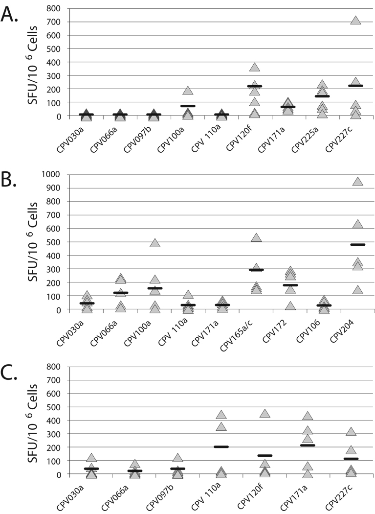
T cell responses were assessed by measuring the number of cells that secreted interferon-γ (IFN- γ) in ELISPOT assays. CD4+ and CD8+ T cell responses to the nine new CPV antigens and homologues of the 4-pox antigens were determined by enumerating the number of splenocytes harvested from immunized mice stimulated with either peptide or protein antigens. Recombinant protein purified as inclusion bodies or overlapping peptides synthesized to span the protein sequence were used. The graphs in Figure 8A and 8B display the number of T cells from individual immune mice that secreted IFNγ in response to peptide antigen, and group averages, from two independently conducted experiments. The experiment displayed in Figure 8B includes the homologues of the VACV vaccine candidates, CPV 165 (A27L), CPV 172 (A33R), CPV 106 (L1R), and CPV 204 (B5R) for comparison. In Figure 8C, ELISpot analysis of IFNγ released from splenocytes stimulated with recombinant protein, or peptides (CPV 100a). In general, protein antigen stimulated more T cells than did the sets of peptide antigen. Immune T cells specifically responded to all nine CPV antigens, as either peptide or protein.
Fig. 8. T cell responses elicited by orthopox subunit vaccine candidates.
A. Survey of peptide-stimulated T cell responses of mice individually immunized with the CPV vaccine candidates. Splenocytes from immunized mice were harvested and stimulated with a peptide pool comprised of 20-mers tiling the ORF-encoded product. IFNγ release of activated T cells was measured by ELISpot. Data are presented as the number of antigen-specific Spot Forming Units (SFU) per million splenocytes. SFU counts were adjusted for background by subtracting the number of spots in wells containing unstimulated splenocytes (no antigen). Triangular icons represent splenocyte results from individual mice. Black bars display group mean SFUs. Error bars reflect the standard deviation between quadruplicate assay wells.
B. Comparison of peptide-stimulated T cell responses of mice immunized with a CPV ORF or a CPV homologue of 4-pox vaccine candidates. Splenocytes from immunized mice were harvested and stimulated as above. CPV homologues of the VACV 4-pox genes are as follow: CPV 165a/c (VACV A27L); CPV172 (VACV A33R); CPV 106 (VACV L1R); CPV 204 (VACV B5R).
C. T cell cytokine release levels of candidate-immunized mice in response to protein-antigen stimulation. Splenocytes were stimulated with 20µg of recombinant protein, except for CPV100a-immune cells which were stimulated with peptide. IFNγ release of activated T cells was measured by ELISpot. Data are presented as the number of antigen-specific Spot Forming Units (SFU) per million splenocytes. SFU counts were adjusted for background by subtracting the number of spots in wells containing unstimulated splenocytes (no antigen). Icons represent splenocyte results from individual mice. Black bars display group mean SFUs. Error bars reflect the standard deviation between quadruplicate assay wells.
DISCUSSION
Non-variola models of smallpox, if not perfect (Precopio et al., 2007), are necessary because humans are the only variola host and natural disease is eradicated. Since efficacy against smallpox cannot be directly tested, a practical approach to subunit discovery is to identify a panel of protective antigens that are most likely to be broadly effective in different hosts and against different orthopoxviruses. To date, several subunits have been identified and characterized as protective predominantly using a vaccinia-BALB/c mouse model of disease. Our rationale for further antigen discovery was the observation that the VACV 4-pox subunit inoculum is not protective in another mouse strain, C57BL/6. By accumulating more antigens we would be prepared for candidate attrition during the vaccine development process.
In the work presented here, an evaluation of all protein subunits of cowpoxvirus as vaccines was conducted in a natural host. To our knowledge, this is the first functional screening of a complete synthetic proteome in vivo. Expression library immunization assumes that individual antigens can be productively immunogenic in the presence of others. This parallels the complexity of a live or killed whole-pathogen vaccine or a pathogen infection, in which the immune system is exposed to a mixture of many foreign components. All antigens are not anticipated to be universally detected in this context but since many can be detected, we have a means of identifying useful immunogens from any class of protein, encoded by any known or unknown gene.
Building all the coding sequences as codon-optimized versions allowed us to normalize antigen expression levels for the screen; placing each ORF in two mammalian expression vectors allowed us to dually target the antigens intracellularly to facilitate broad immune presentation. The relevance of any viral component to disease protection was evaluated in a vaccine assay. All ORF expression constructs were delivered into different groups of mice, and all these genetically immunized animals were challenged with a lethal pulmonary dose of CPV. Survival readouts were used to establish the value of the ORF inocula. From an original library of 789 components, an initial shortlist of 41 ORFs was honed.
This shortlist holds a number of interesting features. The gene sources of these ORFs suggest that the screen was both non-random and complete. In particular, 5 genes were the source of 13 screen-selected ORFs, including a pair of gene duplicates. CPV 008/227 ORF carries an ankyrin repeat motif, indicating a region of protein-protein interaction. This class of proteins has been shown to influence viral pathogenesis, host range, and tissue tropism (McFadden, 2005; Shchelkunov et al., 2000). CPV120, which is the source-gene of three different ORFs on the shortlist, is homologous to VACV protein D1R. It is transcriptional termination factor VTF (Jing et al., 2005) and a subunit of the mRNA capping enzyme (Myette and Niles, 1996). Within D1R, a strong human CD8 epitope has been identified (Jing et al., 2005), and is one of only a few viral proteins recognized by CD8+ T cells across MHC and species boundaries (Oseroff et al., 2008). The other three genes that were repeatedly identified do not encode proteins with high homology to any characterized protein domains in the BLAST databases. The identification by protection assay of more than one region within the same protein may indicate that these are immunoprevalent antigens, potentially serving as rich sources of protective epitopes. Although no function is known, CPV 073 is the homologue of the VACV and VARV gene E6R, which is highly conserved in all currently sequenced poxviruses. This feature would be anticipated for a broadly cross-protective antigen. CPV 066 and CPV 134 correspond to VACV genes F16L and D13L, and to VARV genes E16L and N3L, respectively. A number of the ORFs indicated by the screen have no sequence homology with any known protein or protein domain, and therefore would have been difficult to identify as vaccine candidates by any rational design program. These results corroborate our original hypothesis that a function-based screen for vaccines might reveal candidates that would not have been informatically identified with current algorithms.
Consistent with the lack of protection observed by the 4-pox inoculum in C57BL/6 mice, the CPV homologues of these VACV envelope genes (A27L, A33R, B5R and L1R) are not found on the ELI shortlist. The other antigens previously described as protective in the VACV or ECTV models were also not found. It is possible that these antigens do not translate to the CPV as protective antigens. Alternatively, they may indeed hold some protective capacity for CPV but at a level not detectable in our assay, in which pooled genes were delivered without adding adjuvants or immunomodulators.
From the shortlist, 36 ORFs were tested as individual genes in the protection assay. Nine were found to be protective, and eight of these ORFs were selected for retesting. CPV227 was not retested at this time because its VACV homologue, C9L, is known to be a host range factor. This suggests that it may be relatively unconserved and therefore not cross-protective against other poxviruses nor protective in other animal models of disease, unless homologues are used. In the second independently conducted challenge-protection assay, six of eight tested candidates were confirmed as conferring significant levels of protection. We based our selection of ORFs on identifying those that reside in three positively scored inocula: 1 X, 1 Y, and 1 Z. Given the number of groups calculated as positive from each X, Y, and Z library (17, 8, and 7, respectively) and assuming each selected pool carries one and only one protective antigen, approximately seven positive ORFs is predicted.
Five of the nine ORFs initially found to be protective encode homologues of proteins known to be expressed within the IMV particles of VACV or VARV (CPV 30, CPV 097, CPV 100, CPV 110, and CPV120) but only two of these (CPV 100, CPV 110) are envelope proteins (Chung et al., 2006). Little is known about the location of hypothetical protein CPV 030. CPV 097 (VACV G7L) is homologous to a VACV virion structural core protein (Szajner et al., 2003). Both CPV 100 and CPV 110 carry consensus myristylation sites. This lipid modification is typically involved in directing and anchoring proteins to membranes, and indicates involvement in membrane targeting and signal transduction (Boutin et al., 1997). The previously identified VACV protective antigen L1R is also a myristylated protein (Wolffe, Vijaya, and Moss, 1995). The VACV homologue of CPV 100 (VACV G9R) is an essential component of the entry/fusion complex (Ojeda, Domi, and Moss, 2006; Ojeda, Senkevich, and Moss, 2006). The function of CPV 110 (VACVJ5L) is unknown but it appears to be essential for VACV multiplication (Zajac, Spehner, and Drillien, 1995). CPV 120 is VACV D1R as described above. Among the remaining ORFs, the only other membrane protein is CPV 225. CPV225 (VACV C3L) carries homology to a secreted TNF receptor, Crm-B (Alcami et al., 1999). Secreted TNFR’s have been shown to influence host immune responses to VACV. These immune modifiers are exposed to both intracellular and extracellular environments. VACV deletion of its homologue of CPV 225 lead to decreased virulence (Thornburg et al., 2007). No function has been implicated for CPV066. The VACV homologue of CPV 171 (VACV A32L) is a GTP-ATP binding protein believed to be involved in DNA packaging and virion assembly (Afonso et al., 2000). In sum, a cross section of both surface and non-surface proteins were identified, which suggests that both categories of viral components may serve as vaccine targets.
We screened for individually protective ORFs; we did not screen for combinations of ORFs that may additively or synergistically provide significant protection. Although we tested the top nine candidates as a mix, each ORF was identified from different sub-libraries and tested for its capacity to individually protect. We consider the possibility that some other candidates within the list of screen selected ORFs may require the context of one or more components of their original sub-library to confer protection. For example, genes CPV 073 and CPV 134 were each identified twice in the ELI screen, in which the ORFs were delivered as mixed inocula of ~30 other ORFs. When these two ORFs were tested individually they did not confer protection. CPV 073 and CPV 134 may require immunological help or specific combination with another gene(s) in order to be protective. By contrast, the other genes that were the source of multiple screen-selected ORFs did confer significant partial protection as individual inocula. Since the nine candidates were randomly assembled, with respect to immune cooperation, it is not surprising that they are not synergistic.
All nine molecular vaccine candidates selected in the screen were shown to specifically stimulate both antibody and T cell immune responses. This is consistent with previous work that suggests both humoral and cellular components of adaptive immunity are important against poxviruses (Ferrier-Rembert et al., 2007; Smith and McFadden, 2002). We assayed responses against the antigen itself rather than the whole virus so as to avoid biasing our interpretation of the ORF’s immune potential due to differences in location or stochiometry within the virus. Upcoming studies will characterize these responses to virus and also evaluate responses stimulated against these antigens upon live virus immunization or infection.
Immunity in humans and mice elicited by live virus is characterized by the presence of circulating antiviral antibodies and relatively high frequencies of viral specific memory T and B cells (Demkowicz et al., 1996; Hammarlund et al., 2003; Xu et al., 2008). It is not clear whether these same effectors must be elicited by any successful vaccine, or rather that other mechanisms might provide immunity by an alternative route. The differing performance of the 4-pox inoculum in the Th2-biased BALB/c and Th1-biased C57BL/6 murine models will enable a unique dissection of immune activities associated with protection and non-protection. Whether any of the new candidates, individually or as a pool, also protect BALB/c mice still needs to be determined. Either way, comparison of the immune responses elicited by these inocula in the two genetically diverse host strains may suggest unique correlates of protection.
In conclusion, we searched an expression-normalized synthetic cowpox gene library for sequences that encode protective antigens. Without pre-screening, all sequences were directly evaluated in an optimized animal model for their utility as a vaccine candidate. As is typically observed for subunit vaccines, these single antigens confer partial, not strong, protection. Learning to productively combine the subsets of the nine new orthopox antigens and existing antigens would provide the opportunity to stimulate a breadth of immunity that even a live vaccine might not elicit. The wisdom of emplacing a smallpox vaccination program against a possible biothreat event continues to be controversial (Wiser, Balicer, and Cohen, 2007). If such a policy were ever to be adopted, a subunit-based product is likely to be preferred. Meanwhile, we envision more immediate, non-threat uses of an orthopox vaccine against a number of emerging poxviral diseases.
ACKNOWLEDGEMENTS
The authors are grateful for the support of members of the Center for Innovations in Medicine; in particular we thank Phillip Stafford for statistical analysis and Preston Hunter for work on the figures. We thank Hangil Park for his library efforts. We thank Julie Lovchik for her project management at the UNM Health Sciences Center.
This work was supported by Public Health Service grant U54 AI057156 from the National Institutes of Health to K.S.
Footnotes
Publisher's Disclaimer: This is a PDF file of an unedited manuscript that has been accepted for publication. As a service to our customers we are providing this early version of the manuscript. The manuscript will undergo copyediting, typesetting, and review of the resulting proof before it is published in its final citable form. Please note that during the production process errors may be discovered which could affect the content, and all legal disclaimers that apply to the journal pertain.
REFERENCES
- Afonso CL, Tulman ER, Lu Z, Zsak L, Kutish GF, Rock DL. The Genome of Fowlpox Virus. J. Virol. 2000;74(8):3815–3831. doi: 10.1128/jvi.74.8.3815-3831.2000. [DOI] [PMC free article] [PubMed] [Google Scholar]
- Alcami A, Khanna A, Paul NL, Smith GL. Vaccinia virus strains Lister, USSR and Evans express soluble and cell-surface tumour necrosis factor receptors. J Gen Virol. 1999;80(Pt 4):949–959. doi: 10.1099/0022-1317-80-4-949. [DOI] [PubMed] [Google Scholar]
- Antoine G, Scheiflinger F, Dorner F, Falkner FG. The Complete Genomic Sequence of the Modified Vaccinia Ankara Strain: Comparison with Other Orthopoxviruses. Virology. 1998;244(2):365–396. doi: 10.1006/viro.1998.9123. [DOI] [PubMed] [Google Scholar]
- Barry MA, Howell DPG, Anderson HA, Chen JL, Singh RAK. Expression library immunization to discover and improve vaccine antigens. Immunological Reviews. 2004;199:68–83. doi: 10.1111/j.0105-2896.2004.00143.x. [DOI] [PubMed] [Google Scholar]
- Boutin Y, Leitenberg D, Tao X, Bottomly K. Distinct biochemical signals characterize agonist- and altered peptide ligand-induced differentiation of naive CD4+ T cells into Th1 and Th2 subsets. J Immunol. 1997;159(12):5802–5809. [PubMed] [Google Scholar]
- Buller RM, Palumbo GJ. Poxvirus pathogenesis. Microbiol. Mol. Biol. Rev. 1991;55(1):80–122. doi: 10.1128/mr.55.1.80-122.1991. [DOI] [PMC free article] [PubMed] [Google Scholar]
- Chung CS, Chen CH, Ho MY, Huang CY, Liao CL, Chang W. Vaccinia virus proteome: identification of proteins in vaccinia virus intracellular mature virion particles. J Virol. 2006;80(5):2127–2140. doi: 10.1128/JVI.80.5.2127-2140.2006. [DOI] [PMC free article] [PubMed] [Google Scholar]
- Cornberg M, Sheridan BS, Saccoccio FM, Brehm MA, Selin LK. Protection against vaccinia virus challenge by CD8 memory T cells resolved by molecular mimicry. J Virol. 2007;81(2):934–944. doi: 10.1128/JVI.01280-06. [DOI] [PMC free article] [PubMed] [Google Scholar]
- Dasgupta A, Hammarlund E, Slifka MK, Fruh K. Cowpox Virus Evades CTL Recognition and Inhibits the Intracellular Transport of MHC Class I Molecules. J Immunol. 2007;178(3):1654–1661. doi: 10.4049/jimmunol.178.3.1654. [DOI] [PubMed] [Google Scholar]
- Davies DH, McCausland MM, Valdez C, Huynh D, Hernandez JE, Mu Y, Hirst S, Villarreal L, Felgner PL, Crotty S. Vaccinia Virus H3L Envelope Protein Is a Major Target of Neutralizing Antibodies in Humans and Elicits Protection against Lethal Challenge in Mice. J. Virol. 2005;79(18):11724–11733. doi: 10.1128/JVI.79.18.11724-11733.2005. [DOI] [PMC free article] [PubMed] [Google Scholar]
- Demkowicz WE, Jr, Littaua RA, Wang J, Ennis FA. Human cytotoxic T-cell memory: long-lived responses to vaccinia virus. J Virol. 1996;70(4):2627–2631. doi: 10.1128/jvi.70.4.2627-2631.1996. [DOI] [PMC free article] [PubMed] [Google Scholar]
- Dieli F, Lio D, Sireci G, Salerno A. Genetic control of C3 production by the S region of the mouse MHC. J Immunogenet. 1988;15(5–6):339–343. doi: 10.1111/j.1744-313x.1988.tb00437.x. [DOI] [PubMed] [Google Scholar]
- Drancourt M, Raoult D. Molecular insights into the history of plague. Microbes & Infection. 2002;4(1):105–109. doi: 10.1016/s1286-4579(01)01515-5. [DOI] [PubMed] [Google Scholar]
- Dunlop LR, Oehlberg KA, Reid JJ, Avci D, Rosengard AM. Variola virus immune evasion proteins. Microbes and Infection. 2003;5(11):1049–1056. doi: 10.1016/s1286-4579(03)00194-1. [DOI] [PubMed] [Google Scholar]
- Edghill-Smith Y, Golding H, Manischewitz J, King LR, Scott D, Bray M, Nalca A, Hooper JW, Whitehouse CA, Schmitz JE, Reimann KA, Franchini G. Smallpox vaccine-induced antibodies are necessary and sufficient for protection against monkeypox virus. Nat Med. 2005;11(7):740. doi: 10.1038/nm1261. [DOI] [PubMed] [Google Scholar]
- Ferrier-Rembert A, Tournier JN, Drillien R, Garin D, Crance JM. CD4+ T cells play a pivotal role in the control of cowpox virus infection in mice. Viral Immunol. 2007;20(1):214–220. doi: 10.1089/vim.2006.0091. [DOI] [PubMed] [Google Scholar]
- Flick-Smith HC, Eyles JE, Hebdon R, Waters EL, Beedham RJ, Stagg TJ, Miller J, Alpar HO, Baillie LWJ, Williamson ED. Mucosal or parental administration of mircosphere-associated Bacillus anthracis protective antigen protects against anthrax in mice. Infection & Immunity. 2002a;70(4):2022–2028. doi: 10.1128/IAI.70.4.2022-2028.2002. [DOI] [PMC free article] [PubMed] [Google Scholar]
- Flick-Smith HC, Walker NJ, Gibson P, Bullifent H, Hayward S, Miller J, Titball RW, Williamson ED. A recombinant carboxy-terminal domain of the protective antigen of Bacillus anthracis protects mice against anthrax infection. Infection & Immunity. 2002b;70(3):1653–1656. doi: 10.1128/IAI.70.3.1653-1656.2002. [DOI] [PMC free article] [PubMed] [Google Scholar]
- Golden JW, Hooper JW. Heterogeneity in the A33 protein impacts the cross-protective efficacy of a candidate smallpox DNA vaccine. Virology. 2008;377(1):19–29. doi: 10.1016/j.virol.2008.04.003. [DOI] [PMC free article] [PubMed] [Google Scholar]
- Hammarlund E, Lewis MW, Hansen SG, Strelow LI, Nelson JA, Sexton GJ, Hanifin JM, Slifka MK. Duration of antiviral immunity after smallpox vaccination. Nat Med. 2003;9(9):1131–1137. doi: 10.1038/nm917. [DOI] [PubMed] [Google Scholar]
- Henderson DA. Smallpox: clinical and epidemiologic features. Emerging Infectious Diseases. 1999;5(4):537–539. doi: 10.3201/eid0504.990415. [DOI] [PMC free article] [PubMed] [Google Scholar]
- Heraud J-M, Edghill-Smith Y, Ayala V, Kalisz I, Parrino J, Kalyanaraman VS, Manischewitz J, King LR, Hryniewicz A, Trindade CJ, Hassett M, Tsai W-P, Venzon D, Nalca A, Vaccari M, Silvera P, Bray M, Graham BS, Golding H, Hooper JW, Franchini G. Subunit Recombinant Vaccine Protects against Monkeypox. J Immunol. 2006;177(4):2552–2564. doi: 10.4049/jimmunol.177.4.2552. [DOI] [PubMed] [Google Scholar]
- Hooper JW, Custer DM, Schmaljohn CS, Schmaljohn AL. DNA vaccination with vaccinia virus L1R and A33R genes protects mice against a lethal poxvirus challenge. Virology. 2000;266(2):329–339. doi: 10.1006/viro.1999.0096. [DOI] [PubMed] [Google Scholar]
- Hooper JW, Custer DM, Thompson E. Four-gene-combination DNA vaccine protects mice against a lethal vaccinia virus challenge and elicits appropriate antibody responses in nonhuman primates. Virology. 2003;306(1):181–195. doi: 10.1016/S0042-6822(02)00038-7. [DOI] [PMC free article] [PubMed] [Google Scholar]
- Hooper JW, Thompson E, Wilhelmsen C, Zimmerman M, Ichou MA, Steffen SE, Schmaljohn CS, Schmaljohn AL, Jahrling PB. Smallpox DNA vaccine protects nonhuman primates against lethal monkeypox. Journal of Virology. 2004 doi: 10.1128/JVI.78.9.4433-4443.2004. [DOI] [PMC free article] [PubMed] [Google Scholar]
- Jacobs A. Immunity conferred by smallpox vaccine. How long does immunity last? [letter; comment.] Bmj. 2002;324(7346):1157. doi: 10.1136/bmj.324.7346.1157/a. discussion 1157. [DOI] [PMC free article] [PubMed] [Google Scholar]
- Janeway CPT, Walport M, Schlomchik M. "Immunobiology.". 6th ed. 1 vols. New York; London: Garland Science; 2005. [Google Scholar]
- Jing L, Chong TM, McClurkan CL, Huang J, Story BT, Koelle DM. Diversity in the acute CD8 T cell response to vaccinia virus in humans. J Immunol. 2005;175(11):7550–7559. doi: 10.4049/jimmunol.175.11.7550. [DOI] [PMC free article] [PubMed] [Google Scholar]
- Joklik WK. The purification fo four strains of poxvirus. Virology. 1962;18:9–18. doi: 10.1016/0042-6822(62)90172-1. [DOI] [PubMed] [Google Scholar]
- Karron RA, Wright PF, Belshe RB, Thumar B, Casey R, Newman F, Polack FP, Randolph VB, Deatly A, Hackell J, Gruber W, Murphy BR, Collins PL. Identification of a recombinant live attenuated respiratory syncytial virus vaccine candidate that is highly attenuated in infants. J Infect Dis. 191(7):1093–1104. doi: 10.1086/427813. [DOI] [PubMed] [Google Scholar]
- Kaufman DR, Goudsmit J, Holterman L, Ewald BA, Denholtz M, Devoy C, Giri A, Grandpre LE, Heraud J-M, Franchini G, Seaman MS, Havenga MJE, Barouch DH. Differential Antigen Requirements for Protection against Systemic and Intranasal Vaccinia Virus Challenges in Mice. J. Virol. 2008;82(14):6829–6837. doi: 10.1128/JVI.00353-08. [DOI] [PMC free article] [PubMed] [Google Scholar]
- LeDuc JW, Becher J. Current status of smallpox vaccine. Emerging Infectious Diseases. 1999;5(4):593–594. doi: 10.3201/eid0504.990429. [DOI] [PMC free article] [PubMed] [Google Scholar]
- Lustig S, Fogg C, Whitbeck JC, Eisenberg RJ, Cohen GH, Moss B. Combinations of Polyclonal or Monoclonal Antibodies to Proteins of the Outer Membranes of the Two Infectious Forms of Vaccinia Virus Protect Mice against a Lethal Respiratory Challenge. J. Virol. 2005;79(21):13454–13462. doi: 10.1128/JVI.79.21.13454-13462.2005. [DOI] [PMC free article] [PubMed] [Google Scholar]
- MacNeill AL, Moldawer LL, Moyer RW. The role of the cowpox virus crmA gene during intratracheal and intradermal infection of C57BL/6 mice. Virology. 2009;384(1):151–160. doi: 10.1016/j.virol.2008.10.041. [DOI] [PubMed] [Google Scholar]
- McFadden G. Poxvirus tropism. Nat Rev Micro. 2005;3(3):201–213. doi: 10.1038/nrmicro1099. [DOI] [PMC free article] [PubMed] [Google Scholar]
- Moore ZS, Seward JF, Lane JM. Smallpox. Lancet. 2006;367(9508):425–435. doi: 10.1016/S0140-6736(06)68143-9. [DOI] [PubMed] [Google Scholar]
- Moss B. Vaccinia virus: a tool for research and vaccine development. Science. 1991;252(5013):1662–1667. doi: 10.1126/science.2047875. [DOI] [PubMed] [Google Scholar]
- Moutaftsi M, Peters B, Pasquetto V, Tscharke DC, Sidney J, Bui HH, Grey H, Sette A. A consensus epitope prediction approach identifies the breadth of murine T(CD8+)-cell responses to vaccinia virus. Nat Biotechnol. 2006;24(7):817–819. doi: 10.1038/nbt1215. [DOI] [PubMed] [Google Scholar]
- Myette JR, Niles EG. Domain Structure of the Vaccinia Virus mRNA Capping Enzyme. J. Biol. Chem. 1996;271(20):11936–11944. doi: 10.1074/jbc.271.20.11936. [DOI] [PubMed] [Google Scholar]
- Nelson GE, Sisler JR, Chandran D, Moss B. Vaccinia virus entry/fusion complex subunit A28 is a target of neutralizing and protective antibodies. Virology. 2008;380(2):394–401. doi: 10.1016/j.virol.2008.08.009. [DOI] [PMC free article] [PubMed] [Google Scholar]
- Ojeda S, Domi A, Moss B. Vaccinia virus G9 protein is an essential component of the poxvirus entry-fusion complex. J Virol. 2006;80(19):9822–9830. doi: 10.1128/JVI.00987-06. [DOI] [PMC free article] [PubMed] [Google Scholar]
- Ojeda S, Senkevich TG, Moss B. Entry of vaccinia virus and cell-cell fusion require a highly conserved cysteine-rich membrane protein encoded by the A16L gene. J Virol. 2006;80(1):51–61. doi: 10.1128/JVI.80.1.51-61.2006. [DOI] [PMC free article] [PubMed] [Google Scholar]
- Oseroff C, Peters B, Pasquetto V, Moutaftsi M, Sidney J, Panchanathan V, Tscharke DC, Maillere B, Grey H, Sette A. Dissociation between epitope hierarchy and immunoprevalence in CD8 responses to vaccinia virus western reserve. J Immunol. 2008;180(11):7193–7202. doi: 10.4049/jimmunol.180.11.7193. [DOI] [PMC free article] [PubMed] [Google Scholar]
- Parrino J, Graham BS. Smallpox vaccines: Past, present, and future. Journal of Allergy and Clinical Immunology. 2006;118(6):1320–1326. doi: 10.1016/j.jaci.2006.09.037. [DOI] [PMC free article] [PubMed] [Google Scholar]
- Pickup DJ. Understanding orthopoxvirus interference with host immune responses to inform novel vaccine design. Expert Rev Vaccines. 2007;6(1):87–95. doi: 10.1586/14760584.6.1.87. [DOI] [PubMed] [Google Scholar]
- Precopio ML, Betts MR, Parrino J, Price DA, Gostick E, Ambrozak DR, Asher TE, Douek DC, Harari A, Pantaleo G, Bailer R, Graham BS, Roederer M, Koup RA. Immunization with vaccinia virus induces polyfunctional and phenotypically distinctive CD8(+) T cell responses. J Exp Med. 2007;204(6):1405–1416. doi: 10.1084/jem.20062363. [DOI] [PMC free article] [PubMed] [Google Scholar]
- Pulford DJ, Gates A, Bridge SH, Robinson JH, Ulaeto D. Differential efficacy of vaccinia virus envelope proteins administered by DNA immunisation in protection of BALB/c mice from a lethal intranasal poxvirus challenge. Vaccine. 2004;22(25–26):3358–3366. doi: 10.1016/j.vaccine.2004.02.034. [DOI] [PubMed] [Google Scholar]
- Sakhatskyy P, Wang S, Chou T-hW, Lu S. Immunogenicity and protection efficacy of monovalent and polyvalent poxvirus vaccines that include the D8 antigen. Virology. 2006;355(2):164–174. doi: 10.1016/j.virol.2006.07.017. [DOI] [PMC free article] [PubMed] [Google Scholar]
- Sakhatskyy P, Wang S, Zhang C, Chou T-H, Kishko M, Lu S. Immunogenicity and protection efficacy of subunit-based smallpox vaccines using variola major antigens. Virology. 2008;371(1):98–107. doi: 10.1016/j.virol.2007.09.029. [DOI] [PMC free article] [PubMed] [Google Scholar]
- Shchelkunov SN, Safronov PF, Totmenin AV, Petrov NA, Ryazankina OI, Gutorov VV, Kotwal GJ. The genomic sequence analysis of the left and right species-specific terminal region of a cowpox virus strain reveals unique sequences and a cluster of intact ORFs for immunomodulatory and host range proteins. Virology. 1998;243(2):432–460. doi: 10.1006/viro.1998.9039. [DOI] [PubMed] [Google Scholar]
- Shchelkunov SN, Totmenin AV, Loparev VN, Safronov PF, Gutorov VV, Chizhikov VE, Knight JC, Parsons JM, Massung RF, Esposito JJ. Alastrim Smallpox Variola Minor Virus Genome DNA Sequences. Virology. 2000;266(2):361–386. doi: 10.1006/viro.1999.0086. [DOI] [PubMed] [Google Scholar]
- Slifka MK. The Future of Smallpox Vaccination: is MVA the key? Med Immunol. 2005;4(1):2. doi: 10.1186/1476-9433-4-2. [DOI] [PMC free article] [PubMed] [Google Scholar]
- Smith GL, McFadden G. Smallpox: anything to declare? Nature Reviews. 2002;2:521–527. doi: 10.1038/nri845. [DOI] [PubMed] [Google Scholar]
- Sykes KF, Johnston SA. Genetic live vaccines mimic the antigenicity but not pathogenicity of live viruses. DNA Cell Biol. 1999;18(7):521–531. doi: 10.1089/104454999315079. [DOI] [PubMed] [Google Scholar]
- Szajner P, Jaffe H, Weisberg AS, Moss B. Vaccinia virus G7L protein Interacts with the A30L protein and is required for association of viral membranes with dense viroplasm to form immature virions. J Virol. 2003;77(6):3418–3429. doi: 10.1128/JVI.77.6.3418-3429.2003. [DOI] [PMC free article] [PubMed] [Google Scholar]
- Tang DC, DeVit M, Johnston SA. Genetic immunization is a simple method for eliciting an immune response. Nature. 1992;356(6365):152–154. doi: 10.1038/356152a0. [DOI] [PubMed] [Google Scholar]
- Thornburg NJ, Ray CA, Collier ML, Liao H-X, Pickup DJ, Johnston RE. Vaccination with Venezuelan equine encephalitis replicons encoding cowpox virus structural proteins protects mice from intranasal cowpox virus challenge. Virology. 2007;362(2):441–452. doi: 10.1016/j.virol.2007.01.001. [DOI] [PMC free article] [PubMed] [Google Scholar]
- Tscharke DC, Karupiah G, Zhou J, Palmore T, Irvine KR, Haeryfar SMM, Williams S, Sidney J, Sette A, Bennink JR, Yewdell JW. Identification of poxvirus CD8+ T cell determinants to enable rational design and characterization of smallpox vaccines. J. Exp. Med. 2005;201(1):95–104. doi: 10.1084/jem.20041912. [DOI] [PMC free article] [PubMed] [Google Scholar]
- Vanderplasschen A, Mathew E, Hollinshead M, Sim RB, Smith GL. Extracellular enveloped vaccinia virus is resistant to complement because of incorporation of host complement control proteins into its envelope. Proc Natl Acad Sci U S A. 1998;95(13):7544–7549. doi: 10.1073/pnas.95.13.7544. [DOI] [PMC free article] [PubMed] [Google Scholar]
- Viner KM, Isaacs SN. Activity of vaccinia virus-neutralizing antibody in the sera of smallpox vaccinees. Microbes and Infection. 2005;7(4):579. doi: 10.1016/j.micinf.2005.02.004. [DOI] [PubMed] [Google Scholar]
- Vollmar J, Arndtz N, Eckl KM, Thomsen T, Petzold B, Mateo L, Schlereth B, Handley A, King L, Hulsemann V. Safety and immunogenicity of IMVAMUNE, a promising candidate as a third generation smallpox vaccine. Vaccine. 2006;24(12):2065–2070. doi: 10.1016/j.vaccine.2005.11.022. [DOI] [PubMed] [Google Scholar]
- Vorou RM, Papavassiliou VG, Pierroutsakos IN. Cowpox virus infection: an emerging health threat. Curr Opin Infect Dis. 2008;21(2):153–156. doi: 10.1097/QCO.0b013e3282f44c74. [DOI] [PubMed] [Google Scholar]
- Wang S, Parker C, Taaffe J, Solorzano A, Garcia-Sastre A, Lu S. Heterologous HA DNA vaccine prime--inactivated influenza vaccine boost is more effective than using DNA or inactivated vaccine alone in eliciting antibody responses against H1 or H3 serotype influenza viruses. Vaccine. 2008;26(29–30):3626–3633. doi: 10.1016/j.vaccine.2008.04.073. [DOI] [PMC free article] [PubMed] [Google Scholar]
- Ward JI, Cherry JD, Chang S-J, Partridge S, Lee H, Treanor J, Greenberg DP, Keitel W, Barenkamp S, Bernstein DI, Edelman R, Edwards K the, A. S. G. Efficacy of an Acellular Pertussis Vaccine among Adolescents and Adults. N Engl J Med. 2005;353(15):1555–1563. doi: 10.1056/NEJMoa050824. [DOI] [PubMed] [Google Scholar]
- Weltzin R, Liu J, Pugachev KV, Myers GA, Coughlin B, Blum PS, Nichols R, Johnson C, Cruz J, Kennedy JS, Ennis FA, Monath TP. Clonal vaccinia virus grown in cell culture as a new smallpox vaccine. Nat Med. 2003;9(9):1125–1130. doi: 10.1038/nm916. [DOI] [PubMed] [Google Scholar]
- Wiser I, Balicer RD, Cohen D. An update on smallpox vaccine candidates and their role in bioterrorism related vaccination strategies. Vaccine. 2007;25(6):976–984. doi: 10.1016/j.vaccine.2006.09.046. [DOI] [PubMed] [Google Scholar]
- Wolffe EJ, Vijaya S, Moss B. A myristylated membrane protein encoded by the vaccinia virus L1R open reading frame is the target of potent neutralizing monoclonal antibodies. Virology. 1995;211(1):53–63. doi: 10.1006/viro.1995.1378. [DOI] [PubMed] [Google Scholar]
- Xiao Y, Aldaz-Carroll L, Ortiz AM, Whitbeck JC, Alexander E, Lou H, Davis HL, Braciale TJ, Eisenberg RJ, Cohen GH, Isaacs SN. A protein-based smallpox vaccine protects mice from vaccinia and ectromelia virus challenges when given as a prime and single boost. Vaccine. 2007;25(7):1214–1224. doi: 10.1016/j.vaccine.2006.10.009. [DOI] [PMC free article] [PubMed] [Google Scholar]
- Xu RH, Cohen M, Tang Y, Lazear E, Whitbeck JC, Eisenberg RJ, Cohen GH, Sigal LJ. The orthopoxvirus type I IFN binding protein is essential for virulence and an effective target for vaccination. J Exp Med. 2008;205(4):981–992. doi: 10.1084/jem.20071854. [DOI] [PMC free article] [PubMed] [Google Scholar]
- Zajac P, Spehner D, Drillien R. The vaccinia virus J5L open reading frame encodes a polypeptide expressed late during infection and required for viral multiplication. Virus Research. 1995;37(2):163–173. doi: 10.1016/0168-1702(95)00025-l. [DOI] [PubMed] [Google Scholar]


