Abstract
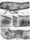
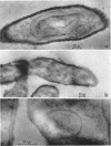
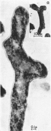
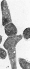
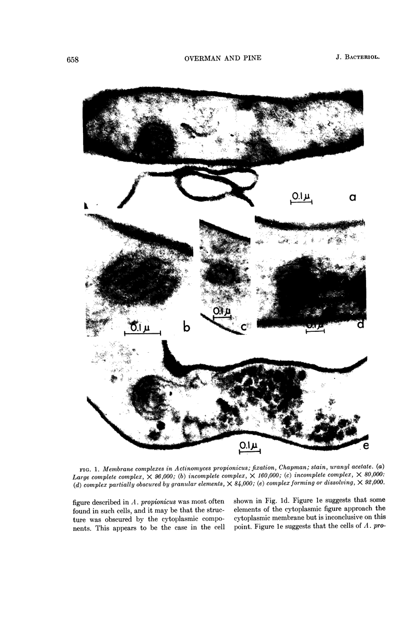
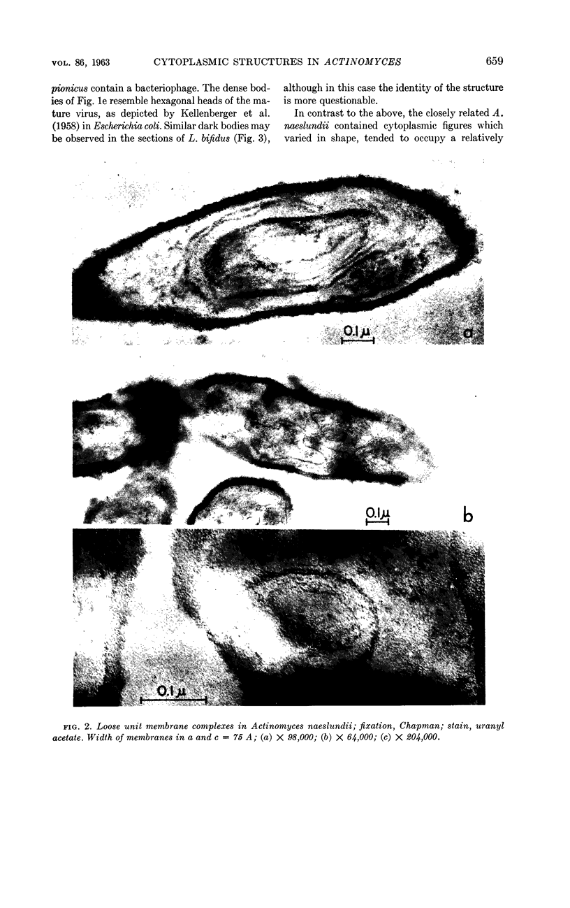
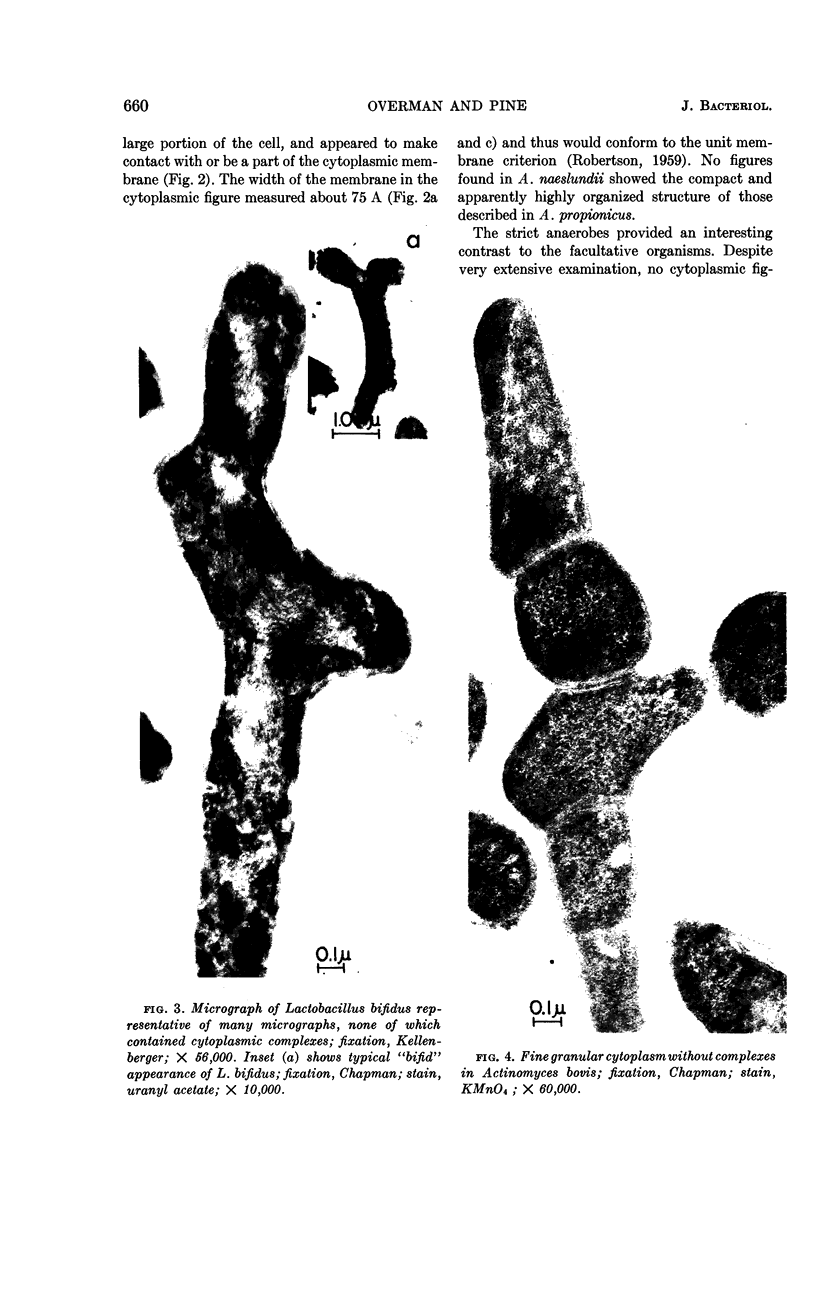
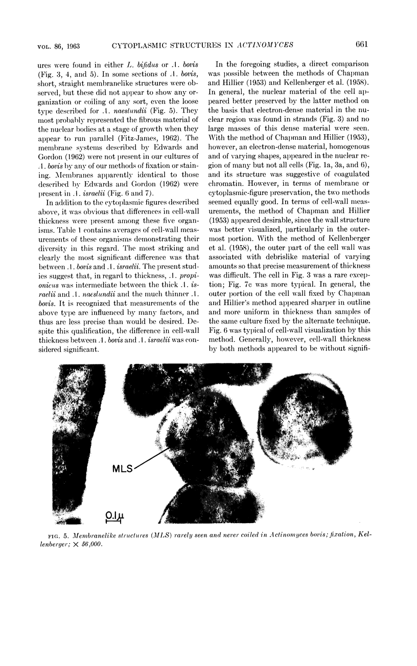
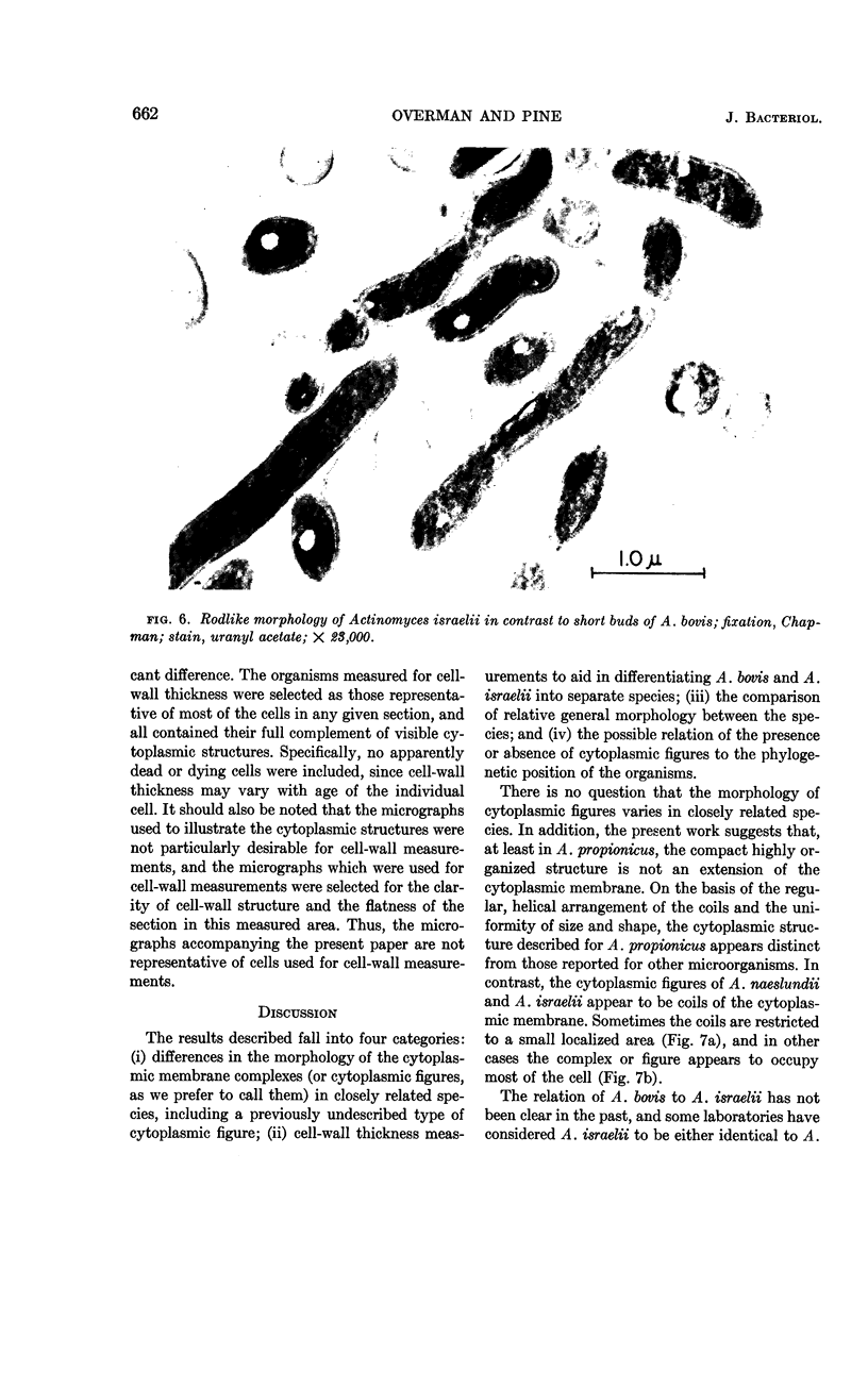
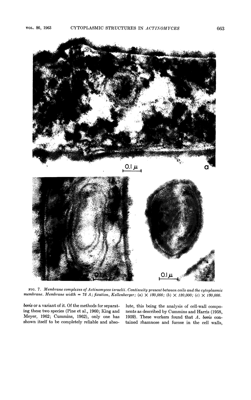
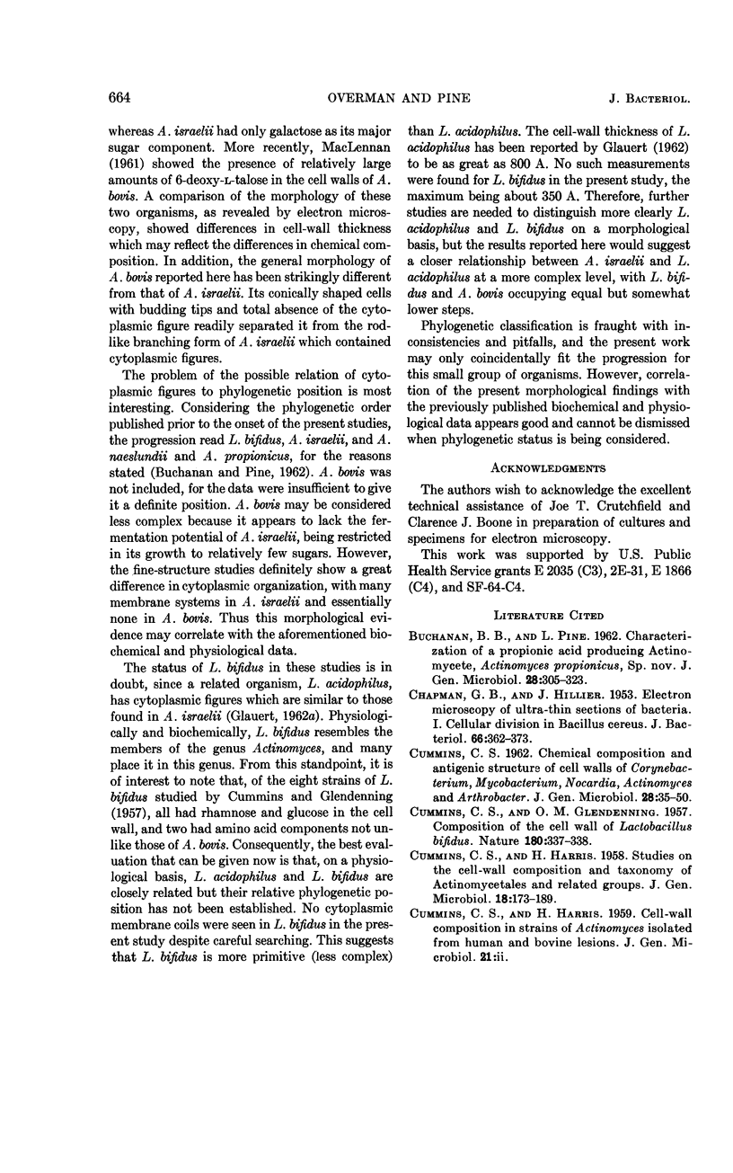
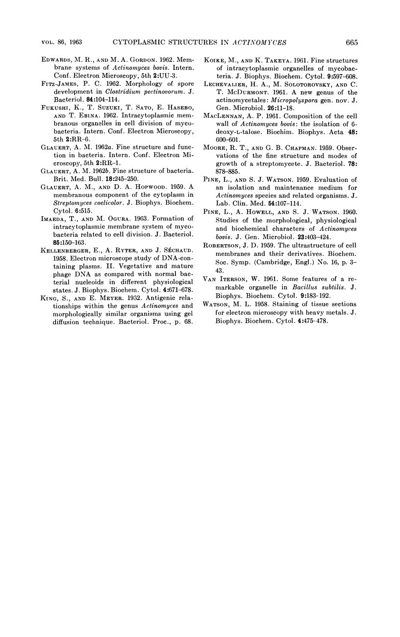
Overman, John R. (Duke University Medical Center, Durham, N.C.) and Leo Pine. Electron microscopy of cytoplasmic structures in facultative and anaerobic Actinomyces. J. Bacteriol. 86:656–665. 1963.—Electron microscopy of cytoplasmic complexes and the cytoplasmic fine structure of Actinomyces bovis, A. israelii, A. naeslundii, and A. propionicus demonstrated marked differences among these four species. Also included in the present study was Lactobacillus bifidus, an organism closely related to the Actinomyces species. A relatively small and compact cytoplasmic membrane complex of A. propionicus was unique in its morphology. Membrane structures of A. naeslundii and A. israelii were relatively large and consisted of coils of various sizes of the cytoplasmic membrane. No membrane complexes were found in L. bifidus or A. bovis. Measurements of cell-wall thickness indicated a significant difference between A. bovis and A. israelii. On the basis of general morphology, cell-wall thickness, and cytoplasmic membrane complexes, A. bovis and A. israelii appear to be distinct species. The relation of the fine structure complexity to phylogenetic position of these organisms is considered.
Full text
PDF
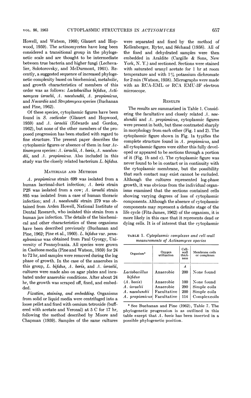
Images in this article
Selected References
These references are in PubMed. This may not be the complete list of references from this article.
- BUCHANAN B. B., PINE L. Characterization of a propionic acid producing actinomycete, Actinomyces propionicus, sp. nov. J Gen Microbiol. 1962 Jun;28:305–323. doi: 10.1099/00221287-28-2-305. [DOI] [PubMed] [Google Scholar]
- CHAPMAN G. B., HILLIER J. Electron microscopy of ultra-thin sections of bacteria I. Cellular division in Bacillus cereus. J Bacteriol. 1953 Sep;66(3):362–373. doi: 10.1128/jb.66.3.362-373.1953. [DOI] [PMC free article] [PubMed] [Google Scholar]
- CUMMINS C. S. Chemical composition and antigenic structure of cell walls of Corynebacterium, Mycobacterium, Nocardia, Actinomyces and Arthrobacter. J Gen Microbiol. 1962 Apr;28:35–50. doi: 10.1099/00221287-28-1-35. [DOI] [PubMed] [Google Scholar]
- CUMMINS C. S., GLENDENNING O. M. Composition of the cell wall of Lactobacillus bifidus. Nature. 1957 Aug 17;180(4581):337–338. doi: 10.1038/180337b0. [DOI] [PubMed] [Google Scholar]
- CUMMINS C. S., HARRIS H. Studies on the cell-wall composition and taxonomy of Actinomycetales and related groups. J Gen Microbiol. 1958 Feb;18(1):173–189. doi: 10.1099/00221287-18-1-173. [DOI] [PubMed] [Google Scholar]
- Fitz-James P. C. MORPHOLOGY OF SPORE DEVELOPMENT IN CLOSTRIDIUM PECTINOVORUM. J Bacteriol. 1962 Jul;84(1):104–114. doi: 10.1128/jb.84.1.104-114.1962. [DOI] [PMC free article] [PubMed] [Google Scholar]
- GLAUERT A. M., HOPWOOD D. A. A membranous component of the cytoplasm in Streptomyces coelicolor. J Biophys Biochem Cytol. 1959 Dec;6:515–516. doi: 10.1083/jcb.6.3.515. [DOI] [PMC free article] [PubMed] [Google Scholar]
- GLAUERT A. M. The fine structure of bacteria. Br Med Bull. 1962 Sep;18:245–250. doi: 10.1093/oxfordjournals.bmb.a069988. [DOI] [PubMed] [Google Scholar]
- IMAEDA T., OGURA M. Formation of intracytoplasmic membrane system of mycobacteria related to cell division. J Bacteriol. 1963 Jan;85:150–163. doi: 10.1128/jb.85.1.150-163.1963. [DOI] [PMC free article] [PubMed] [Google Scholar]
- KELLENBERGER E., RYTER A., SECHAUD J. Electron microscope study of DNA-containing plasms. II. Vegetative and mature phage DNA as compared with normal bacterial nucleoids in different physiological states. J Biophys Biochem Cytol. 1958 Nov 25;4(6):671–678. doi: 10.1083/jcb.4.6.671. [DOI] [PMC free article] [PubMed] [Google Scholar]
- KOIKE M., TAKEYA K. Fine structures of intracytoplasmic organelles of mycobacteria. J Biophys Biochem Cytol. 1961 Mar;9:597–608. doi: 10.1083/jcb.9.3.597. [DOI] [PMC free article] [PubMed] [Google Scholar]
- LECHEVALIER H. A., SOLOTOROVSKY M., McDURMONT C. I. A new genus of the Actinomycetales: Micropolyspora gen. nov. J Gen Microbiol. 1961 Sep;26:11–18. doi: 10.1099/00221287-26-1-11. [DOI] [PubMed] [Google Scholar]
- MACLENNAN A. P. Composition of the cell wall of Actinomyces bovis: the isolation of 6-deoxy-L-talose. Biochim Biophys Acta. 1961 Apr 15;48:600–601. doi: 10.1016/0006-3002(61)90062-2. [DOI] [PubMed] [Google Scholar]
- MOORE R. T., CHAPMAN G. B. Observations of the fine structure and modes of growth of a streptomycete. J Bacteriol. 1959 Dec;78:878–885. doi: 10.1128/jb.78.6.878-885.1959. [DOI] [PMC free article] [PubMed] [Google Scholar]
- PINE L., HOWELL A., Jr, WATSON S. J. Studies of the morphological, physiological, and biochemical characters of Actinomyces bovis. J Gen Microbiol. 1960 Dec;23:403–424. doi: 10.1099/00221287-23-3-403. [DOI] [PubMed] [Google Scholar]
- PINE L., WATSON S. J. Evaluation of an isolation and maintenance medium for Actinomyces species and related organisms. J Lab Clin Med. 1959 Jul;54(1):107–114. [PubMed] [Google Scholar]
- ROBERTSON J. D. The ultrastructure of cell membranes and their derivatives. Biochem Soc Symp. 1959;16:3–43. [PubMed] [Google Scholar]
- VAN ITERSON W. Some features of a remarkable organelle in Bacillus subtilis. J Biophys Biochem Cytol. 1961 Jan;9:183–192. doi: 10.1083/jcb.9.1.183. [DOI] [PMC free article] [PubMed] [Google Scholar]
- WATSON M. L. Staining of tissue sections for electron microscopy with heavy metals. J Biophys Biochem Cytol. 1958 Jul 25;4(4):475–478. doi: 10.1083/jcb.4.4.475. [DOI] [PMC free article] [PubMed] [Google Scholar]


