

Abstract
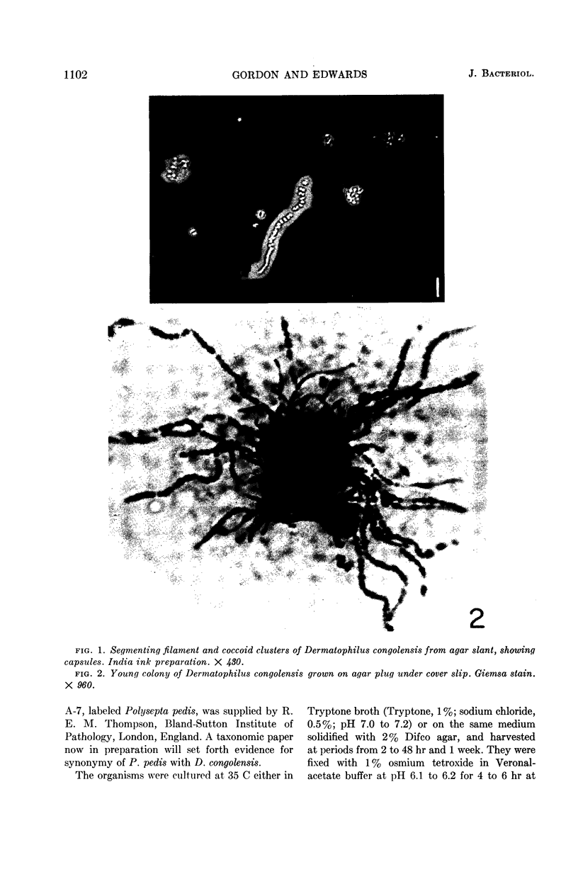
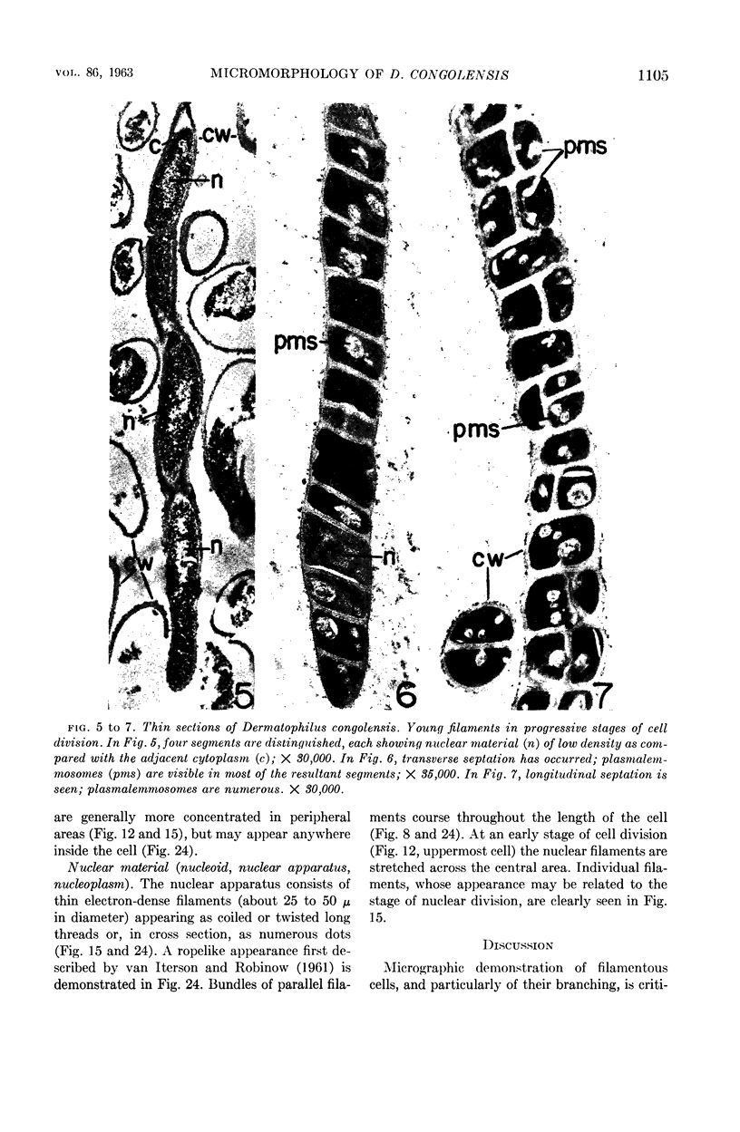
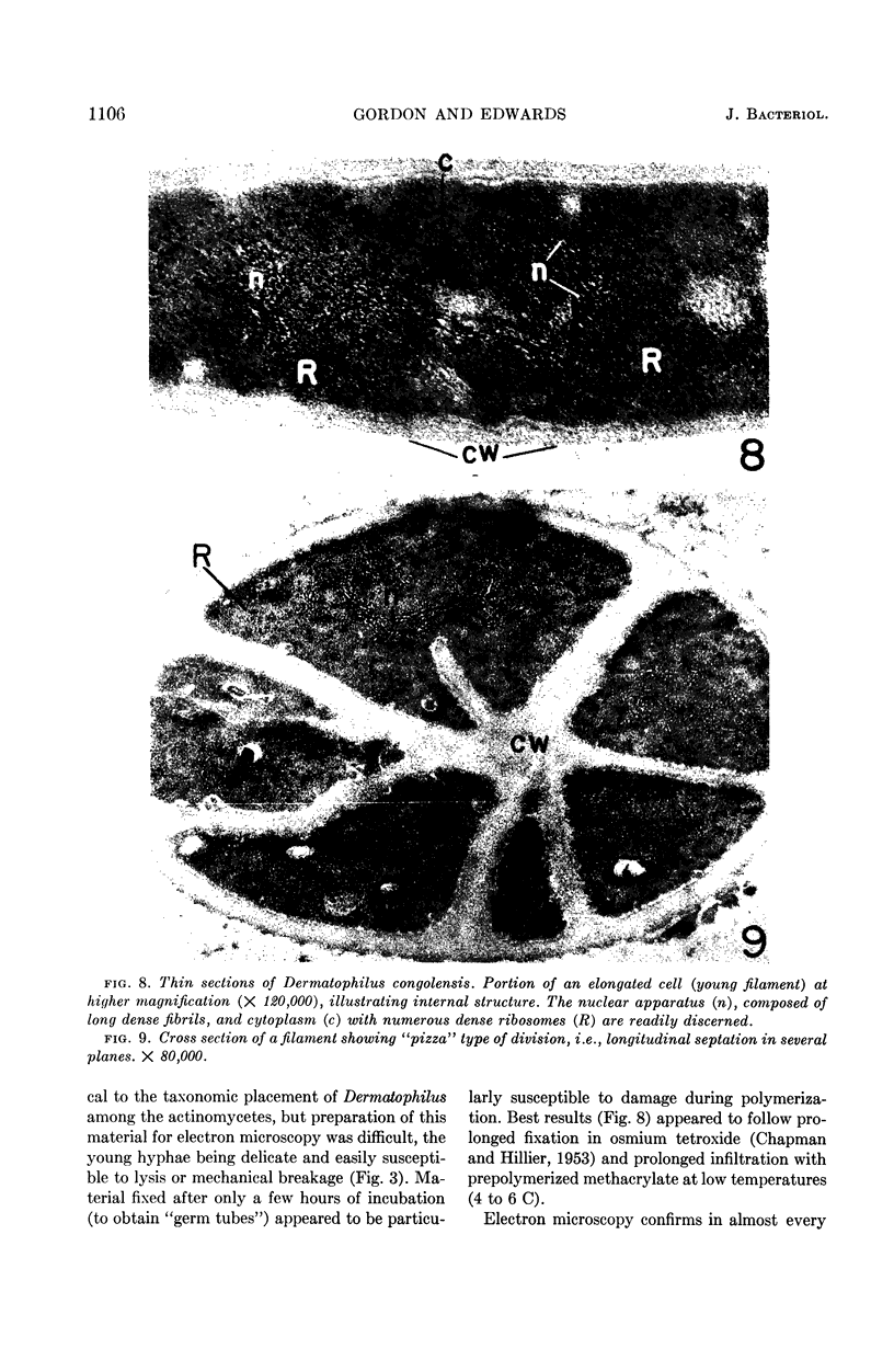
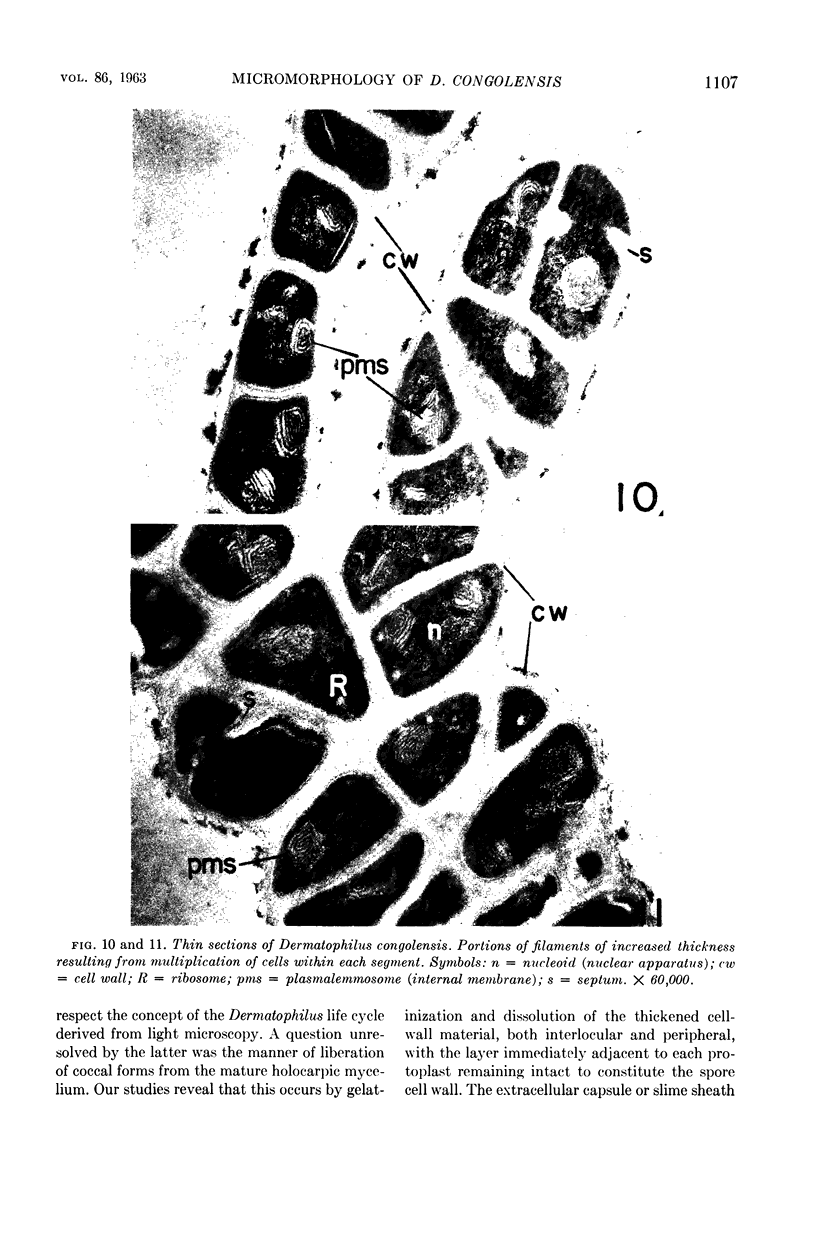
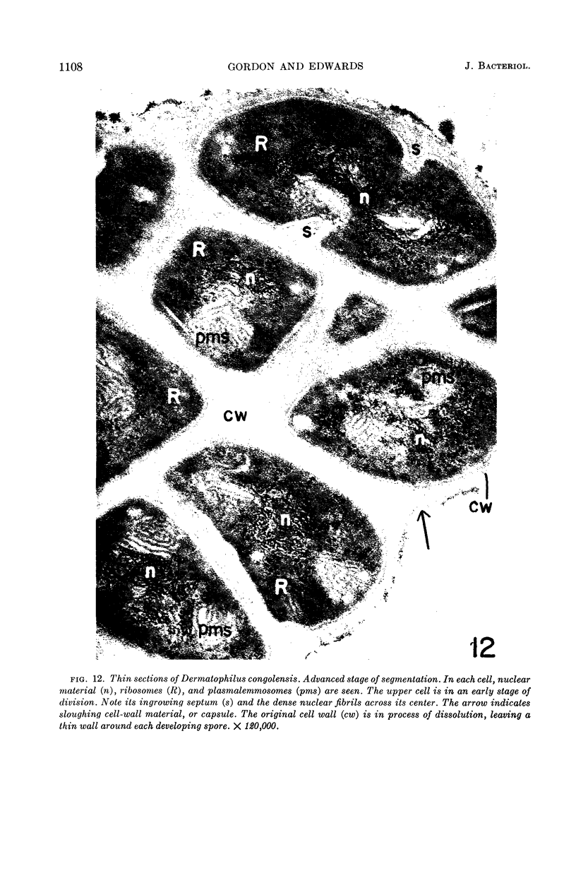
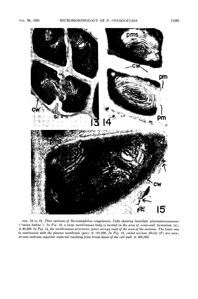
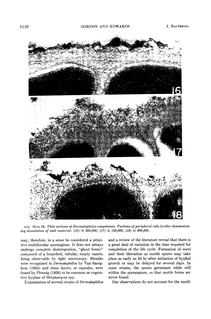
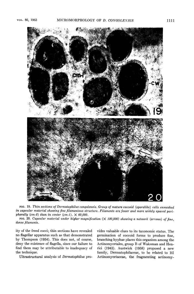
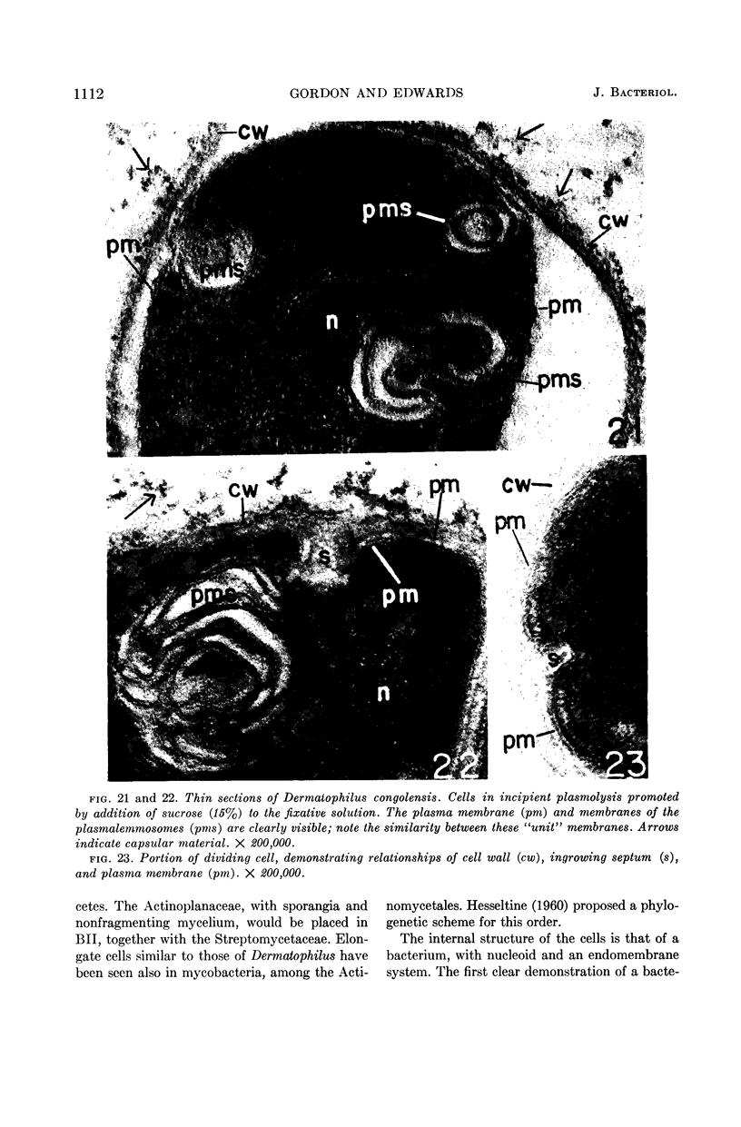
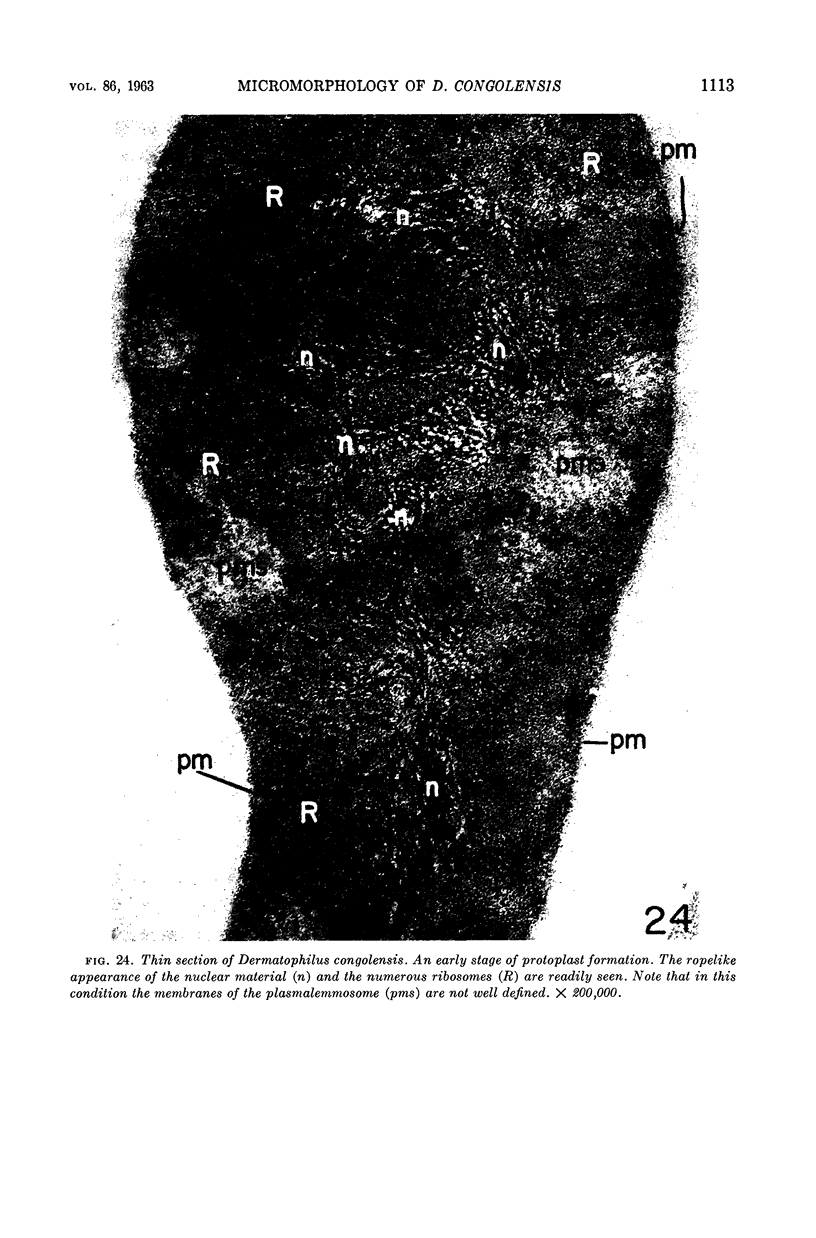
Gordon, Morris A. (Division of Laboratories and Research, New York State Department of Health, Albany), and Mercedes R. Edwards. Micromorphology of Dermatophilus congolensis. J. Bacteriol. 86:1101–1115. 1963.—As seen in electron micrographs of thin sections, Dermatophilus congolensis is a holocarpic actinomycete that fragments, after formation of septa in several planes, into Sarcina-like packets and then into individual cocci. Release of coccal forms from the filaments and packets is by dissolution of a capsular matrix, which is a product of degradation of the cell wall. The plasmalemma is a “unit membrane.” Regularly occurring plasmalemmosomes (“onion bodies”) of uniform structure are apparently related to septum formation. A typical bacterial nucleoid is seen in most sections, and ribosomes are scattered throughout the cytoplasm. Specimens for electron microscopy were prepared by a modification of Kellenberger's method.
Full text
PDF
Images in this article
Selected References
These references are in PubMed. This may not be the complete list of references from this article.
- BLADEN H. A., Jr Demonstration of unusual ultrastructure found in Bacteroides: a conjugatory bridge? J Bacteriol. 1963 Jan;85:250–253. doi: 10.1128/jb.85.1.250-253.1963. [DOI] [PMC free article] [PubMed] [Google Scholar]
- CHAPMAN G. B., HILLIER J. Electron microscopy of ultra-thin sections of bacteria I. Cellular division in Bacillus cereus. J Bacteriol. 1953 Sep;66(3):362–373. doi: 10.1128/jb.66.3.362-373.1953. [DOI] [PMC free article] [PubMed] [Google Scholar]
- EDWARDS M. R., STEVENS R. W. FINE STRUCTURE OF LISTERIA MONOCYTOGENES. J Bacteriol. 1963 Sep;86:414–428. doi: 10.1128/jb.86.3.414-428.1963. [DOI] [PMC free article] [PubMed] [Google Scholar]
- FITZ-JAMES P. C. Participation of the cytoplasmic membrane in the growth and spore fromation of bacilli. J Biophys Biochem Cytol. 1960 Oct;8:507–528. doi: 10.1083/jcb.8.2.507. [DOI] [PMC free article] [PubMed] [Google Scholar]
- GLAUERT A. M., HOPWOOD D. A. A membranous component of the cytoplasm in Streptomyces coelicolor. J Biophys Biochem Cytol. 1959 Dec;6:515–516. doi: 10.1083/jcb.6.3.515. [DOI] [PMC free article] [PubMed] [Google Scholar]
- GLAUERT A. M. The fine structure of bacteria. Br Med Bull. 1962 Sep;18:245–250. doi: 10.1093/oxfordjournals.bmb.a069988. [DOI] [PubMed] [Google Scholar]
- IMAEDA T., OGURA M. Formation of intracytoplasmic membrane system of mycobacteria related to cell division. J Bacteriol. 1963 Jan;85:150–163. doi: 10.1128/jb.85.1.150-163.1963. [DOI] [PMC free article] [PubMed] [Google Scholar]
- KELLENBERGER E., RYTER A., SECHAUD J. Electron microscope study of DNA-containing plasms. II. Vegetative and mature phage DNA as compared with normal bacterial nucleoids in different physiological states. J Biophys Biochem Cytol. 1958 Nov 25;4(6):671–678. doi: 10.1083/jcb.4.6.671. [DOI] [PMC free article] [PubMed] [Google Scholar]
- KOIKE M., TAKEYA K. Fine structures of intracytoplasmic organelles of mycobacteria. J Biophys Biochem Cytol. 1961 Mar;9:597–608. doi: 10.1083/jcb.9.3.597. [DOI] [PMC free article] [PubMed] [Google Scholar]
- ROBERT D. S. The life cycle of Dermatophilus dermatonomus, the causal agent of ovine mycotic dermatitis. Aust J Exp Biol Med Sci. 1961 Oct;39:463–476. [PubMed] [Google Scholar]
- ROBERTSON J. D. The ultrastructure of cell membranes and their derivatives. Biochem Soc Symp. 1959;16:3–43. [PubMed] [Google Scholar]
- ROBINOW C. F. On the plasma membrane of some bacteria and fungi. Circulation. 1962 Nov;26:1092–1104. doi: 10.1161/01.cir.26.5.1092. [DOI] [PubMed] [Google Scholar]
- RYTER A., KELLENBERGER E., BIRCHANDERSEN A., MAALOE O. Etude au microscope électronique de plasmas contenant de l'acide désoxyribonucliéique. I. Les nucléoides des bactéries en croissance active. Z Naturforsch B. 1958 Sep;13B(9):597–605. [PubMed] [Google Scholar]
- STUART D. C., Jr Fine structure of the nucleoid and internal membrane systems of Streptomyces. J Bacteriol. 1959 Aug;78:272–281. doi: 10.1128/jb.78.2.272-281.1959. [DOI] [PMC free article] [PubMed] [Google Scholar]
- THOMPSON R. E. A species of Rhizobium isolated from strawberry foot-rot in the sheep. J Pathol Bacteriol. 1954 Oct;68(2):445–452. doi: 10.1002/path.1700680218. [DOI] [PubMed] [Google Scholar]
- THOMPSON R. E., BISSET K. A. Polysepta: a new genus and sub-order of bacteria. Nature. 1957 Mar 16;179(4559):590–591. doi: 10.1038/179590a0. [DOI] [PubMed] [Google Scholar]
- VAN ITERSON W., ROBINOW C. F. Observations with the electron microscope on the fine structure of the nuclei of two spherical bacteria. J Biophys Biochem Cytol. 1961 Jan;9:171–181. doi: 10.1083/jcb.9.1.171. [DOI] [PMC free article] [PubMed] [Google Scholar]
- Waksman S. A., Henrici A. T. The Nomenclature and Classification of the Actinomycetes. J Bacteriol. 1943 Oct;46(4):337–341. doi: 10.1128/jb.46.4.337-341.1943. [DOI] [PMC free article] [PubMed] [Google Scholar]