Abstract
The purpose of this study was to examine the effect of DOTA (1,4,7,10-tetraazacyclododecane-1,4,7,10-tetraacetic acid) position on melanoma targeting and pharmacokinetics of radiolabeled lactam bridge-cyclized alpha-melanocyte stimulating hormone (α-MSH) peptide.
Method:
A novel lactam bridge-cyclized α-MSH peptide, Ac-GluGlu-CycMSH[DOTA] {Ac-Glu-Glu-c[Lys(DOTA)-Nle-Glu-His-DPhe-Arg-Trp-Gly-Arg-Pro-Val-Asp]}, was synthesized using standard 9-fluorenylmethyloxycarbonyl (Fmoc) chemistry. DOTA was directly attached to the alpha amino group of Lys in the cyclic ring while the N-terminus of the peptide was acetylated to generate Ac-GluGlu-CycMSH[DOTA]. The MC1 receptor binding affinity of Ac-GluGlu-CycMSH[DOTA] was determined in B16/F1 melanoma cells. Melanoma targeting and pharmacokinetic properties of Ac-GluGlu-CycMSH[DOTA]-111In were determined in B16/F1 melanoma-bearing C57 mice and compared to that of 111In-DOTA-Gly-Glu-c[Lys-Nle-Glu-His-DPhe-Arg-Trp-Gly-Arg-Pro-Val-Asp] (111In-DOTA-GlyGlu-CycMSH, DOTA was coupled to the N-terminus of the peptide).
Results:
Ac-GluGlu-CycMSH[DOTA] displayed 0.6 nM MC1 receptor binding affinity in B16/F1 cells. Ac-GluGlu-CycMSH[DOTA]-111In was readily prepared with greater than 95% radiolabeling yield. Ac-GluGlu-CycMSH[DOTA]-111In exhibited high tumor uptake (11.42 ± 2.20% ID/g 2 h post-injection) and prolonged tumor retention (9.42 ± 2.41% ID/g 4 h post-injection) in B16/F1 melanoma-bearing C57 mice. The uptake values for non-target organs were generally low (<1.3% ID/g) except for the kidneys 2, 4 and 24 h post-injection.
Conclusions:
DOTA position exhibited profound effect on melanoma targeting and pharmacokinetic properties of Ac-GluGlu-CycMSH[DOTA]-111In, providing a new insight into the design of lactam bridge-cyclized peptide for melanoma imaging and therapy.
Keywords: Melanoma detection, lactam bridge cyclization, alpha-melanocyte stimulating hormone peptide, melanocortin-1 receptor
Introduction
Malignant melanoma is the most lethal form of skin cancer and the most commonly diagnosed malignancy among young adults with an increasing incidence in the United States. It is predicted that there will be 68,720 cases of malignant melanoma newly reported and 8,650 fatalities in the year 2009 (1). Unfortunately, no curative treatment currently exists for metastatic melanoma due to its resistance to conventional chemotherapy and immunotherapy. It is highly desirable to develop novel effective diagnostic and therapeutic approaches to fulfill the desperate need in melanoma treatment. G protein-coupled melanocortin-1 (MC1) receptors are distinct molecular targets for developing peptide radiopharmaceuticals due to their over-expression on human and mouse melanoma cells (2-6). Alpha-melanocyte stimulating hormone (α-MSH) peptides can selectively target diagnostic and therapeutic radionuclides to MC1 receptors through specific peptide-receptor interaction for melanoma imaging and therapy (7-19).
Recently, we have reported a novel class of 111In-labeled lactam bridge-cyclized α-MSH peptides as effective imaging probes for melanoma detection (20, 21). Unique lactam bridge-cyclization made the radiolabeled α-MSH peptides stable both in vitro and in vivo. Metal chelator DOTA was coupled to the N-terminus of the lactam bridge-cyclized α-MSH peptides for radiolabeling. 111In-DOTA-GlyGlu-CycMSH exhibited high receptor-mediated melanoma uptake (10.40 ± 1.40% ID/g at 2 h post-injection) in B16/F1 melanoma-bearing C57 mice (20). Both primary melanoma and pulmonary melanoma metastases were clearly visualized by small animal SPECT/CT using 111In-DOTA-GlyGlu-CycMSH as an imaging agent (20, 21), highlighting the potential applications of radiolabeled lactam bridge-cyclized α-MSH peptides as effective diagnostic and therapeutic agents for melanoma.
DOTA position (conjugating to N-terminus or towards C-terminus) of the radiolabeled linear α-MSH peptide has displayed profound effect on melanoma and kidney uptakes of 111In-labeled linear α-MSH peptide derivatives (13-15). For instance, DOTA was coupled to the N-terminus or towards C-terminus to generate 111In-DOTA-MSHoct {111In-DOTA-[β-Ala3, Nle4, Asp5, D-Phe7, Lys10]-α-MSH3-10 and 111In-DOTA-NAPamide {[Nle4, Asp5, D-Phe7, Lys11(DOTA)-111In]-α-MSH4-11}, respectively. 111In-DOTA-NAPamide exhibited 75% higher melanoma uptake and 63% less renal uptake than 111In-MSHoct 4 h post-injection in B16/F1 melanoma-bearing C57 mice (14). In this report, we further examined the effect of DOTA position on the melanoma and renal uptakes of 111In-labeled lactam bridge-cyclized α-MSH peptide. A novel DOTA-conjugated lactam bridge-cyclized α-MSH peptide, namely Ac-GluGlu-CycMSH[DOTA] was synthesized using standard 9-fluorenylmethyloxycarbonyl (Fmoc) chemistry. DOTA was directly attached to the alpha amino group of Lys (toward C-terminus) in the cyclic ring while the N-terminus of the peptide was acetylated to generate Ac-GluGlu-CycMSH[DOTA]. The melanoma targeting and pharmacokinetic properties of Ac-GluGlu-CycMSH[DOTA]-111In were determined in B16/F1 melanoma-bearing C57 mice. Meanwhile, the effect of L-lysine co-injection on the renal uptake of Ac-GluGlu-CycMSH[DOTA]-111In was examined in B16/F1 melanoma-bearing C57 mice.
Experimental Procedures
Chemicals and Reagents
Amino acid and resin were purchased from Advanced ChemTech Inc. (Louisville, KY) and Novabiochem (San Diego, CA). DOTA-tri-t-butyl ester was purchased from Macrocyclics Inc. (Richardson, TX). 111InCl3 was purchased from Trace Life Sciences, Inc. (Dallas, TX). 125I-Tyr2-[Nle4, D-Phe7]-α-MSH {125I-(Tyr2)-NDP-MSH} was obtained from PerkinElmer, Inc. (Shelton, CT). All other chemicals used in this study were purchased from Thermo Fischer Scientific (Waltham, MA) and used without further purification. B16/F1 murine melanoma cells were obtained from American Type Culture Collection (Manassas, VA).
Peptide Synthesis
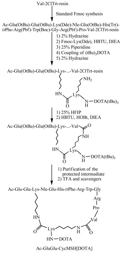
Intermediate scaffold of Ac-Glu(OtBu)-Glu(OtBu)-Lys(Dde)-Nle-Glu(OtBu)-His(Trt)-DPhe-Arg(Pbf)-Trp(Boc)-Gly-Arg(Pbf)-Pro-Val was synthesized on Val-2-Chlorotrityl Chloride (Val-2ClTrt) resin using standard Fmoc chemistry by an Advanced ChemTech multiple-peptide synthesizer (Louisville, KY). A small aliquot of the scaffold material was cleaved and characterized by liquid chromatography-mass spectrometry (LC-MS) prior to further peptide synthesis and cyclization. The protecting group (Dde) of the Lys in the intermediate scaffold was removed by 2% Hydrazine and the branch moiety of the peptide was generated by coupling Fmoc-Lys(Dde) to the epsilon amino group of the Lys in the intermediate scaffold manually. (tBu)3DOTA was coupled to the alpha amino group of Lys(Dde) in the peptide branch manually using standard Fmoc chemistry. The protecting group (Dde) of the Lys in peptide branch was removed by 2% Hydrazine. After the removal of (Dde) protecting group, the protected branched peptide was cleaved from the resin treating with 25% Hexafluoroisopropanol (HFIP) and 5% triisopropylsilane (TIS) in dichloromethane (DCM) and characterized by LC-MS. Peptide cyclization between the acid moiety of Val and the epsilon amino group of Lys in the peptide branch was achieved by overnight reaction in DMF in the presence of a mixture of 1 mM 1-hydroxybenzotriazole (HOBT), 2-(1H-benzotriazole-1-yl)-1,1,3,3-tetranethyluronium hexafluorophosphate (HBTU) and N,N-diisopropylethylamine (DIEA). The protecting groups were totally removed by treating with a mixture of trifluoroacetic acid (TFA), thioanisole, phenol, water, ethanedithiol and triisopropylsilane (87.5:2.5:2.5:2.5:2.5:2.5) for 2 h at room temperature (25°C). The peptide was precipitated and washed with ice-cold ether for four times, purified by reverse phase-high performance liquid chromatography (RP-HPLC) and characterized by LC-MS.
In vitro Competitive Binding Assay
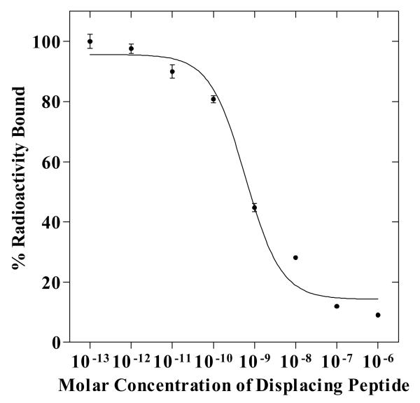
The IC50 value of Ac-GluGlu-CycMSH[DOTA] was determined by in vitro competitive binding assay according to the method described previously (20). B16/F1 cells were harvested and seeded into a 24-well cell culture plate (5×10⁵ cells/well) and incubated at 37°C overnight. After being washed once with binding medium [Dulbecco's Modified Eagle's Medium with 25 mM N-(2-hydroxyethyl)-piperazine-N'-(2-ethanesulfonic acid), 0.2% BSA and 0.3 mM 1,10-phenathroline], the cells were incubated at 25°C for 2 h with approximately 50,000 cpm of 125I-(Tyr2)-NDP-MSH in the presence of increasing concentrations (10−13 to 10−6 M) of Ac-GluGlu-CycMSH[DOTA] in 0.3 ml of binding medium. The reaction medium was aspirated after incubation. Cells were rinsed with 0.5 ml of ice-cold pH 7.4, 0.2% BSA / 0.01 M PBS twice and lysed in 0.5 ml of 1 N NaOH for 5 min. The activities associated with cells were measured in a Wallac 1480 automated gamma counter (PerkinElmer, NJ). The IC50 value of Ac-GluGlu-CycMSH[DOTA] was calculated by using Prism software (GraphPad Software, La Jolla, CA).
Complexation of the Peptide with 111In
Ac-GluGlu-CycMSH[DOTA]-111In was prepared using a 0.5 M NH4OAc-buffered solution at pH 5.4. Briefly, 50 μl of 111InCl3 (18.5-37.0 MBq in 0.05 M HCl), 10 μl of 1 mg/ml Ac-GluGlu-CycMSH[DOTA] aqueous solution and 400 μl of 0.5 M NH4OAc (pH 5.4) were added into a reaction vial and incubated at 75°C for 45 min. After the incubation, 20 μl of 0.5% EDTA aqueous solution was added into the reaction vial to quench the reaction. The radiolabeled complexes were purified to single species by Waters RP-HPLC (Milford, MA) on a Grace Vadyc C-18 reverse phase analytical column (Deerfield, IL) using the following gradient at a flowrate of 1 ml/min. The mobile phase consisted of solvent A (20 mM HCl aqueous solution) and solvent B (100% CH3CN). The gradient was initiated and kept at 82:18 A/B for 3 min followed by a linear gradient of 82:18 A/B to 72:28 A/B over 20 min. Then, the gradient was changed from 72:28 A/B to 10:90 A/B over 3 mins followed by an additional 5 min at 10:90 A/B. Thereafter, the gradient was changed from 10:90 A/B to 82:18 A/B over 3 mins.. Purified peptide sample was purged with N2 gas for 20 minutes to remove the acetonitrile. The pH of final solution was adjusted to 7.4 with 0.1 N NaOH and normal saline for animal studies. Serum stability of Ac-GluGlu-CycMSH[DOTA]-111In was determined according to the published procedure (20) by incubation in mouse serum at 37°C for 24 h, and monitored for degradation by RP-HPLC.
Cellular Internalization and Efflux of Ac-GluGlu-CycMSH[DOTA]-111In
Cellular internalization and efflux of Ac-GluGlu-CycMSH[DOTA]-111In were evaluated in B16/F1 cells as described by Miao et al (20). After being washed once with binding medium, B16/F1 cells seeded in cell culture plates were incubated at 25°C for 20, 40, 60, 90 and 120 min (n=3) in the presence of approximately 160,000 counts per minute (cpm) of HPLC purified Ac-GluGlu-CycMSH[DOTA]-111In. After incubation, the reaction medium was aspirated and cells were rinsed with 2×0.5 ml of ice-cold pH 7.4, 0.2% BSA / 0.01 M PBS. Cellular internalization of Ac-GluGlu-CycMSH[DOTA]-111In was assessed by washing the cells with acidic buffer [40 mM sodium acetate (pH 4.5) containing 0.9% NaCl and 0.2% BSA] to remove the membrane-bound radioactivity. The remaining internalized radioactivity was obtained by lysing the cells with 0.5 ml of 1N NaOH for 5 min. Membrane-bound and internalized 111In activities were counted in a gamma counter. Cellular efflux was determined by incubating B16/F1 cells with Ac-GluGlu-CycMSH[DOTA]-111In for 2 h, removing non-specific-bound activity with 2×0.5 ml of ice-cold pH 7.4, 0.2% BSA / 0.01 M PBS rinse, and monitoring radioactivity released into cell culture medium. At time points of 20, 40, 60, 90 and 120 min, the radioactivities on cell surface and in cells were separately collected and counted in a gamma counter.
Biodistribution Studies
All the animal studies were conducted in compliance with Institutional Animal Care and Use Committee approval. The biodistribution properties of Ac-GluGlu-CycMSH[DOTA]-111In were determined in B16/F1 melanoma-bearing C57 female mice (Harlan, Indianapolis, IN). C57 mice were inoculated subcutaneously with 1×10⁶ B16/F1 melanoma cells in the right flank for each mouse. After 10 days, when the weight of tumors reached approximately 0.2 g, 0.037 MBq of Ac-GluGlu-CycMSH[DOTA]-111In was injected into each mouse through the tail vein. Groups of 5 mice were sacrificed at 0.5, 2, 4 and 24 h post-injection, and tumors and organs of interest were harvested, weighed and counted. Blood values were taken as 6.5% of the whole-body weight. The specificity of the tumor uptake of Ac-GluGlu-CycMSH[DOTA]-111In was determined by blocking the tumor uptake with the co-injection of 10 μg (6.07 nmol) of unlabeled NDP-MSH, a linear α-MSH peptide analogue with picomolar affinity for the MC1 receptor present on melanoma cells.
The effect of L-lysine co-injection on the renal uptake of Ac-GluGlu-CycMSH[DOTA]-111In was examined in B16/F1 melanoma-bearing C57 as well. A group of 5 mice were injected with a mixture of 0.037 MBq of Ac-GluGlu-CycMSH[DOTA]-111In and 15 mg of L-lysine. The mice were sacrificed at 2 h post-injection, and tumors and organs of interest were harvested, weighed and counted. Blood values were taken as 6.5% of the whole body weight.
Imaging Melanoma with Ac-GluGlu-CycMSH[DOTA]-111In
One B16/F1 melanoma-bearing C57 mouse was injected with 7.4 MBq of Ac-GluGlu-CycMSH[DOTA]-111In via the tail vein. The mouse was anesthetized with 1.5% isoflurane for small animal SPECT/CT (Nano-SPECT/CT®, Bioscan) imaging 2 h post-injection. The 9-min CT imaging was immediately followed by the SPECT imaging of whole body. The SPECT scans of 24 projections were acquired and total acquisition time was 45 min. Reconstructed data from SPECT and CT were visualized and co-registered using InVivoScope (Bioscan, Washington DC).
Urinary Metabolites of Ac-GluGlu-CycMSH[DOTA]-111In
One hundred microliter of HPLC purified Ac-GluGlu-CycMSH[DOTA]-111In (3.7 MBq) was injected into a B16/F1 melanoma-bearing C57 mouse through the tail vein. At 2 h after dose administration, the mouse was sacrificed and the urine was collected. The radioactive metabolites in the urine were analyzed by injecting aliquots of the urine into HPLC. A 20-minute gradient of 18-28% acetonitrile / 20 mM HCl was used for the urine analysis.
Statistical Methods
Statistical analysis was performed using the Student's t-test for unpaired data to determine the significant differences between Ac-GluGlu-CycMSH[DOTA]-111In with or without NDP-MSH co-injection, and Ac-GluGlu-CycMSH[DOTA]-111In with or without L-lysine co-injection described in biodistribution studies above. Differences at the 95% confidence level (p<0.05) were considered significant.
Results
To examine the effect of DOTA position on the melanoma and renal uptakes of 111In-labeled lactam bridge-cyclized α-MSH peptide, a novel peptide of Ac-GluGlu-CycMSH[DOTA] was synthesized and purified by RP-HPLC. The identity of Ac-GluGlu-CycMSH[DOTA] was confirmed by electrospray ionization mass spectrometry (EIMS MW: 2220.5; Calculated MW: 2220.0). Ac-GluGlu-CycMSH[DOTA] displayed greater than 95% purity with 30% overall synthetic yield. The schematic structure of Ac-GluGlu-CycMSH[DOTA] is shown in Figure 1. Figure 2 illustrates the synthetic scheme of Ac-GluGlu-CycMSH[DOTA]. The competitive binding curve of Ac-GluGlu-CycMSH[DOTA] is presented in Figure 3. The IC50 value of Ac-GluGlu-CycMSH[DOTA] was 0.6 nM in B16/F1 melanoma cells.
Figure 1.
Schematic structures of Ac-GluGlu-CycMSH[DOTA] and DOTA-GlyGlu-CycMSH.
Figure 2.
Synthetic scheme of Ac-GluGlu-CycMSH[DOTA].
Figure 3.
The competitive binding curve of Ac-GluGlu-CycMSH[DOTA] in B16/F1 murine melanoma cells. The IC50 value of Ac-GluGlu-CycMSH[DOTA] was 0.6 nM.
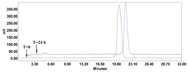
The peptide was readily labeled with 111In in 0.5 M ammonium acetate at pH 5.4 with greater than 95% radiolabeling yield. Ac-GluGlu-CycMSH[DOTA]-111In was completely separated from its excess non-labeled peptide by RP-HPLC. The retention times of Ac-GluGlu-CycMSH[DOTA]-111In and its non-labeled peptide were 20.1 (Fig. 4) and 15.5 min, respectively. The specific activity of Ac-GluGlu-CycMSH[DOTA]-111In was 7.41×10⁸ MBq/g. Ac-GluGlu-CycMSH[DOTA]-111In showed greater than 98% radiochemical purity after the HPLC purification. Ac-GluGlu-CycMSH[DOTA]-111In was stable in mouse serum at 37°C. Only the Ac-GluGlu-CycMSH[DOTA]-111In was detected by RP-HPLC after 24 h of incubation (Fig. 4).
Figure 4.
HPLC profile of radioactive Ac-GluGlu-CycMSH[DOTA]-111In (T=0) and mouse serum stability of Ac-GluGlu-CycMSH[DOTA]-111In (T=24 h) after 24 h incubation at 37°C. The retention time of Ac-GluGlu-CycMSH[DOTA]-111In was 20.1 min.
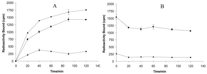
Cellular internalization and efflux of Ac-GluGlu-CycMSH[DOTA]-111In were evaluated in B16/F1 cells. Figure 5 illustrates cellular internalization and efflux of Ac-GluGlu-CycMSH[DOTA]-111In. Ac-GluGlu-CycMSH[DOTA]-111In exhibited rapid cellular internalization and extended cellular retention. There was 72.54 ± 3.10% of the cellular uptake of Ac-GluGlu-CycMSH[DOTA]-111In activity internalized in the B16/F1 cells 40 min post incubation. There was 81.12 ± 0.28% of the cellular uptake of Ac-GluGlu-CycMSH[DOTA]-111In activity internalized in the cells after 2 h incubation. Cellular efflux results demonstrated that 68.60±1.43% of the internalized Ac-GluGlu-CycMSH[DOTA]-111In activity remained inside the cells 2 h after incubating cells in culture medium.
Figure 5.
Cellular internalization (A) and efflux (B) of Ac-GluGlu-CycMSH[DOTA]-111In in B16/F1 murine melanoma cells at 25°C. Total bound radioactivity (◆), internalized activity (■) and cell membrane activity (▲) were presented as counts per minute (cpm).
The melanoma targeting and pharmacokinetic properties of Ac-GluGlu-CycMSH[DOTA]-111In were determined in B16/F1 melanoma-bearing C57 mice. The biodistribution results of Ac-GluGlu-CycMSH[DOTA]-111In are shown in Table 1. Ac-GluGlu-CycMSH[DOTA]-111In exhibited rapid and high melanoma uptake. The tumor uptake value was 12.11 ± 1.21% ID/g 0.5 h post-injection. There was 9.42 ± 2.41% ID/g of Ac-GluGlu-CycMSH[DOTA]-111In activity remained in the tumors 4 h post-injection. The tumor uptake value of Ac-GluGlu-CycMSH[DOTA]-111In gradually decreased to 3.64 ± 0.74% ID/g 24 h post-injection. In the blocking study, the tumor uptake value of Ac-GluGlu-CycMSH[DOTA]-111In with 10 μg (6.07 nmol) of non-radiolabeled NDP-MSH co-injection were only 4.8% of the tumor uptake value without NDP-MSH co-injection at 2 h post-injection (p<0.05), demonstrating that the tumor uptake was specific and receptor-mediated. Whole-body clearance of Ac-GluGlu-CycMSH[DOTA]-111In was rapid, with approximately 78% of the injected radioactivity cleared through the urinary system by 2 h post-injection (Table 1). Normal organ uptakes of Ac-GluGlu-CycMSH[DOTA]-111In were generally very low (<1.3% ID/g) except for the kidneys 2, 4 and 24 h post-injection. High tumor/blood and tumor/normal organ uptake ratios were demonstrated as early as 0.5 h post-injection (Table 1). The renal uptake of Ac-GluGlu-CycMSH[DOTA]-111In was 51.52 ± 6.17% ID/g 0.5 h post-injection, reached its peak value of 67.21 ± 8.63% ID/g 4 h post-injection and gradually decreased to 21.09 ± 6.59% ID/g 24 h post-injection. Co-injection of 15 mg of L-lysine reduced the kidney uptake value of Ac-GluGlu-CycMSH[DOTA]-111In to 18.35 ± 5.92% ID/g (70% reduction) without affecting the tumor uptake value 2 h post-injection (Table 1).
Table 1.
Biodistribution of Ac-GluGlu-CycMSH[DOTA]-111In in B16/F1 melanoma-bearing C57 mice. The data were presented as percent injected dose/gram or as percent injected dose (Mean±SD, n=5).
| Percent Injected Dose/Gram (%ID/g) | ||||||
|---|---|---|---|---|---|---|
| Tissues | 0.5 h | 2 h | 2 h NDP blockade |
2 h L-lysine co-injection |
4 h | 24 h |
| Tumor | 12.11±1.21 | 11.42±2.20 | 0.49±0.15* | 11.86±2.18 | 9.42±2.41 | 3.64±0.74 |
| Brain | 0.14±0.01 | 0.01±0.01 | 0.04±0.02 | 0.02±0.01 | 0.03±0.01 | 0.01±0.00 |
| Blood | 2.03±0.32 | 0.21±0.05 | 0.16±0.13 | 0.32±0.04* | 0.16±0.11 | 0.03±0.02 |
| Heart | 0.69±0.17 | 0.13±0.11 | 0.17±0.13 | 0.38±0.12* | 0.26±0.15 | 0.03±0.03 |
| Lung | 2.49±0.25 | 0.64±0.16 | 0.33±0.05* | 0.32±0.04* | 0.34±0.10 | 0.07±0.02 |
| Liver | 1.85±0.14 | 1.02±0.18 | 0.62±0.13* | 0.58±0.14* | 1.28±0.24 | 1.01±0.28 |
| Spleen | 0.79±0.24 | 0.47±0.05 | 0.48±0.12 | 0.16±0.14* | 0.64±0.36 | 0.27±0.10 |
| Stomach | 0.69±0.09 | 0.32±0.19 | 0.05±0.02* | 0.25±0.06 | 0.18±0.07 | 0.07±0.04 |
| Kidneys | 51.52±6.17 | 60.48±8.37 | 47.02±13.14 | 18.35±5.92* | 67.21±8.63 | 21.09±6.59 |
| Muscle | 0.37±0.13 | 0.12±0.02 | 0.36±0.18 | 0.08±0.10 | 0.04±0.03 | 0.04±0.02 |
| Pancreas | 0.53±0.08 | 0.12±0.01 | 0.08±0.01* | 0.37±0.29 | 0.07±0.05 | 0.02±0.02 |
| Bone | 0.43±0.22 | 0.41±0.17 | 0.21±0.14 | 0.35±0.31 | 0.24±0.14 | 0.05±0.02 |
| Skin | 1.75±0.06 | 0.39±0.03 | 0.25±0.03* | 0.46±0.11 | 0.26±0.15 | 0.18±0.02 |
| Percent injected dose (%ID) | ||||||
| Intestines | 0.78±0.13 | 1.39±0.87 | 0.63±0.30 | 0.55±0.09 | 0.44±0.14 | 0.18±0.07 |
| Urine | 51.65±5.38 | 77.59±1.52 | 87.32±2.53 | 90.77±1.78 | 78.84±2.34 | 90.82±2.05 |
| Uptake Ratio of Tumor/Normal Organ | ||||||
| Tumor/Blood | 5.97 | 54.38 | 3.06 | 37.06 | 58.88 | 121.33 |
| Tumor/Kidneys | 0.24 | 0.19 | 0.01 | 0.65 | 0.14 | 0.17 |
| Tumor/Lung | 4.86 | 17.84 | 1.48 | 37.06 | 27.71 | 52.00 |
| Tumor/Liver | 6.55 | 11.20 | 0.79 | 20.45 | 7.36 | 3.60 |
| Tumor/Muscle | 32.73 | 95.17 | 1.36 | 148.25 | 235.50 | 91.00 |
| Tumor/Skin | 6.92 | 29.28 | 1.96 | 25.78 | 36.23 | 20.22 |
p<0.05 significance comparison of Ac-GluGlu-CycMSH[DOTA]-111In with or without peptide blockade, and with or without L-lysine co-injection 2 h post-injection.
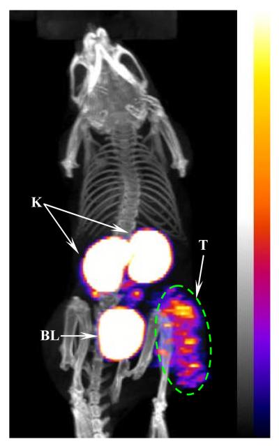
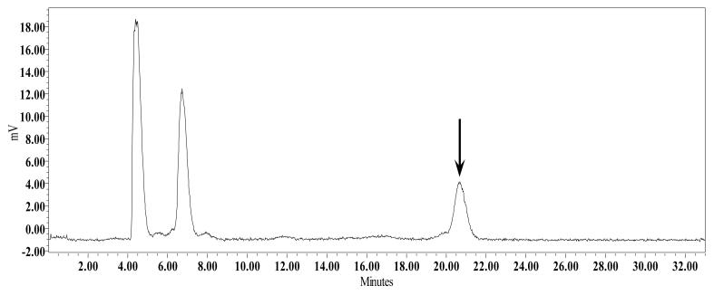
One B16/F1 melanoma-bearing C57 mouse was injected with 7.4 MBq of Ac-GluGlu-CycMSH[DOTA]-111In through the tail vein to visualize the tumors at 2 h after dose administration. The whole-body SPECT image of the mouse was fused with CT image. The whole-body SPECT/CT image is presented in Fig. 6. Flank melanoma tumors were visualized clearly using Ac-GluGlu-CycMSH[DOTA]-111In as an imaging probe 2 h post-injection. Ac-GluGlu-CycMSH[DOTA]-111In exhibited high tumor to normal organ uptake ratios except for the kidney, which was coincident with the biodistribution results. In view of the substantial renal uptake value of Ac-GluGlu-CycMSH[DOTA]-111In in the biodistribution results, the urinary metabolites of Ac-GluGlu-CycMSH[DOTA]-111In were analyzed by RP-HPLC at 2 h post-injection. The HPLC elution profile of Ac-GluGlu-CycMSH[DOTA]-111In is shown in Fig. 7. Eighty-three percent of Ac-GluGlu-CycMSH[DOTA]-111In was transformed to two more polar metabolites and only 17% of Ac-GluGlu-CycMSH[DOTA]-111In remained intact at 2 h post-injection.
Figure 6.
Whole-body image of Ac-GluGlu-CycMSH[DOTA]-111In in B16/F1 flank melanoma-bearing C57 mice at 2 h post-injection. Tumor (T), kidneys (K) and bladder (BL) are highlighted with arrows on the image.
Figure 7.
HPLC profile of radioactive urine sample of a B16/F1 melanoma-bearing C57 mouse at 2 h post-injection of Ac-GluGlu-CycMSH[DOTA]-111In. The arrow indicates the retention time of the original compound of Ac-GluGlu-CycMSH[DOTA]-111In prior to the tail vein injection.
Discussion
Both radiolabeled linear and cyclic α-MSH peptides have been reported to target the MC1 receptors for melanoma imaging and therapy (4-22). Cyclization of α-MSH peptides has been successfully used to improve the binding affinities and in vivo stability of peptides (14-16). In comparison with linear peptide, stabilization of secondary structures such as beta turns makes the cyclic peptide less flexible in conformation and better fit receptor binding pocket, enhancing the binding affinities of the cyclic peptides. Among the 111In-labeled metal-cyclized, lactam bridge-cyclized and disulfide bridge-cyclized α-MSH peptides, 111In-labeled metal-cyclized and lactam bridge-cyclized α-MSH peptides displayed higher melanoma uptake values and less renal uptake values in B16/F1 melanoma-bearing C57 mice (4, 20). We have recently developed a novel class of DOTA-conjugated lactam bridge-cyclized α-MSH peptides where the metal chelator DOTA was attached to the N-terminus of the peptide for radiolabeling. Both primary melanoma and pulmonary melanoma metastases were clearly imaged by small animal SPECT/CT using 111In-DOTA-GlyGlu-CycMSH as an imaging probe (20, 21).
Eberle and coworkers reported the profound effect of DOTA position on melanoma and kidney uptakes of 111In-labeled linear α-MSH peptide derivatives (14, 15). Initially, DOTA was directly attached to the N-terminus of the peptide to yield 111In-DOTA-MSHoct {111In-DOTA-[β-Ala3, Nle4, Asp5, D-Phe7, Lys10]-α-MSH3-10}, which exhibited 4.31 ± 0.30% ID/g tumor uptake 4 h post-injection in B16/F1 melanoma-bearing C57 mice (14). Thereafter, another 111In-labeled linear α-MSH peptide derivative, namely 111In-DOTA-NAPamide ([Nle4, Asp5, D-Phe7, Lys11(DOTA)-111In]-α-MSH4-11), was developed by coupling DOTA to the epsilon amino group of Lys in the C-terminus. Interestingly, coupling of DOTA towards the C-terminus of the linear peptide improved the tumor uptake and reduced renal uptake of 111In-DOTA-NAPamide. At 4 h post-injection, 111In-DOTA-NAPamide displayed 75% higher tumor uptake and 63% less renal uptake than 111In-DOTA-MSHoct (14), indicating that the DOTA position could dramatically affect the melanoma uptake and renal uptake of the radiolabeled α-MSH peptide. Hence, we examined the effect of DOTA position on the biodistribution properties of 111In-labeled lactam bridge-cyclized α-MSH peptide in this study. DOTA was conjugated to the Lys in the cyclic ring while the N-terminus of the peptide was acetylated to generate Ac-GluGlu-CycMSH[DOTA]. A Lys was used to connect DOTA (at alpha amino group of the Lys) as well as to cyclize the peptide (through the epsilon amino group of the Lys). To eliminate the potential influence of the overall positive charge on the renal uptake of the radiolabeled α-MSH peptide,one more negatively-charged Glu was introduced to replace the Gly at the N-terminus besides the acetylation of the N-terminus (Fig. 1) to make the overall charge of Ac-GluGlu-CycMSH[DOTA] equal to that of DOTA-GlyGlu-CycMSH.
The structural change didn't sacrifice the nanomolar binding affinity of Ac-GluGlu-CycMSH[DOTA] compared to DOTA-GlyGlu-CycMSH. Ac-GluGlu-CycMSH[DOTA] displayed 0.6 nM receptor binding affinity in B16/F1 cells, which was similar to that of DOTA-GlyGlu-CycMSH (0.8 nM). Ac-GluGlu-CycMSH[DOTA]-111In exhibited rapid cellular internalization and extended cellular retention in B16/F1 cells (Fig. 5), making Ac-GluGlu-CycMSH[DOTA]-111In suitable for melanoma imaging. The shift of DOTA from the N-terminus toward the lactam bridge-cyclic ring did increase the melanoma uptake and retention. The tumor uptake values of Ac-GluGlu-CycMSH[DOTA]-111In were 1.1, 1.3 and 1.5 times the tumor uptake values of 111In-DOTA-GlyGlu-CycMSH 2, 4 and 24 h post-injection, respectively (20). Compared to the linear α-MSH peptide 111In-DOTA-NAPamide, Ac-GluGlu-CycMSH[DOTA]-111In exhibited 24.6% and 56.9% higher tumor uptake values 4 and 24 h post-injection, respectively. Flank melanoma tumors were clearly visualized by SPECT/CT using Ac-GluGlu-CycMSH[DOTA]-111In as an imaging probe at 2 h post-injection (Fig. 6). The SPECT imaging of tumors accurately matched the anatomical information from CT images. Ac-GluGlu-CycMSH[DOTA]-111In displayed high tumor to normal organ uptake ratios except for the kidney. The high receptor-mediated tumor uptake and tumor to normal organ uptake ratios highlighted the potential of radiolabeled lactam bridge-cyclized α-MSH peptides as a novel class of peptide radiopharmaceuticals for melanoma imaging.
The liver uptake values of Ac-GluGlu-CycMSH[DOTA]-111In were 3.8, 6.1 and 4.2 times the liver uptake values of 111In-DOTA-GlyGlu-CycMSH 2, 4 and 24 h post-injection, respectively. The higher liver uptakes of Ac-GluGlu-CycMSH[DOTA]-111In were likely due to its higher lipophilicity. Under the same HPLC gradient, Ac-GluGlu-CycMSH[DOTA]-111In was washed out of the C18 analytical column later than 111In-DOTA-GlyGlu-CycMSH. As shown in biodsitribution results (Table 1) and SPECT/CT image (Fig. 6), Ac-GluGlu-CycMSH[DOTA]-111In was primarily excreted through the kidneys. Surprisingly, the renal uptake values of Ac-GluGlu-CycMSH[DOTA]-111In were 4.6, 5.5 and 2.3 times the renal uptake values of 111In-DOTA-GlyGlu-CycMSH 2, 4 and 24 h post-injection, respectively (20).. The strategy of infusing basic amino acid such as lysine has been successfully employed to decrease the renal uptakes of radiolabeled metal-cyclized α-MSH peptides by shielding the electrostatic interaction between positively-charged peptides and negatively-charged surface of tubule cells (5, 17, 22). Therefore, 15 mg of L-lysine was co-injected with Ac-GluGlu-CycMSH[DOTA]-111In to examine its effect in reducing the renal uptake of Ac-GluGlu-CycMSH[DOTA]-111In. As we anticipated, co-injection of L-lysine dramatically reduced the renal uptake of Ac-GluGlu-CycMSH[DOTA]-111In by 70% (p<0.05) 2 h post-injection, demonstrating that the electrostatic interaction played a key role in the non-specific renal uptake of Ac-GluGlu-CycMSH[DOTA]-111In. The urine analysis showed that Ac-GluGlu-CycMSH[DOTA]-111In was metabolized into two moieties that might be responsible for the activity retention in kidneys. Another way to decrease the renal uptake of the radiolabeled α-MSH peptide is to increase the overall negative charge of the peptide by introducing a negatively-charged aspartic acid or glutamic acid into the peptide sequence. Introduction of a glutamic acid as a linker has successfully reduced the renal uptake values of 111In-DOTA-GlyGlu-CycMSH by 44% compared to 111In-DOTA-CycMSH {111In-DOTA-c[Lys-Nle-Glu-His-DPhe-Arg-Trp-Gly-Arg-Pro-Val-Asp]} (20). Since we have demonstrated that shielding the electrostatic interaction dramatically reduced the renal uptake of Ac-GluGlu-CycMSH[DOTA]-111In, it is likely that introducing a negatively-charged amino acid between the DOTA and Lys in Ac-GluGlu-CycMSH[DOTA]-111In will decrease the non-specific renal uptake. Further decrease of the renal uptake will facilitate the potential applications of radiolabeled lactam bridge-cyclized α-MSH peptides for melanoma imaging and therapy.
A potential application of this MC1 receptor-targeting peptide construct is to develop melanoma-specific dual-modality imaging agents. As demonstrated in this study and our previous work (20), no matter that the DOTA chelator was coupled to the N-terminus of 111In-DOTA-GlyGlu-CycMSH or to the amino group of Lys in Ac-GluGlu-CycMSH[DOTA]-111In, both 111In-conjugates maintained high MC1 receptor binding affinities in vitro and in vivo. These results indicated that both amino groups at the N-terminus and at the Lys in the lactam bridge-cyclized peptide ring could be used to attach two different imaging tags for dual-modality imaging. For instance, both optical tag and radionuclide could be attached to the same lactam bridge-cyclized α-MSH peptide simultaneously to yield a dual-modality imaging peptide. The development of dual-modality imaging peptide would provide a robust imaging tool combining the advantages of both optical imaging and radioimaging for melanoma detection.
In conclusion, the shift of DOTA from the N-terminus toward the lactam bridge-cyclic ring increased the melanoma uptake and retention. DOTA position exhibited profound effect on melanoma targeting and pharmacokinetic properties of Ac-GluGlu-CycMSH[DOTA]-111In, providing a new insight into the design of lactam bridge-cyclized peptide for melanoma imaging and therapy.
Acknowledgments
We thank Mr. Benjamin M. Gershman for his technical assistance. This work was supported in part by the Southwest Melanoma SPORE Developmental Research Program, the Stranahan Foundation, the Oxnard Foundation, the DOD grant W81XWH-09-1-0105 and the NIH grant NM-INBRE P20RR016480. The image in this article was generated by the Keck-UNM Small Animal Imaging Resource established with funding from the W.M. Keck Foundation and the University of New Mexico Cancer Research and Treatment Center (NIH P30 CA118100).
Grant support: This work was supported in part by the Southwest Melanoma SPORE Developmental Research Program, the Stranahan Foundation, the Oxnard Foundation, the DOD grant W81XWH-09-1-0105 and the NIH grant NM-INBRE P20RR016480. The image in this article was generated by the Keck-UNM Small Animal Imaging Resource established with funding from the W.M. Keck Foundation and the University of New Mexico Cancer Research and Treatment Center (NIH P30 CA118100).
References
- 1.Jemal A, Siegel R, Ward E, Hao Y, Xu J, Thun MJ. Cancer statistics, 2009. CA. Cancer J. Clin. 2009;59:225–249. doi: 10.3322/caac.20006. [DOI] [PubMed] [Google Scholar]
- 2.Tatro JB, Reichlin S. Specific receptors for alpha-melanocyte-stimulating hormone are widely distributed in tissues of rodents. Endocrinology. 1987;121:1900–1907. doi: 10.1210/endo-121-5-1900. [DOI] [PubMed] [Google Scholar]
- 3.Siegrist W, Solca F, Stutz S, Giuffre L, Carrel S, Girard J, Eberle AN. Characterization of receptors for alpha-melanocyte-stimulating hormone on human melanoma cells. Cancer Res. 1989;49:6352–6358. [PubMed] [Google Scholar]
- 4.Chen J, Cheng Z, Hoffman TJ, Jurisson SS, Quinn TP. Melanoma-targeting properties of 99mTechnetium-labeled cyclic α-melanocyte-stimulating hormone peptide analogues. Cancer Res. 2000;60:5649–5658. [PubMed] [Google Scholar]
- 5.Miao Y, Owen NK, Whitener D, Gallazzi F, Hoffman TJ, Quinn TP. In vivo evaluation of 188Re-labeled alpha-melanocyte stimulating hormone peptide analogs for melanoma therapy. Int. J. Cancer. 2002;101:480–487. doi: 10.1002/ijc.10640. [DOI] [PubMed] [Google Scholar]
- 6.Miao Y, Whitener D, Feng W, Owen NK, Chen J, Quinn TP. Evaluation of the human melanoma targeting properties of radiolabeled alpha-melanocyte stimulating hormone peptide analogues. Bioconjugate Chem. 2003;14:1177–1184. doi: 10.1021/bc034069i. [DOI] [PubMed] [Google Scholar]
- 7.Miao Y, Owen NK, Fisher DR, Hoffman TJ, Quinn TP. Therapeutic efficacy of a 188Re labeled α-melanocyte stimulating hormone peptide analogue in murine and human melanoma-bearing mouse models. J. Nucl. Med. 2005;46:121–129. [PubMed] [Google Scholar]
- 8.Miao Y, Benwell K, Quinn TP. 99mTc- and 111In-labeled α-melanocyte-stimulating hormone peptides as imaging probes for primary and pulmonary metastatic melanoma detection. J. Nucl. Med. 2007;48:73–80. [PubMed] [Google Scholar]
- 9.Miao Y, Hylarides M, Fisher DR, Shelton T, Moore H, Wester DW, Fritzberg AR, Winkelmann CT, Hoffman TJ, Quinn TP. Melanoma therapy via peptide-targeted α-radiation. Clin. Cancer Res. 2005;11:5616–5621. doi: 10.1158/1078-0432.CCR-05-0619. [DOI] [PubMed] [Google Scholar]
- 10.Miao Y, Figueroa SD, Fisher DR, Moore HA, Testa RT, Hoffman TJ, Quinn TP. 203Pb-labeled alpha-melanocyte stimulating hormone peptide as an imaging probe for melanoma detection. J. Nucl. Med. 2008;49:823–829. doi: 10.2967/jnumed.107.048553. [DOI] [PMC free article] [PubMed] [Google Scholar]
- 11.Miao Y, Hoffman TJ, Quinn TP. Tumor targeting properties of 90Y and 177Lu labeled alpha-melanocyte stimulating hormone peptide analogues in a murine melanoma model. Nucl. Med. Biol. 2005;32:485–493. doi: 10.1016/j.nucmedbio.2005.03.007. [DOI] [PubMed] [Google Scholar]
- 12.Miao Y, Shelton T, Quinn TP. Therapeutic efficacy of a 177Lu labeled DOTA conjugated α-melanocyte stimulating hormone peptide in a murine melanoma-bearing mouse model. Cancer Biother. Radiopharm. 2007;22:333–341. doi: 10.1089/cbr.2007.376.A. [DOI] [PubMed] [Google Scholar]
- 13.Froidevaux S, Calame-Christe M, Tanner H, Sumanovski L, Eberle AN. A novel DOTA-α-melanocyte-stimulating hormone analog for metastatic melanoma diagnosis. J. Nucl. Med. 2002;43:1699–1706. [PubMed] [Google Scholar]
- 14.Froidevaux S, Calame-Christe M, Schuhmacher J, Tanner H, Saffrich R, Henze M, Eberle AN. A Gallium-labeled DOTA-α-melanocyte-stimulating hormone analog for PET imaging of melanoma metastases. J. Nucl. Med. 2004;45:116–123. [PubMed] [Google Scholar]
- 15.Froidevaux S, Calame-Christe M, Tanner H, Eberle AN. Melanoma targeting with DOTA-alpha-melanocyte-stimulating hormone analogs: structural parameters affecting tumor uptake and kidney uptake. J. Nucl. Med. 2005;46:887–895. [PubMed] [Google Scholar]
- 16.McQuade P, Miao Y, Yoo J, Quinn TP, Welch MJ, Lewis JS. Imaging of melanoma using 64Cu and 86Y-DOTA-ReCCMSH(Arg11), a cyclized peptide analogue of α-MSH. J. Med. Chem. 2005;48:2985–2992. doi: 10.1021/jm0490282. [DOI] [PubMed] [Google Scholar]
- 17.Wei L, Butcher C, Miao Y, Gallazzi F, Quinn TP, Welch MJ, Lewis JS. Synthesis and Biological Evaluation of Cu-64 labeled Rhenium-Cyclized α-MSH Peptide Analog Using a Cross-Bridged Cyclam Chelator. J. Nucl. Med. 2007;48:64–72. [PubMed] [Google Scholar]
- 18.Wei L, Miao Y, Gallazzi F, Quinn TP, Welch MJ, Vavere AL, Lewis JS. Ga-68 labeled DOTA-rhenium cyclized α-MSH Analog for imaging of malignant melanoma. Nucl. Med. Biol. 2007;34:945–953. doi: 10.1016/j.nucmedbio.2007.07.003. [DOI] [PMC free article] [PubMed] [Google Scholar]
- 19.Cheng Z, Xiong Z, Subbarayan M, Chen X, Gambhir SS. 64Cu-labeled alpha-melanocyte-stimulating hormone analog for MicroPET imaging of melanocortin 1 receptor expression. Bioconjugate Chem. 2007;18:765–772. doi: 10.1021/bc060306g. [DOI] [PMC free article] [PubMed] [Google Scholar]
- 20.Miao Y, Gallazzi F, Guo H, Quinn TP. 111In-labeled lactam bridge-cyclized alpha-melanocyte stimulating hormone peptide analogues for melanoma imaging. Bioconjugate Chem. 2008;19:539–547. doi: 10.1021/bc700317w. [DOI] [PMC free article] [PubMed] [Google Scholar]
- 21.Guo H, Shenoy N, Gershman BM, Yang J, Sklar LA, Miao Y. Metastatic melanoma imaging with an 111In-labeled lactam bridge-cyclized alpha-melanocyte stimulating hormone peptide. Nucl. Med. Biol. 2009;36:267–276. doi: 10.1016/j.nucmedbio.2009.01.003. [DOI] [PMC free article] [PubMed] [Google Scholar]
- 22.Miao Y, Fisher DR, Quinn TP. Reducing renal uptake of 90Y and 177Lu labeled alpha-melanocyte stimulating hormone peptide analogues. Nucl. Med. Biol. 2006;33:723–733. doi: 10.1016/j.nucmedbio.2006.06.005. [DOI] [PubMed] [Google Scholar]


