Abstract
Merkel cell polyomavirus (MCV) is a recently discovered human polyomavirus causing the majority of human Merkel cell carcinomas. We mapped a 71-bp minimal MCV replication core origin sufficient for initiating eukaryotic DNA replication in the presence of wild-type MCV large T protein (LT). The origin includes a poly(T)-rich tract and eight variably oriented, GAGGC-like pentanucleotide sequences (PS) that serve as LT recognition sites. Mutation analysis shows that only four of the eight PS are required for origin replication. A single point mutation in one origin PS from a naturally occurring, tumor-derived virus reduces LT assembly on the origin and eliminates viral DNA replication. Tumor-derived LT having mutations truncating either the origin-binding domain or the helicase domain also prevent LT-origin assembly. Optimal MCV replication requires coexpression of MCV small T protein (sT), together with LT. An intact DnaJ domain on the LT is required for replication but is dispensable on the sT. In contrast, PP2A targeting by sT is required for enhanced replication. The MCV origin provides a novel model for eukaryotic replication from a defined DNA element and illustrates the selective pressure within tumors to abrogate independent MCV replication.
Unlike human cellular DNA replication origins, polyomavirus replication origins are discrete and well defined and yet retain many features of eukaryotic cellular origins. For this reason, polyomavirus replication origins, particularly the simian virus 40 (SV40) origin, have been used as easily tractable models to define eukaryotic replication requirements (1).
Polyomaviruses are small, double-stranded DNA viruses with circular genomes functionally divided into coding and noncoding regions (10). The early coding region for all polyomaviruses encodes large tumor (LT) and small T (sT) antigens that serve as viral oncoproteins and a late coding region that produces viral structural proteins. Aside from LT and sT, other T-antigen isoforms, such as middle T antigen (MT) and 17kT/57kT, may be present and are virus specific. LT has pleiotropic functions that include initiation and maintenance of viral DNA replication, regulation of early and late genes transcription, and virion assembly (11, 21, 36, 43, 51, 52, 54). Expression of LT also leads to the transformation of susceptible cell lines mediated in part by functional regions such as the DnaJ, pocket protein binding, and p53 binding domains that target growth-suppressing and cell cycle regulatory proteins (53). In addition, sT has been shown to play an important role in LT mediated cell transformation in SV40 (3, 6, 7, 24, 38) and has been reported to increase virus replication efficiency in JC virus (JCV) (39).
Merkel cell polyomavirus (MCV) was recently identified by digital transcriptome subtraction (23) as a new human polyomavirus present in ∼80% of Merkel cell carcinoma (MCC) (22). Preferential detection of MCV in MCC has subsequently been confirmed in a variety of different settings (4, 17, 29, 58). Similar to JCV and BK virus, this new polyomavirus appears to be a near-ubiquitous infection of humans (30, 55). Monoclonality studies (22, 45), mutation analyses (45, 50), in situ T-antigen expression studies (42, 49), and serologic studies (55) independently support the notion that this virus plays a causal role in most cases of MCC. Tumor-derived MCV strains are integrated into the MCC genome, analogous to high-risk human papillomavirus integration in cervical cancer, and have a distinctive tumor-specific mutation signature that truncates the C-terminal LT helicase domain while leaving intact LT DnaJ and retinoblastoma protein-family binding domains (45, 50). These tumor-specific LT protein mutations eliminate virus replication and may prevent DNA replication from an adventitious, integrated viral origin (50).
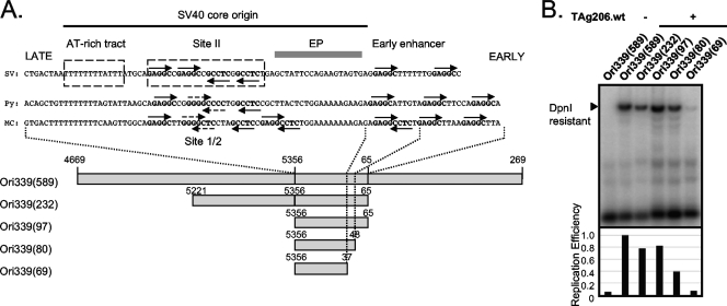
Polyomavirus origins are situated within a noncoding site, located between early and late viral coding regions, which also contains promoters for early and late transcriptional units and enhancers that mediate cis activation of early gene transcription (10) (Fig. 1A). Polyomavirus replication origins contain a central region, referred to as site 1/2 in murine polyomavirus (Py) and site II in SV40, with variable numbers of LT-binding pentanucleotide sequences. Since MCV is more closely related to Py, we use the Py nomenclature in the present study. Site 1/2 is flanked by AT-rich regions: the homopolymeric T tract on the late side of MCV site 1/2 is called the AT-rich tract and is an initial site for polyomavirus DNA melting during replication (15). In contrast for SV40, the initial site of DNA melting occurs on the early side of site II in the early palindromic region. Although this region is also AT-rich in the MCV origin, no palindrome is present (22).
FIG. 1.
Mapping of the MCV core origin. (A) Comparison of the origin sequences among polyomavirus family. The straight and dotted arrow heads indicate complete (GAGGC) or incomplete pentanucleotide sequences (GXGGC and GAGXC) serving for LT binding sites, respectively. The origin of SV40 subgroup (GenBank accession no. EF59667.1) consists of highly conserved region, including the AT-rich tract on the late gene side, site II GAGGC repeats, the early palindrome (EP), and early enhancer region on the early gene side. The MCV origin (GenBank accession no. EU375804) is closely related to the origin of Py (GenBank accession no. J02288). MCV origin constructs of different lengths (589 to 69 bp) were cloned into pCR2.1 vector (Invitrogen) to identify the core origin sequence. (B) Deletion analysis of MCV origin. The origin constructs were cotransfected into 293 cells with either TAg206.wt or empty vector as a negative control, and the replication from the MCV origin was analyzed by Southern blotting. The autoradiogram was quantified by using ImageQuant software (GE Healthcare). The DpnI-sensitive DNA band was used as an internal standard for the amount of input DNA.
An initial step in polyomavirus DNA replication is seeding of LT at consensus DNA 5′-G(A/G)GGC-3′ pentanucleotide sequences (PS) in the LT binding site 1/2 region followed by oligomerization of double LT hexamers in a head-to-head orientation (10). Three structural domains of SV40 LT, the DnaJ domain, the origin-binding domain (OBD), and the helicase domain, have all been subjects of structural studies. Although precise mechanistic details remain unclear, it is known that the helicase domain works in concert with the OBD to initially melt double-stranded DNA in the AT-rich tracts. In SV40, the melted DNA is believed to be threaded through the double hexamer of LT such that the two 3′ ends leaving site II (site 1/2 in Py) pass through the centers of two opposing hexameric rings formed by the LT helicase domain. Once loaded, LT functions as an ATPase-driven DNA pump that also recruits cellular machinery (replication protein A, topoisomerase, and the polymerase alpha complex) for DNA replication (27, 31-35, 44, 57). Sequence analysis shows that the MCV origin has an arrangement of sequence motifs comparable to that of the replication origins of Py subgroup polyomaviruses (22). In contrast to the SV40 origin, which has a minimum, definable boundary, the replication functions of the origin of Py are more diffusely distributed (12, 13, 41, 47). Despite similarities between MCV and SV40 origins, neither MCV nor SV40 T antigen initiate replication of the heterologous origins from the other virus (50).
We identify here an MCV core origin and define the requirement for individual PS in LT-mediated replication that suggests the possibility of a more complex orchestration of LT seeding for MCV than the head-to-head double hexameric arrangement for SV40. Further, a single origin point mutation in a tumor-derived MCV strain at a critical PS outside of the canonical LT interaction region (site II for SV40 and site 1/2 for Py) abrogates MCV origin replication. We show that optimal LT initiation of MCV replication requires both viral sT protein expression and intact cellular Hsc70 binding to fully support efficient viral DNA replication.
MATERIALS AND METHODS
Plasmids.
Construction of genomic (TAg206.wt, TAg350, and TAg339) and cDNA (57kT.wt) TAg expression plasmids were described previously (49, 50). For the LT cDNA construct, cDNA was first amplified by PCR (49), followed by site-directed mutagenesis to delete the 57kT exon 3 splice acceptor site using the PCR primers 5′-CAC TTT TCC CCA AAA GCA AAT CTA AGA GAT TCC C-3′ and 5′-GGG AAT CTC TTA GAT TTG CTT TTG GGG AAA AGT G-3′. To generate sT-eGFP, the open reading frame of sT was amplified from MCC350 (22) by using the primers LT.EcoRV.S (CCG ATA TCA TGG ATT TAG TCC TAA ATA GG) and sT.XhoI.AS (GGG CTC GAG TAG AAA AGG TGC AGA TGC AG) and cloned into pcDNA6/V5-HisB (Invitrogen) using EcoRV and XhoI restriction sites. The sT fragment was then transferred into pEGFP-N1 (Clontech) by using the NheI and SacII sites.
For DnaJ domain mutants of LT genomic constructs, D44H and D44N were generated by a QuikChange Lightning site-directed mutagenesis kit (Stratagene) using TAg206.wt (50) as a template and the following primer pairs: T.D44H(S) (5′-GCT TAA AGC ATC ACC CTC ATA AAG GGG GAA ATC C-3′)/T.D44H(AS) (5′-GGA TTT CCC CCT TTA TGA GGG TGA TGC TTT AAG C-3′) and T.D44N(S) (5′-GCT TAA AGC ATC ACC CTA ATA AAG GGG GAA ATC C-5′)/T.D44N(AS) (5′-GGA TTT CCC CCT TTA TTA GGG TGA TGC TTT AAG C-3′). For LXCXK.D44N, the primers T.D44N(S) and T.D44N(AS) were used for PCR using TAg206.wt.LXCXK (50) as a template.
MCV replication origin plasmids were amplified by PCR from MCC339 genomic DNA (GenBank accession no. EU375804) with the primer sets listed in supplemental Table S1 in the supplemental material and cloned into pCR2.1 (Invitrogen).
Generation of antibodies.
Monoclonal antibodies CM8E6 and CM2B4 (49) were generated by standard methods of immunizing mice with keyhole limpet hemocyanin-derivatized MDLVLNRKERREALC or CSRSRKPSSNASRGA peptide that correspond to the MCV TAg exons 1 and 2, respectively (Epitope Recognition Immunoreagent Core facility, University of Alabama).
MCV origin replication assay.
Human embryonic kidney (HEK) 293 cells were maintained in Dulbecco modified Eagle medium supplemented with 10% fetal bovine serum, 2 mM glutamine, and penicillin-streptomycin. 293 cells were transfected with MCV origin constructs and either empty vector or TAg expression vector by Lipofectamine 2000 (Invitrogen). The cells were harvested 48 h after transfection, lysed with Tris-EDTA-0.6% sodium dodecyl sulfate (SDS) buffer (10 mM Tris-HCl [pH 8.0], 1 mM EDTA, and 0.6% SDS) for 10 min, and then NaCl was added at a final concentration of 1 M. Lysates were centrifuged at 14,000 rpm for 30 min at 4°C to collect episomal DNA. After phenol-chloroform extraction, 5 μg of the collected DNA was double digested overnight with 10 U each of DpnI and BamHI. Digested DNA was loaded on 0.8% agarose gel, transferred onto nitrocellulose membrane, and subjected to Southern hybridization (50). Replication efficiency was detected by Southern hybridization with 32P-labeled probe produced by PCR with the primer pair Rep-S (5′-GCC GCC AAG GAT CTG ATG-3′) and pCR2.1-AS (5′-CTG CGC AAG GAA CGC CCG TCG-3′) and analyzed by using a PhosphorImager (Typhoon 9400; GE Healthcare) and ImageQuant software (GE Healthcare). All replication assays were performed at least twice, and the results of a representative experiment are shown. The expression levels for LT and sT-eGFP were confirmed in each transfection by immunoblotting with anti-V5 (Invitrogen), CM2B4, or anti-GFP (B-2; Santa Cruz).
Immunoprecipitation.
293 cells were cotransfected with pCMV-myc/Hsc70 (5) and either pcDNA6/V5-His/lacZ plasmid (Invitrogen) or TAg plasmid (TAg206.wt, D44H, and D44N) using Lipofectamine 2000 (Invitrogen). Cells were harvested 48 h after transfection and suspended in lysis buffer (50 mM Tris-HCl, 0.15 M NaCl, 1% Triton X-100 [pH 7.4]) supplemented with protease inhibitors. Precleared lysates were immunoprecipitated with either mouse monoclonal anti-c-myc (9E10; Santa Cruz) or rabbit anti-V5 (Bethyl) overnight at 4°C. Lysates were incubated with protein A-Sepharose beads (Amersham) for 3 h at 4°C, collected, and washed with lysis buffer. Beads were resuspended in 2× SDS loading buffer, and proteins were separated by SDS-polyacrylamide gel electrophoresis. Immunoblotting was performed with either anti-c-myc (Santa Cruz) or anti-V5 (Invitrogen) antibody.
siRNA knockdown.
Small interfering RNA (siRNA) targeting TAg (siT [AAGAGAGGCTCTCTGCAAGCT]) and sT (sisT [AAGTTGTCTCGCCAGCATTGT]) alone were designed and synthesized (Qiagen). A nonsilencing siRNA plasmid (siCtrl; Qiagen) was used as control siRNA. For origin replication assay, 1 μg of TAg expression construct, 1 μg of origin construct was cotransfected with 50 nM siRNA into 293 cells grown in a six-well plate by using Lipofectamine 2000 (Invitrogen). Cells were harvested and processed as described above.
Chromatin immunoprecipitation (ChIP).
TAg expression plasmids (TAg206.wt, TAg339, and TAg350) or pcDNA6/V5-His empty vector (Invitrogen) were transfected with Ori339(97) or Ori350(97) plasmid into 293 cells using Lipofectamine 2000 (Invitrogen). Cells were cross-linked with 1% formaldehyde in phosphate-buffered saline, and DNA was sheared by sonication in lysis buffer (50 mM Tris [pH 8.0], 1% SDS, and 10 mM EDTA [pH 8.0] with protease inhibitors). Cell lysates were diluted 10 times with dilution buffer (20 mM Tris [pH 8.0], 1% Triton X-100, 2 mM EDTA [pH 8.0], and 150 mM NaCl with protease inhibitors), and one-third of the diluted lysates was precleared with protein A-Sepharose (GE Healthcare) and 2 μg of sonicated salmon sperm DNA (Stratagene) for 2 h at 4°C. Cell lysates were incubated overnight at 4°C with anti-V5 (Bethyl) antibody and then with protein A-Sepharose beads and 2 μg of salmon sperm DNA for 2 h at 4°C. Beads were washed four times with buffer I (20 mM Tris-HCl [pH 8.0], 150 mM NaCl, 1% Triton X-100, 2 mM EDTA, 0.1% SDS), buffer II (20 mM Tris-HCl [pH 8.0], 500 mM NaCl, 1% Triton X-100, 2 mM EDTA, 0.1% SDS), buffer III (10 mM Tris-HCl [pH 8.0], 500 mM LiCl, 1% NP-40, 1% deoxycholate, 1 mM EDTA), and TE (10 mM Tris-HCl [pH 8.0], 1 mM EDTA). DNA was extracted with buffer (100 mM NaHCO3, 1% SDS) and incubated at 65°C overnight to reverse cross-linking. DNA was purified and resuspended in distilled water, followed by real-time quantitative PCR performed with the primers Rep-S (5′-GCC GCC AAG GAT CTG ATG-3′) and Rep-AS (5′-GAG AAC CTG CGT GCA ATC-3′) using Smart Cycler 5RX4Z01 (Cepheid) and SYBR GreenER qPCR SuperMix reagents (Invitrogen) according to the manufacturer's instruction.
Molecular modeling.
The model of the MCV OBDs on DNA was generated in COOT (18). A homology model of the MCV TAg OBD on a fragment of double-stranded GAGGC-containing DNA was generated by using the SV40 OBD structure (PDB code 2NCG) as a template. Copies of the MCV OBD model were placed on idealized B-form DNA with the MCV origin sequence by superimposing the GAGGC sequence within the homology model onto each of the GAGGC-like sites in the model of the origin. The figure shown was generated in Pymol (http://www.pymol.org/).
RESULTS
The minimum MCV replication origin is defined by a 71-nucleotide genomic region.
MCV is more closely related to the Py subgroup of polyomaviruses than to other human polyomaviruses or SV40 (22). We aligned the sequences of the MCV origin with presumptive origin sequences of SV40 and Py (Fig. 1A). The common features of the core origin in the SV40 subgroup are the presence of the AT-rich tracts contributing to DNA melting (2, 15), flanking a site 1/2 region containing six GAGGC pentanucleotide repeats. An early promoter region containing additional GAGGC binding sites is also present (termed site A in Py). This region is required for Py virus replication but is dispensable for SV40 origin replication (37, 56, 59). The entire origin of MCV contains 10 potential LT binding sequences (PS1 to PS10, see Fig. 3A): six on one strand and four on the opposite strand. Eight of these pentanucleotides match the most common LT binding sequence 5′-GAGGC-3′, while two, PS2 and PS3, in site 1/2 have imperfect consensus sequences: 5′-GGGGC-3′ and 5′-GAGCC-3′, respectively.
FIG. 3.
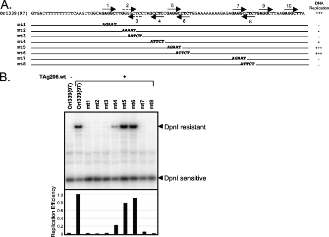
Pentanucleotide requirements for MCV origin replication. (A) Mutation of each pentanucleotide in the 97-bp origin was generated by PCR-based direct mutagenesis (AT to GC) to define the essential pentanucleotides for MCV origin replication. (B) Replication efficiency was examined by Southern blotting.
To define the elements required for LT-mediated replication of MCV, deletions were generated for a tumor-derived MCV origin construct [Ori339(589), nucleotides 4669 to 269] from MCV clone 339 (GenBank accession no. EU375804) (Fig. 1A). This origin fragment is perfectly conserved in most tumor and all non-tumor-derived MCV strains thus far sequenced. Origin constructs were cloned into pCR2.1 (Invitrogen) and cotransfected together with a full-length wild-type T-antigen genomic expression construct (TAg206.wt, isolated from intestine tissue of a patient without MCC) into 293 cells. In the present study, we refer to the genomic T antigen cassette as TAg, which expresses sT and 57kT proteins from alternatively spliced mRNAs in addition to LT protein (50). Origin replication was determined by Southern hybridization in which DpnI-resistant (replicated) signal is normalized to DpnI-sensitive (unreplicated) signal. In contrast to Py but similar to SV40, the MCV origin can be reduce to a discrete region such that constructs encompassing only an 80-bp origin show efficient replication by MCV TAg (Fig. 1B). Replication is completely abolished for a shorter 69-bp origin construct. The 80-bp fragment (nucleotides 5356 to 48) contains, in addition to the AT-rich tract, eight of the identifiable LT binding PS (Fig. 1A), indicating that PS9 and PS10 are not required for origin replication.
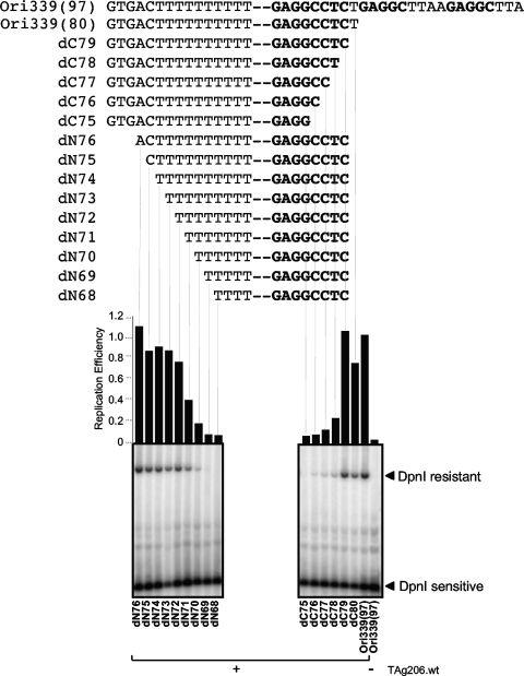
To define a minimal core origin, we successively truncated single base pairs from the early and late sides of the 80-bp origin until replication was completely abolished (Fig. 2). A significant decline in MCV replication efficiency occurs only after deletion of four of the ten thymidines in the poly(T) tract (dN70). This is in contrast to the SV40 origin, which requires an intact poly(T) tract for efficient replication (15). MCV origin replication also declines sharply starting at the dC78 deletion that truncates PS8 on the early side of the origin. Thus, unlike Py, a minimal MCV core origin consisting of the 71-bp region from nucleotides 5364 through 47 can efficiently sustain LT-mediated DNA replication.
FIG. 2.
Definition of MCV minimal core origin. Single-base-pair deletions from both sides of the origin sequence (late, nucleotide 5356; early, nucleotide 48) were performed to define a minimal core origin. The expression level of TAg was observed by Western blotting (data not shown), and an autoradiogram of replication was quantified as in Fig. 1B (middle panel).
Pentanucleotide requirements for MCV replication.
Four LT binding sites (Fig. 1A) are present in the SV40 core origin, and all four pentanucleotides are necessary for origin DNA unwinding and replication events with pentanucleotides 1 and 3 needed for double hexamer formation (14, 16). These pentanucleotides are arranged on opposite strands, allowing the double SV40 LT hexamer to form in a head-to-head orientation (28). To determine whether a similar pentanucleotide arrangement is needed for MCV replication, mutations were introduced that disrupted nucleotides in each of the pentanucleotide sequences from PS1 to PS8 in the 97-bp origin (Fig. 3A). Mutations in three pentanucleotides within the region most analogous to site II (PS1 to PS4) (Fig. 1A) and two pentanucleotides (PS7 and PS8) near the early enhancer region abolish replication. The PS4 mutant significantly diminishes replication. In contrast, PS5 and PS6 are dispensable (Fig. 3B).
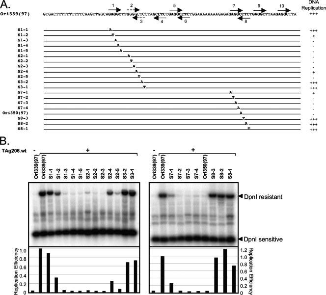
Since PS2 and PS3 partially overlap, as do PS7 and PS8, complete pentanucleotide substitutions affect overlapping elements because of shared nucleotides. We therefore investigated critical nucleotides in these four elements (PS2, PS3, PS7, and PS8) by introducing single mutations to determine which of the overlapping PS sequences are essential (Fig. 4A), as well as critical nucleotides in PS1. Figure 4B shows that single base pair mutations in three pentanucleotides (PS1, PS2, and PS7) impair efficient replication. The three mutations in PS8 that do not overlap PS7 do not impair replication (Fig. 4B). In the nonconsensus PS2 element, a single substitution mutation that reconstitutes a canonical GAGGC site remains competent for replication (see Fig. S1 in the supplemental material). Single mutations unique to the PS3 site that do not overlap with the opposite strand PS2 have no effect on replication, suggesting that PS2 is the critical LT binding element in this overlapping pair. A PS3 (S3-2C) mutation that creates the same head-to-head LT binding site as in the Py origin (GGGGCCCC) (Fig. 1A) has no replication capacity (see Fig. S1 in the supplemental material), indicating that the MCV LT replication complex may form assemblies on the DNA that are distinct from Py and SV40.
FIG. 4.
Single mutational analysis of pentanucleotides. (A) Each sequence of pentanucleotides 1, 2, 3, 7, and 8 was mutated by AT-to-GC substitutions to disrupt the TAg binding site. To avoid reconstitution of the T-antigen binding site [5′-G(A/G)GGC-3′] by mutation, AG-to-T substitutions were introduced in S7-2, S8-2, S2-2, and S3-2. (B) Southern blot results demonstrate that single base mutations in pentanucleotides 1, 2, and 7 can abolish replication efficiently, indicating that these pentanucleotides sites are critical for MCV origin replication as a functional unit.
The importance of PS7 to MCV replication is illustrated by naturally arising mutations at the viral origin [Ori350(97)] lane of Fig. 4B. We sequenced MCV origins from one nontumor tissue, eight MCC tumors and two cell lines (22). Only the origin sequence from one tumor, MCV350 (GenBank accession no. EU375803), has a C-to-A substitution in position 5 of PS7 that also alters the sequence of the overlapping PS8. This natural mutation completely abrogates origin replication in the presence of TAg (Fig. 4B).
Effects of tumor-derived LT mutations on origin binding and replication.
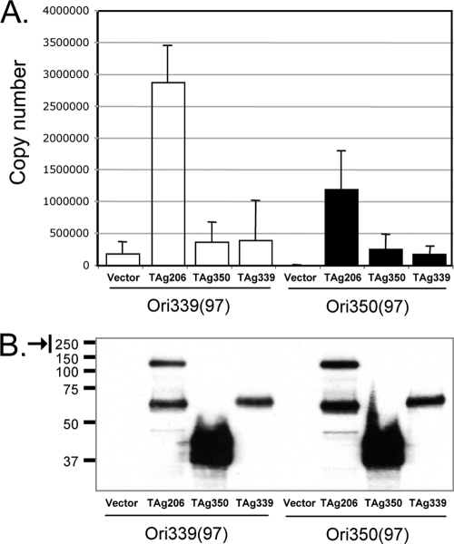
As previously described (50), tumor-derived MCV TAg is unable to initiate MCV origin replication due to truncating mutations in the LT protein. We compared origin-binding properties for wild-type LT protein (TAg206.wt) on the wild-type origin [Ori339(97)] and the mutated Ori350 [Ori350(97)], as well as mutant LT proteins from tumor-derived viral strains (MCV339 and MCV350) (Fig. 5). MCV339 LT protein retains the OBD but has a truncated helicase domain, whereas the MCV350 LT mutation eliminates both the OBD and the helicase domains (50). Quantitative ChIP assays demonstrate that wild-type TAg206 efficiently binds to the wild-type origin Ori339 but binding to Ori350, possessing the PS7/PS8 mutation, is reduced ∼50%. Origin binding by MCV339 and MCV350 TAg proteins is comparable to vector alone controls for both viral origins, suggesting that structural or enzymatic features of the MCV LT helicase domain are required for efficient recognition of the DNA element. Relative equal expression levels of wild-type and mutant LT proteins are shown by Western blotting (Fig. 5B).
FIG. 5.
ChIP analysis. (A) The binding efficiency of both non-tumor- and tumor-derived TAg to MCV origin in vivo was examined by using a ChIP assay. The 97-bp origin-containing plasmid [Ori339(97) or Ori350(97)] and the TAg constructs from nontumor (TAg206.wt) or tumor (TAg350 and TAg339) tissues (50) were cotransfected into 293 cells, and the binding of LT to the origin was investigated by quantitative PCR. All experiments were repeated at least three times. Error bars indicate the standard deviations. (B) The relative expression levels of wild-type and mutant LT proteins are demonstrated by Western blotting.
Role of intrinsic activities of T antigens that influence MCV replication.
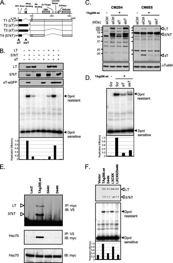
Four major TAg transcripts (T1 to T4) have been identified by using RACE and Northern blotting that correspond to LT, sT, and 57kT (50) (Fig. 6A). The sT of MCV shares exon 1 with LT, which contains the conserved CR1 (LXXLL motif) and DnaJ (HPDK motif) domains but lacks the LT OBD required for DNA recognition. The sT also encodes a PP2A binding site (CXCXXC) that is spliced out of LT and 57kT cDNAs.
FIG. 6.
Intrinsic activities of T antigens that influence MCV replication. (A) Alternative spliced products (T1 to T4) of the early region give rise to LT, sT, and 57kT. Arrows indicate the positions of siRNAs, siT, and sisT. siT targets all TAg transcripts, while sisT targets only sT transcripts. An asterisk indicates the epitope site for CM8E6, and a star indicates the epitope site of CM2B4 antibodies. These antibodies detect LT/57kT/sT or LT/57kT proteins, respectively. The predicted MCV LT gene sequence retains all major conserved features of other polyomavirus LTs, including DnaJ, Rb-targeting, origin-binding, and helicase/ATPase domains. (B) sT expression significantly contributes to the efficiency of origin replication. LT, 57kT, and sT expression plasmids were cotransfected in various combinations into 293 cell. For sT expression, a green fluorescent protein (GFP) fusion construct was used. Western blotting was performed to monitor the expression level of each transcript product using V5 antibody (Invitrogen) for LT and 57kT and anti-GFP (SantaCruz) antibody for sT. A representative experiment out of three repeats is shown. (C) siRNA knockdown of LT, 57kT, and/or sT expression confirms the contribution of sT to efficient origin replication (D). A nonsilencing siRNA (siCtrl) was used as a control. Three specific bands (120, 57, and 19 kDa) are detected by Western blotting with CM8E6 antibody in 293 cells transfected with genomic TAg expression constructs (TAg206.wt) (C, right panel). CM2B4 detected both ∼120- and ∼57-kDa bands corresponding to the full-length LT and 57kT protein products, respectively (49) (C, left panel). (E) To examine the interaction between TAg and Hsc70 and its role in origin replication, Myc-tagged Hsc70 construct (pCMV-myc-Hsc70) was transfected into 293 cells with either V5-tagged wild-type TAg (TAg206.wt) or Hsc70 mutant TAg (D44H, D44N). Lysates were immunoprecipitated with anti-myc or anti-V5 antibody and immunoblotted with anti-V5 or anti-myc antibody in a reciprocal way, respectively. The LacZ gene-expressing vector, pcDNA6/V5-His/LacZ (Invitrogen), was used as a negative control for binding and 2% of the lysate was used for input control. (F) Hsc70 binding to TAg promotes efficient viral replication. For the replication assay, TAg206.wt or mutants (Hsc70 binding mutant, D44N; Rb binding mutant, LXCXK; Rb/Hsc70 binding mutant, LXCXK/D44N) were cotransfected with MCV origin plasmid [Ori339(97)] into 293 cells. Replication efficiency was measured and quantified by using ImageQuant (GE Healthcare).
To test the effect of sT and other transcripts on viral origin replication, each cDNA transcript was cloned to express a unique TAg isoform. Each cDNA construct, singly and in combination, was then transfected into 293 cells to assess effects on origin replication. As seen in Fig. 6B, LT cDNA expression alone is not sufficient for fully efficient replication, but replication efficiency is restored to that of the wild-type genomic TAg by coexpression of LT with sT. Coexpression of LT with 57kT does not increase replication over LT alone, and neither MCV sT nor 57kT individually have replication capacity.
To confirm the importance of sT in replication, we used two siRNAs: siT targets all TAg transcripts, and sisT targets only sT transcripts (Fig. 6A) (50). We also generated a monoclonal antibody, CM8E6, directed against an exon 1 peptide shared by all TAg isoforms. Western blotting shows that siT downregulates LT, sT, and 57kT protein expression as expected, whereas sisT only reduces sT expression (19 kDa) (Fig. 6C). Knockdown of TAg proteins by either siT and sisT results in inhibition of origin replication compared to siCtrl control, confirming the functional importance of sT expression to MCV origin replication (Fig. 6D). Similar results were obtained with vector-based short hairpin targeting of TAg (data not shown).
Both MCV LT and sT contain the DnaJ motif encoded by exon 1 that is conserved among polyomaviruses and which recruits chaperone heat shock proteins (25). SV40 LT association with Hsc70 (46) is mediated through the HPDK motif within the DnaJ domain (5, 48), which is required for efficient origin replication. We confirmed that this motif in MCV is a site for Hsc70 interaction by coimmunoprecipitation of V5-tagged wild-type (TAg206.wt) and DnaJ domain mutant (D44H and D44N) proteins with myc-tagged Hsc70 (Fig. 6E).
To test whether Hsc70 interaction with MCV TAg is required for efficient viral DNA replication, the wild type (TAg 206.wt) and the LT DnaJ mutant D44N were tested for origin replication using Ori339(589) (Fig. 6F). Mutation of the LT DnaJ domain markedly decreases origin replication compared to wild-type MCV TAg, whereas mutation of the MCV LT retinoblastoma-binding motif (LXCXE) does not, results similar to those found for SV40 TAg (5). The MCV D44N LT mutant is less stable than wild-type MCV LT but enhanced expression to generate comparable levels of both proteins does not increase replication with the DnaJ-binding-deficient protein (Fig. 6F). In contrast, mutation of the DnaJ domain in sT has no effect on the ability of sT to enhance wild-type LT replication efficiency. In addition, mutation in the PP2A domain of sT abolishes its ability to enhance LT-mediated origin replication (see Fig. S3 in the supplemental material).
DISCUSSION
We found that a minimal MCV 71-bp core origin sequence is sufficient for TAg-directed DNA replication. Like other polyomaviruses, this MCV core origin contains three subdomains: an AT-rich tract, a LT-binding domain, and an early enhancer region. The AT-rich tract for SV40 directs DNA bending that may be a structural signal for LT binding (15). Adjacent to the AT-rich tract, pentanucleotides sequences are arranged as two pairs of imperfect repeat sequences (PS1 to PS4) inverted with respect to each other. These repeats are similar but more closely spaced than those in SV40. Of these four, only three (PS1, PS2, and PS4) are required for origin replication (Fig. 7). In addition to these three PS elements, the MCV origin has an additional LT binding site at PS7, which is required for origin replication and, if disrupted, as found in a naturally occurring tumor-derived strain, markedly reduces LT binding to the origin element in our quantitative ChIP assay and completely ablates origin replication. We found that PS3, PS8, PS9, PS10 are dispensable (Fig. 7), but mutational substitution analysis shows that even single nucleotide alterations in essential pentanucleotides, e.g., PS1, PS2, PS3, and PS7, disrupt origin replication.
FIG. 7.
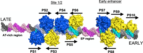
Model of the MCV OBDs on the MCV origin. A model of the MCV OBD was placed on each of the putative binding sites. OBDs positioned at the required PS1, PS2, PS4, and PS7 are shown in blue. The remainder are yellow. The GAGGC sequences are shown in cyan. The AT-rich tracts of DNA that flank site 1/2 are shown in light magenta.
The OBDs of MCV, Py, and SV40 are similar in sequence, and residues shown to bind GAGGC pentamers are highly conserved in the three viruses. The MCV OBD is 49% identical to Py and 48% identical to SV40 (see Fig. S2 in the supplemental material) To visualize the spatial arrangement of GAGGC binding sites of MCV, the crystal structure of the SV40 OBD on DNA was used to model the interaction of MCV TAg with the MCV origin. As shown in Fig. 7, the closely spaced PS sites also position the MCV LT OBD sites close to one another. Given this proximity, it is likely that OBD-OBD interactions occur while apposing LT molecules are bound to the DNA. Such interactions are not observed in SV40, where the GAGGC sequences are farther separated, but they do occur in the more distantly related bovine papillomavirus (BPV) system (19, 20). In BPV, the analogous domain is part of the E1 protein and is not capable of recognizing its binding site without the help of the virally encoded E2 protein. MCV origin recognition may share features of both SV40 and BPV. MCV is similar to SV40 in that replication does not require an E2-like protein but, like BPV, interactions among the OBDs may help stabilize the complex on the DNA, perhaps alleviating the need for well-formed binding sites at all positions.
Based on our modeling, two sets of OBD-OBD interactions may occur on the DNA, both of which are head-to-head interactions. The first is between the OBDs bound to PS2 and PS3, and the putative protein-protein interface involves amino acids within the major groove of the DNA. The second potential interaction involves residues well outside of the DNA-binding cleft and could occur between OBDs on PS1 and PS3 and between OBDs on PS2 and PS4.
Aside from direct LT binding to the origin, other viral factors and motifs were found to be critical for efficient MCV DNA replication. Several studies using SV40, Py and JCV sT defective viruses and constructs demonstrate an auxiliary role for sT in origin replication (8, 9, 39). We found that sT, but not 57kT, enhances the origin replication activity of MCV LT. The mechanism for this enhancement remains poorly understood, but it is likely that sT does not act directly in the LT-origin replication complex. A mutation the MCV sT PP2A-binding domain (26) completely abrogates sT-enhanced LT-origin replication (see Fig. S3 in the supplemental material). It is reasonable to speculate that MCV sT stimulates cell cycle entry and thereby increases origin replication efficiency via PP2A targeting or that PP2A targeting by sT selectively affects the phosphorylation state of LT to enhance its binding affinity to the origin DNA binding site (40, 61). In contrast, whereas both MCV LT and sT possess DnaJ domains encoded from exon 1 that recruit Hsp70, this domain is dispensable from sT but is required for LT-mediated origin replication. As seen for other polyomaviruses (60), site-directed mutagenesis of LT DnaJ domain residues reduces the protein's stability. This is unlikely to account for the reduction in replication for LT DnaJ domain mutants, however, since replication activity remains reduced even when the mutant LT protein is expressed at comparable levels to the wild-type protein. Surprisingly, while SV40 17kT protein has been proposed to play a role in SV40 replication, we found no evidence for a role of the analogous 57kT MCV protein in in vitro MCV replication. Since 57kT is highly conserved among MCV strains, there is little doubt that it plays a role in MCV survival and propagation, but at present we have no clues as to what this role might be.
MCV provides a novel model for a defined DNA replication system in eukaryotic cells. We have characterized an MCV minimal replication origin, as well as the viral components of the replication complex for this virus so that it may be amenable to structural studies. Our results already suggest intriguing similarities and differences from the better-studied SV40 replication origin, and additional comparisons between these viruses are likely to provide insights that cannot be obtained from either virus individually.
The importance of MCV replication studies is also illustrated by the MCV350 origin mutation that eliminates TAg-mediated replication. The MCV350 strain encodes both a truncated T-antigen protein and an origin mutation at PS7, either one of which is sufficient to prevent viral DNA replication, illustrating the strong selective pressure against replication-competent viruses in human MCC tumors. It is possible that as additional tumor-derived MCV strains are described, viruses with intact T antigens but defective replication origins may be found. Dissection of the individual components and steps for both wild-type and tumor-derived virus replication may point toward unique antiviral chemotherapies for this devastating cancer.
Supplementary Material
Acknowledgments
We thank Huichen Feng and Ole Gjoerup for helpful comments, Mary Ann Accavitti for production of CM2B4 and CM8E6 antibodies, and Xi Liu for PP2A/small T interaction studies. We thank Marc Mellits and John Adams for help with the manuscript.
This study was supported in part by awards from the Al Copeland Foundation, the University of Pittsburgh EXPLORER program, and NIH grants CA136363 and CA120726. A.B. and G.M. are supported by NIH grant R21 AI082496. Y.C. and P.S.M. are funded as American Cancer Society Research Professors.
Footnotes
Published ahead of print on 16 September 2009.
Supplemental material for this article may be found at http://jvi.asm.org/.
REFERENCES
- 1.Benbow, R. M., J. Zhao, and D. D. Larson. 1992. On the nature of origins of DNA replication in eukaryotes. Bioessays 14:661-670. [DOI] [PubMed] [Google Scholar]
- 2.Borowiec, J. A., and J. Hurwitz. 1988. Localized melting and structural changes in the SV40 origin of replication induced by T-antigen. EMBO J. 7:3149-3158. [DOI] [PMC free article] [PubMed] [Google Scholar]
- 3.Bouck, N., N. Beales, T. Shenk, P. Berg, and G. di Mayorca. 1978. New region of the simian virus 40 genome required for efficient viral transformation. Proc. Natl. Acad. Sci. USA 75:2473-2477. [DOI] [PMC free article] [PubMed] [Google Scholar]
- 4.Buck, C. B., and D. R. Lowy. 2009. Getting stronger: the relationship between a newly identified virus and Merkel cell carcinoma. J. Investig. Dermatol. 129:9-11. [DOI] [PMC free article] [PubMed] [Google Scholar]
- 5.Campbell, K. S., K. P. Mullane, I. A. Aksoy, H. Stubdal, J. Zalvide, J. M. Pipas, P. A. Silver, T. M. Roberts, B. S. Schaffhausen, and J. A. DeCaprio. 1997. DnaJ/hsp40 chaperone domain of SV40 large T antigen promotes efficient viral DNA replication. Genes Dev. 11:1098-1110. [DOI] [PubMed] [Google Scholar]
- 6.Chang, L. S., S. Pan, M. M. Pater, and G. Di Mayorca. 1985. Differential requirement for SV40 early genes in immortalization and transformation of primary rat and human embryonic cells. Virology 146:246-261. [DOI] [PubMed] [Google Scholar]
- 7.Chang, L. S., M. M. Pater, N. I. Hutchinson, and G. di Mayorca. 1984. Transformation by purified early genes of simian virus 40. Virology 133:341-353. [DOI] [PubMed] [Google Scholar]
- 8.Chen, M. C., D. Redenius, F. Osati-Ashtiani, and M. M. Fluck. 1995. Enhancer-mediated role for polyomavirus middle T/small T in DNA replication. J. Virol. 69:326-333. [DOI] [PMC free article] [PubMed] [Google Scholar]
- 9.Cicala, C., M. L. Avantaggiati, A. Graessmann, K. Rundell, A. S. Levine, and M. Carbone. 1994. Simian virus 40 small-t antigen stimulates viral DNA replication in permissive monkey cells. J. Virol. 68:3138-3144. [DOI] [PMC free article] [PubMed] [Google Scholar]
- 10.Cole, C. N., and S. D. Conzen. 2001. Polyomavirinae: the viruses and their replication, p. 2141-2174. In D. M. Knipe and P. M. Howley (ed.), Fields virology, 4th ed. Lippincott/The Williams & Wilkins Co., Philadelphia, PA.
- 11.Cowan, K., P. Tegtmeyer, and D. D. Anthony. 1973. Relationship of replication and transcription of simian virus 40 DNA. Proc. Natl. Acad. Sci. USA 70:1927-1930. [DOI] [PMC free article] [PubMed] [Google Scholar]
- 12.Cowie, A., and R. Kamen. 1986. Guanine nucleotide contacts within viral DNA sequences bound by polyomavirus large T antigen. J. Virol. 57:505-514. [DOI] [PMC free article] [PubMed] [Google Scholar]
- 13.Cowie, A., and R. Kamen. 1984. Multiple binding sites for polyomavirus large T antigen within regulatory sequences of polyomavirus DNA. J. Virol. 52:750-760. [DOI] [PMC free article] [PubMed] [Google Scholar]
- 14.Dean, F. B., P. Bullock, Y. Murakami, C. R. Wobbe, L. Weissbach, and J. Hurwitz. 1987. Simian virus 40 (SV40) DNA replication: SV40 large T antigen unwinds DNA containing the SV40 origin of replication. Proc. Natl. Acad. Sci. USA 84:16-20. [DOI] [PMC free article] [PubMed] [Google Scholar]
- 15.Deb, S., A. L. DeLucia, A. Koff, S. Tsui, and P. Tegtmeyer. 1986. The adenine-thymine domain of the simian virus 40 core origin directs DNA bending and coordinately regulates DNA replication. Mol. Cell. Biol. 6:4578-4584. [DOI] [PMC free article] [PubMed] [Google Scholar]
- 16.Deb, S., S. Tsui, A. Koff, A. L. DeLucia, R. Parsons, and P. Tegtmeyer. 1987. The T-antigen-binding domain of the simian virus 40 core origin of replication. J. Virol. 61:2143-2149. [DOI] [PMC free article] [PubMed] [Google Scholar]
- 17.Duncavage, E. J., B. A. Zehnbauer, and J. D. Pfeifer. 2009. Prevalence of Merkel cell polyomavirus in Merkel cell carcinoma. Mod. Pathol. 22:516-521. [DOI] [PubMed] [Google Scholar]
- 18.Emsley, P., and K. Cowtan. 2004. Coot: model-building tools for molecular graphics. Acta Crystallogr. D Biol. Crystallogr. 60:2126-2132. [DOI] [PubMed] [Google Scholar]
- 19.Enemark, E. J., G. Chen, D. E. Vaughn, A. Stenlund, and L. Joshua-Tor. 2000. Crystal structure of the DNA binding domain of the replication initiation protein E1 from papillomavirus. Mol. Cell 6:149-158. [PubMed] [Google Scholar]
- 20.Enemark, E. J., A. Stenlund, and L. Joshua-Tor. 2002. Crystal structures of two intermediates in the assembly of the papillomavirus replication initiation complex. EMBO J. 21:1487-1496. [DOI] [PMC free article] [PubMed] [Google Scholar]
- 21.Fanning, E., and R. Knippers. 1992. Structure and function of simian virus 40 large tumor antigen. Annu. Rev. Biochem. 61:55-85. [DOI] [PubMed] [Google Scholar]
- 22.Feng, H., M. Shuda, Y. Chang, and P. S. Moore. 2008. Clonal integration of a polyomavirus in human Merkel cell carcinoma. Science 319:1096-1100. [DOI] [PMC free article] [PubMed] [Google Scholar]
- 23.Feng, H., J. L. Taylor, P. V. Benos, R. Newton, K. Waddell, S. B. Lucas, Y. Chang, and P. S. Moore. 2007. Human transcriptome subtraction by using short sequence tags to search for tumor viruses in conjunctival carcinoma. J. Virol. 81:11332-11340. [DOI] [PMC free article] [PubMed] [Google Scholar]
- 24.Hahn, W. C., S. K. Dessain, M. W. Brooks, J. E. King, B. Elenbaas, D. M. Sabatini, J. A. DeCaprio, and R. A. Weinberg. 2002. Enumeration of the simian virus 40 early region elements necessary for human cell transformation. Mol. Cell. Biol. 22:2111-2123. [DOI] [PMC free article] [PubMed] [Google Scholar]
- 25.Hartl, F. U. 1996. Molecular chaperones in cellular protein folding. Nature 381:571-579. [DOI] [PubMed] [Google Scholar]
- 26.Janssens, V., and J. Goris. 2001. Protein phosphatase 2A: a highly regulated family of serine/threonine phosphatases implicated in cell growth and signaling. Biochem. J. 353:417-439. [DOI] [PMC free article] [PubMed] [Google Scholar]
- 27.Jiang, X., V. Klimovich, A. I. Arunkumar, E. B. Hysinger, Y. Wang, R. D. Ott, G. D. Guler, B. Weiner, W. J. Chazin, and E. Fanning. 2006. Structural mechanism of RPA loading on DNA during activation of a simple pre-replication complex. EMBO J. 25:5516-5526. [DOI] [PMC free article] [PubMed] [Google Scholar]
- 28.Joo, W. S., X. Luo, D. Denis, H. Y. Kim, G. J. Rainey, C. Jones, K. R. Sreekumar, and P. A. Bullock. 1997. Purification of the simian virus 40 (SV40) T antigen DNA-binding domain and characterization of its interactions with the SV40 origin. J. Virol. 71:3972-3985. [DOI] [PMC free article] [PubMed] [Google Scholar]
- 29.Kassem, A., A. Schopflin, C. Diaz, W. Weyers, E. Stickeler, M. Werner, and A. Zur Hausen. 2008. Frequent detection of Merkel cell polyomavirus in human Merkel cell carcinomas and identification of a unique deletion in the VP1 gene. Cancer Res. 68:5009-5013. [DOI] [PubMed] [Google Scholar]
- 30.Kean, J. M., S. Rao, M. Wang, and R. L. Garcea. 2009. Seroepidemiology of human polyomaviruses. PLoS Pathog. 5:e1000363. [DOI] [PMC free article] [PubMed] [Google Scholar]
- 31.Kumar, A., G. Meinke, D. K. Reese, S. Moine, P. J. Phelan, A. Fradet-Turcotte, J. Archambault, A. Bohm, and P. A. Bullock. 2007. Model for T-antigen-dependent melting of the simian virus 40 core origin based on studies of the interaction of the beta-hairpin with DNA. J. Virol. 81:4808-4818. [DOI] [PMC free article] [PubMed] [Google Scholar]
- 32.Li, D., R. Zhao, W. Lilyestrom, D. Gai, R. Zhang, J. A. DeCaprio, E. Fanning, A. Jochimiak, G. Szakonyi, and X. S. Chen. 2003. Structure of the replicative helicase of the oncoprotein SV40 large tumour antigen. Nature 423:512-518. [DOI] [PubMed] [Google Scholar]
- 33.Liu, H., Y. Shi, X. S. Chen, and A. Warshel. 2009. Simulating the electrostatic guidance of the vectorial translocations in hexameric helicases and translocases. Proc. Natl. Acad. Sci. USA 106:7449-7454. [DOI] [PMC free article] [PubMed] [Google Scholar]
- 34.Meinke, G., P. A. Bullock, and A. Bohm. 2006. Crystal structure of the simian virus 40 large T-antigen origin-binding domain. J. Virol. 80:4304-4312. [DOI] [PMC free article] [PubMed] [Google Scholar]
- 35.Meinke, G., P. Phelan, S. Moine, E. Bochkareva, A. Bochkarev, P. A. Bullock, and A. Bohm. 2007. The crystal structure of the SV40 T-antigen origin binding domain in complex with DNA. PLoS Biol. 5:e23. [DOI] [PMC free article] [PubMed] [Google Scholar]
- 36.Myers, R. M., D. C. Rio, A. K. Robbins, and R. Tjian. 1981. SV40 gene expression is modulated by the cooperative binding of T antigen to DNA. Cell 25:373-384. [DOI] [PubMed] [Google Scholar]
- 37.Peng, Y. C., and N. H. Acheson. 1998. Polyomavirus large T antigen binds cooperatively to its multiple binding sites in the viral origin of DNA replication. J. Virol. 72:7330-7340. [DOI] [PMC free article] [PubMed] [Google Scholar]
- 38.Porras, A., J. Bennett, A. Howe, K. Tokos, N. Bouck, B. Henglein, S. Sathyamangalam, B. Thimmapaya, and K. Rundell. 1996. A novel simian virus 40 early-region domain mediates transactivation of the cyclin A promoter by small-t antigen and is required for transformation in small-t antigen-dependent assays. J. Virol. 70:6902-6908. [DOI] [PMC free article] [PubMed] [Google Scholar]
- 39.Prins, C., and R. J. Frisque. 2001. JC virus T' proteins encoded by alternatively spliced early mRNAs enhance T antigen-mediated viral DNA replication in human cells. J. Neurovirol. 7:250-264. [DOI] [PubMed] [Google Scholar]
- 40.Prives, C. 1990. The replication functions of SV40 T antigen are regulated by phosphorylation. Cell 61:735-738. [DOI] [PubMed] [Google Scholar]
- 41.Prives, C., Y. Murakami, F. G. Kern, W. Folk, C. Basilico, and J. Hurwitz. 1987. DNA sequence requirements for replication of polyomavirus DNA in vivo and in vitro. Mol. Cell. Biol. 7:3694-3704. [DOI] [PMC free article] [PubMed] [Google Scholar]
- 42.Pulitzer, M. P., B. D. Amin, and K. J. Busam. 2009. Merkel cell carcinoma: review. Adv. Anat. Pathol. 16:135-144. [DOI] [PubMed] [Google Scholar]
- 43.Rio, D., A. Robbins, R. Myers, and R. Tjian. 1980. Regulation of simian virus 40 early transcription in vitro by a purified tumor antigen. Proc. Natl. Acad. Sci. USA 77:5706-5710. [DOI] [PMC free article] [PubMed] [Google Scholar]
- 44.San Martin, M. C., C. Gruss, and J. M. Carazo. 1997. Six molecules of SV40 large T antigen assemble in a propeller-shaped particle around a channel. J. Mol. Biol. 268:15-20. [DOI] [PubMed] [Google Scholar]
- 45.Sastre-Garau, X., M. Peter, M. F. Avril, H. Laude, J. Couturier, F. Rozenberg, A. Almeida, F. Boitier, A. Carlotti, B. Couturaud, and N. Dupin. 2009. Merkel cell carcinoma of the skin: pathological and molecular evidence for a causative role of MCV in oncogenesis. J. Pathol. 218:48-56. [DOI] [PubMed] [Google Scholar]
- 46.Sawai, E. T., and J. S. Butel. 1989. Association of a cellular heat shock protein with simian virus 40 large T antigen in transformed cells. J. Virol. 63:3961-3973. [DOI] [PMC free article] [PubMed] [Google Scholar]
- 47.Scheller, A., and C. Prives. 1985. Simian virus 40 and polyomavirus large tumor antigens have different requirements for high-affinity sequence-specific DNA binding. J. Virol. 54:532-545. [DOI] [PMC free article] [PubMed] [Google Scholar]
- 48.Sheng, Q., D. Denis, M. Ratnofsky, T. M. Roberts, J. A. DeCaprio, and B. Schaffhausen. 1997. The DnaJ domain of polyomavirus large T antigen is required to regulate Rb family tumor suppressor function. J. Virol. 71:9410-9416. [DOI] [PMC free article] [PubMed] [Google Scholar]
- 49.Shuda, M., R. Arora, H. J. Kwun, H. Feng, R. Sarid, M. Fernández-Figueras, Y. Tolstov, O. Gjoerup, M. M. Mansukhani, S. H. Swerdlow, P. M. Chaudhary, J. M. Kirkwood, M. A. Nalesnik, J. A. Kant, L. M. Weiss, P. S. Moore, and Y. Chang. 2009. Human Merkel cell polyomavirus infection I. MCV T antigen expression in Merkel cell carcinoma, lymphoid tissues and lymphoid tumors. Int. J. Cancer 125:1243-1249. [DOI] [PMC free article] [PubMed] [Google Scholar]
- 50.Shuda, M., H. Feng, H. J. Kwun, S. T. Rosen, O. Gjoerup, P. S. Moore, and Y. Chang. 2008. T antigen mutations are a human tumor-specific signature for Merkel cell polyomavirus. Proc. Natl. Acad. Sci. USA 105:16272-16277. [DOI] [PMC free article] [PubMed] [Google Scholar]
- 51.Spence, S. L., and J. M. Pipas. 1994. Simian virus 40 large T antigen host range domain functions in virion assembly. J. Virol. 68:4227-4240. [DOI] [PMC free article] [PubMed] [Google Scholar]
- 52.Spence, S. L., and J. M. Pipas. 1994. SV40 large T antigen functions at two distinct steps in virion assembly. Virology 204:200-209. [DOI] [PubMed] [Google Scholar]
- 53.Srinivasan, A., A. J. McClellan, J. Vartikar, I. Marks, P. Cantalupo, Y. Li, P. Whyte, K. Rundell, J. L. Brodsky, and J. M. Pipas. 1997. The amino-terminal transforming region of simian virus 40 large T and small t antigens functions as a J domain. Mol. Cell. Biol. 17:4761-4773. [DOI] [PMC free article] [PubMed] [Google Scholar]
- 54.Tjian, R. 1981. T antigen binding and the control of SV40 gene expression. Cell 26:1-2. [DOI] [PubMed] [Google Scholar]
- 55.Tolstov, Y. L., D. V. Pastrana, H. Feng, J. C. Becker, F. J. Jenkins, S. Moschos, Y. Chang, C. B. Buck, and P. S. Moore. 2009. Human Merkel cell polyomavirus infection II. MCV is a common human infection that can be detected by conformational capsid epitope immunoassays. Int. J. Cancer 125:1250-1256. [DOI] [PMC free article] [PubMed] [Google Scholar]
- 56.Triezenberg, S. J., and W. R. Folk. 1984. Essential nucleotides in the polyomavirus origin region. J. Virol. 51:437-444. [DOI] [PMC free article] [PubMed] [Google Scholar]
- 57.Valle, M., X. S. Chen, L. E. Donate, E. Fanning, and J. M. Carazo. 2006. Structural basis for the cooperative assembly of large T antigen on the origin of replication. J. Mol. Biol. 357:1295-1305. [DOI] [PubMed] [Google Scholar]
- 58.Varga, E., M. Kiss, K. Szabo, and L. Kemeny. 2009. Detection of Merkel cell polyomavirus DNA in Merkel cell carcinomas. Br. J. Dermatol. 161:930-932. [DOI] [PubMed] [Google Scholar]
- 59.Weichselbraun, I., G. Haider, and E. Wintersberger. 1989. Optimal replication of plasmids carrying polyomavirus origin regions requires two high-affinity binding sites for large T antigen. J. Virol. 63:961-964. [DOI] [PMC free article] [PubMed] [Google Scholar]
- 60.Whalen, K. A., R. de Jesus, J. A. Kean, and B. S. Schaffhausen. 2005. Genetic analysis of the polyomavirus DnaJ domain. J. Virol. 79:9982-9990. [DOI] [PMC free article] [PubMed] [Google Scholar]
- 61.Yang, S. I., R. L. Lickteig, R. Estes, K. Rundell, G. Walter, and M. C. Mumby. 1991. Control of protein phosphatase 2A by simian virus 40 small-t antigen. Mol. Cell. Biol. 11:1988-1995. [DOI] [PMC free article] [PubMed] [Google Scholar]
Associated Data
This section collects any data citations, data availability statements, or supplementary materials included in this article.


