Abstract
For Bovine viral diarrhea virus (BVDV), the type species of the genus Pestivirus in the family Flaviviridae, cytopathogenic (cp) and noncytopathogenic (ncp) viruses are distinguished according to their effect on cultured cells. It has been established that cytopathogenicity of BVDV correlates with efficient production of viral nonstructural protein NS3 and with enhanced viral RNA synthesis. Here, we describe generation and characterization of a temperature-sensitive (ts) mutant of cp BVDV strain CP7, termed TS2.7. Infection of bovine cells with TS2.7 and the parent CP7 at 33°C resulted in efficient viral replication and a cytopathic effect. In contrast, the ability of TS2.7 to cause cytopathogenicity at 39.5°C was drastically reduced despite production of high titers of infectious virus. Further experiments, including nucleotide sequencing of the TS2.7 genome and reverse genetics, showed that a Y1338H substitution at residue 193 of NS2 resulted in the temperature-dependent attenuation of cytopathogenicity despite high levels of infectious virus production. Interestingly, TS2.7 and the reconstructed mutant CP7-Y1338H produced NS3 in addition to NS2-3 throughout infection. Compared to the parent CP7, NS2-3 processing was slightly decreased at both temperatures. Quantification of viral RNAs that were accumulated at 10 h postinfection demonstrated that attenuation of the cytopathogenicity of the ts mutants at 39.5°C correlated with reduced amounts of viral RNA, while the efficiency of viral RNA synthesis at 33°C was not affected. Taken together, the results of this study show that a mutation in BVDV NS2 attenuates viral RNA replication and suppresses viral cytopathogenicity at high temperature without altering NS3 expression and infectious virus production in a temperature-dependent manner.
The pestiviruses Bovine viral diarrhea virus-1 (BVDV-1), BVDV-2, Classical swine fever virus (CSFV), and Border disease virus (BDV) are causative agents of economically important livestock diseases. Together with the genera Flavivirus, including several important human pathogens like Dengue fever virus, West Nile virus, Yellow fever virus, and Tick-borne encephalitis virus, and Hepacivirus (human Hepatitis C virus [HCV]), the genus Pestivirus constitutes the family Flaviviridae (8, 20). All members of this family are enveloped viruses with a single-stranded positive-sense RNA genome encompassing one large open reading frame (ORF) flanked by 5′ and 3′ nontranslated regions (NTR) (see references 8 and 28 for reviews). The ORF encodes a polyprotein which is co- and posttranslationally processed into the mature viral proteins by viral and cellular proteases. For BVDV, the RNA genome is about 12.3 kb in length and encodes a polyprotein of about 3,900 amino acids. The first third of the ORF encodes a nonstructural (NS) autoprotease and four structural proteins, while the remaining part of the genome encodes NS proteins which share many common characteristics and functions with the corresponding NS proteins encoded by the HCV genome (8, 28). NS2 of BVDV represents a cysteine autoprotease which is distantly related to the HCV NS2-3 protease (26). NS3, NS4A, NS4B, NS5A, and NS5B are essential components of the pestivirus replicase (7, 10, 49). NS3 possesses multiple enzymatic activities, namely serine protease (48, 52, 53), NTPase (46), and helicase activity (51). NS4A acts as an essential cofactor for the NS3 proteinase. NS5B represents the RNA-dependent RNA polymerase (RdRp) (22, 56). The functions of NS4B and NS5A remain to be determined. NS5A has been shown to be a phosphorylated protein that is associated with cellular serine/threonine kinases (44).
According to their effects in tissue culture, two biotypes of pestiviruses are distinguished: cytopathogenic (cp) and noncytopathogenic (ncp) viruses (17, 27). The occurrence of cp BVDV in cattle persistently infected with ncp BVDV is directly linked to the induction of lethal mucosal disease in cattle (12, 13). Previous studies have shown that cp BVDV strains evolved from ncp BVDV strains by different kinds of mutations. These include RNA recombination with various cellular mRNAs, resulting in insertions of cellular protein-coding sequences into the viral genome, as well as insertions, duplications, and deletions of viral sequences, and point mutations (1, 2, 9, 24, 33, 36, 37, 42). A common consequence of all these genetic changes in cp BVDV genomes is the efficient production of NS3 at early and late phases of infection. In contrast, NS3 cannot be detected in cells at late time points after infection with ncp BVDV. An additional major difference is that the cp viruses produce amounts of viral RNA significantly larger than those of their ncp counterparts (7, 32, 50). While there is clear evidence that cell death induced by cp BVDV is mediated by apoptosis, the molecular mechanisms involved in pestiviral cytopathogenicity are poorly understood. In particular, the role of NS3 in triggering apoptosis remains unclear. It has been hypothesized that the NS3 serine proteinase might be involved in activation of the apoptotic proteolytic cascade (21, 55). Furthermore, it has been suggested that the NS3-mediated, enhanced viral RNA synthesis of cp BVDV and subsequently larger amounts of viral double-stranded RNAs may play a crucial role in triggering apoptosis (31, 54).
In this study, we describe generation and characterization of a temperature-sensitive (ts) cp BVDV mutant whose ability to cause viral cytopathogenicity at high temperature is strongly attenuated. Our results demonstrate that a single amino acid substitution in NS2 attenuates BVDV cytopathogenicity at high temperature without affecting production of infectious viruses and expression of NS3 in a temperature-dependent manner.
MATERIALS AND METHODS
Cells and viruses.
Madin-Darby bovine kidney (MDBK) cells and sheep fetal thymus cells were obtained from the American Type Culture Collection (Rockville, MD) and the Friedrich Loeffler Institute (Isle of Riems, Germany), respectively. Cells were grown in Dulbecco's modified Eagle's medium supplemented with 10% horse serum. Cells were tested regularly for the absence of pestiviruses by reverse transcription (RT)-PCR and immunofluorescence (IF) (3). The cytopathogenic BVDV-1 strain CP7 and its isogenic ncp derivative NCP7 were generated from the authentic full-length infectious BVDV cDNA clones pCP7-388 and pNCPC-5A, which have been described previously (1, 39). The first passage after transfection of pCP7-388- and pNCP7-5A-derived RNAs was used throughout this study.
Chemical mutagenesis and plaque purification of ts mutants.
To generate ts mutants, a set of MDBK cells was infected with BVDV CP7 at a multiplicity of infection (MOI) of 1 PFU/cell. After 1 h adsorption, media that contained various concentrations of proflavin (4.0 to 1,000 μg/ml) were laid over the cells, and the infected cells were incubated at 37°C for 48 h. To determine the effects of proflavin on virus replication and survival, progeny viruses from the supernatants of infected cells were titrated. The decrease in the yield of infectious virus was assessed by comparison with the titer produced in untreated cells. The supernatants from cell cultures that had been treated with 36 μg/ml proflavin showed about 2% survival compared to the control. Serial dilutions of this supernatant were used for infection of MDBK cells. After adsorption for 1 h, the cells were washed with phosphate-buffered saline (PBS) and overlaid with semisolid medium containing 0.6% low-melting-point agarose (Gibco-BRL) and 5% horse serum. After 3 days of incubation at 33°C, the plaques were circled and the plates were incubated at 39.5°C for 2 days. Plaques that failed to enlarge at 39.5°C were considered potentially ts and picked for further analysis.
RNA preparation, RT-PCR, and molecular cloning.
Total cellular RNA was prepared using the QIAshredder and RNeasy MinElute cleanup kits (Qiagen). RT of heat-denatured RNA, PCR, molecular cloning, and nucleotide sequencing were essentially done as described previously (3). Primers were deduced from the BVDV CP7 sequence reported previously (5, 34). For determination of the 5′ and 3′ terminal sequences of BVDV TS2.7, an RNA ligation method was employed as described elsewhere (4, 5). The cDNA fragments obtained after RT-PCR were separated by agarose gel electrophoresis, purified using the Qiaex DNA purification kit, and cloned into vector pDrive (Qiagen).
Nucleotide sequencing and sequence analysis.
Nucleotide sequences were determined by cycle sequencing using the Thermo Sequenase kit (Amersham Buchler, Braunschweig, Germany) and dye (IRD 800)-labeled primers (MWG Biotech). Analysis of sequencing gels was carried out with the DNA sequencer Li-Cor 4000 L (MWG Biotech). Computer analysis of sequence data was performed using HUSAR (DKFZ, Heidelberg, Germany), which provides the GCG software package (16). Multiple sequence alignments of the amino acid sequences were generated with the program PILEUP.
Construction of BVDV mutant CP7-Y1338H.
All nucleotide numberings included in this study refer to pCP7-388 (39). For construction of CP7-Y1338H, the point mutation identified in the genome of ts mutant TS2.7 at position 4394 was introduced into the CP7 cDNA by QuikChange PCR (Stratagene, Heidelberg, Germany) using a subclone of pCP7-388, which lacks nucleotides (nt) 5883 (SacI) to 11076 (ClaI) of the CP7 sequence. Finally, the XhoI (nt 222)/AgeI (5356) fragment carrying the mutation was cloned into pCP7-388 precut with XhoI and AgeI. The presence of the mutation at position 4394 and absence of additional mutations were confirmed by nucleotide sequencing of the genomic region encompassing nt 222 to 5356. Further details of the cloning strategies as well as primer sequences are available upon request.
In vitro synthesis of RNA.
SmaI (12294)-linearized plasmids of full-length BVDV clones were treated with phenol-chloroform, precipitated with ethanol, and then used as DNA templates to generate in vitro transcripts using the SP6 RNA polymerase (Takara, Shiga, Japan), as described previously (5). After transcription, a DNase I (Ambion, Austin, TX) digestion was performed for 15 min at 37°C. Photometric quantification of the transcribed RNA was carried out by using a GeneQuantII photometer (Pharmacia). The quality and the calculated amount of RNA were assessed by ethidium bromide staining of samples after agarose gel electrophoresis. The RNA transcripts used for transfection contained >80% of full-length RNA.
Transfection of RNA.
The confluent MDBK cells from a dish 10 cm in diameter were trypsinized, washed, resuspended in 0.4 ml of PBS without Ca2+ and Mg2+, and mixed with 2 μg of in vitro-transcribed RNA immediately before the pulse (950 μF and 180 V). For electroporation, Gene Pulser II (Bio-Rad, Munich, Germany) was used. The electroporated cells were resuspended in 6 ml of medium containing 10% horse serum, and this cell suspension was then distributed to three wells of a six-well plate immediately posttransfection. Cells were checked 2 and 3 days posttransfection by light microscopy for the appearance of a cytopathic effect (CPE) and by IF analysis using monoclonal antibody 8.12.7 (directed against NS3), kindly provided by E. J. Dubovi (Cornell University, Ithaca, NY) (15).
Plaque assay and immunostaining of infected cells.
MDBK cells (2 × 10⁶) were infected with 10-fold serial dilutions of infectious supernatants obtained from the second cell culture passage of BVDV CP7, TS2.7, and CP7-Y1338H. After adsorption for 1 h, the cells were washed with PBS and overlaid with semisolid medium containing 0.6% low-melting-point agarose (Gibco-BRL) and 5% horse serum. After 6 or 7 days of incubation at 33°C or 39.5°C, 2% paraformaldehyde (wt/vol) was used for fixation of the cells. After removal of the agarose overlays, the cells were washed with PBS, air dried, and subjected to immunostaining using an anti-BVDV polyclonal antiserum and a peroxidase-linked goat anti-bovine immunoglobulin antibody (5).
Determination of growth kinetics and indirect IF.
MDBK cells (2 × 10⁶) were infected with infectious supernatants obtained from the second cell culture passage of BVDV CP7, NCP7, TS2.7, and CP7-Y1338H at an MOI of 0.1. After adsorption for 1 hour at 33°C or 39.5°C, the cells were washed six times with PBS, overlaid with medium containing 10% horse serum, and incubated over a 3-day period. At the indicated time points, aliquots (200 μl) of the supernatant were removed, and the 50% tissue culture infectious dose (TCID50) of progeny virus was determined. In addition to microscopic examination of a CPE, indirect IF with monoclonal antibody 8.12.7 directed against NS3 was used to monitor the presence of viral antigen (15) as previously described (3).
Immunoblot analysis.
Sodium dodecyl sulfate-polyacrylamide gel electrophoresis, transfer to nitrocellulose, and detection of NS2-3 and NS3 with monoclonal antibody 8.12.7 (15) was described previously (2).
Quantitative real-time RT-PCR.
For comparative quantification of viral genomic RNAs, duplicate sets of MDBK cells were infected with transcript-derived virus of BVDV strains CP7, TS2.7, and CP7-Y1338H at an MOI of 1.0 and incubated at 33°C and 39.5°C. Total cellular RNA was prepared 10 h postinfection (p.i.). Subsequently, photometric analyses were performed in order to determine the RNA concentration for each sample; 1 μl of each sample (corresponding to 0.45 to 0.68 μg) was then subjected to real-time RT-PCR analysis. After RT using the reverse primer pv03R (5′-TCCATGTGCCATGTACAGCAG-3′; nt 367 through 387) and Superscript II reverse transcriptase (Invitrogen, Karlsruhe, Germany), the quantitative real-time PCR was run using the AbiPrism 7000 sequence detection system (Applied Biosystems, Branchburg, NJ) in the “absolute quantification” mode and the TaqMan universal PCR master mix (Applied Biosystems) without MgCl2. For PCR, the BVDV-1-specific probe pvtaq01 (6-carboxyfluorescein [FAM]-ACAGTCTGATAGGATGCTGCAGAGGCCC-6-carboxytetramethylrhodamine [TAMRA]; nt 317 through 344) and the primer pair pv02 (5′-GTGGACGAGGGCATGCC-3′; nt 235 through 251; sense primer)/pv03R were used. Cycling conditions were 1 cycle (2 min at 50°C, 10 min at 95°C) followed by 38 cycles (15 s at 95°C, 1 min at 60°C). To compare the amounts of viral genomic RNA that accumulated in cells infected with the BVDV mutant viruses TS2.7 and CP7-Y1338H to those in cells infected by parental BVDV CP7, the formula % RNA = 1/(1.98ΔΔCT) × 100 was used. Finally, the results were normalized to the different amounts of input RNA. Three replicates were assayed for each sample.
RESULTS
Generation of ts BVDV mutant TS2.7.
For generation of ts mutants of BVDV, various concentrations of the chemical mutagen proflavin were applied to MDBK cells infected with cp BVDV strain CP7. To minimize initial genetic variation of the virus population prior to mutagenesis, the first passage of BVDV strain CP7 derived from the infectious cDNA clone pCP7-388 was used for infections (39). The percentage of progeny virus survival was calculated by comparison of mutated progeny virus titers to progeny virus titers without mutagen (data not shown). The yield of infectious virus produced decreased with increasing amounts of proflavin. It has been reported that the optimal mutagen concentration for production of ts mutants results in a 1 to 10% survival of viral progeny (41). This condition was met when the BVDV infection was performed in the presence of 36 μg proflavin per ml. Using serial dilutions of the mutagenized virus stock, plaque assays were performed and incubated at 33°C (chosen as the permissive temperature for this study). After an incubation period of 72 h, the plaques were circled and the plates were placed at 39.5°C (chosen as the restrictive temperature for this study) for an additional 48 h. One out of about 120 examined plaques failed to enlarge after transfer to 39.5°C, and thus, the respective virus represented a potential ts mutant. This plaque was picked, subjected to two rounds of plaque purification, and amplified through two passages at the permissive temperature.
Growth characteristics of TS2.7.
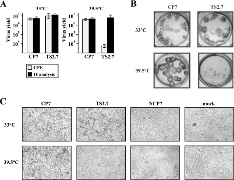
The temperature sensitivity of the obtained virus stock, termed TS2.7, was first examined by comparing its abilities to cause cytopathogenicity at permissive and restrictive temperatures. For this analysis, the virus stocks of TS2.7 and the parental BVDV strain CP7 were titrated in duplicate 96-well plates and incubated at 33°C and 39.5°C for 3 days. Microscopic examination revealed that CPEs, including cell lysis, were found after infection with comparable dilutions of the unmutagenized CP7 strain at both temperatures (Fig. 1A). In contrast, the amount of virus required by TS2.7 to cause cytopathogenicity at the restrictive temperature was about 1,000-fold more than that required for induction of cytopathogenicity at the permissive temperature (Fig. 1A). After examination of CPEs, the 96-well plates were subjected to IF analysis in order to detect viral antigen. For CP7, titers determined by IF analysis were identical to titers obtained by examination of a CPE. Surprisingly, IF analysis of cells infected with TS2.7 at 39.5°C revealed a titer almost 1,000-fold higher than the one previously determined by examination of CPE (Fig. 1A). Accordingly, the phenotype of mutant TS2.7 is characterized by a significant attenuation of cytopathogenicity at 39.5°C without reducing the yield of infectious virus titers. In contrast, the ability of TS2.7 to cause a CPE at 33°C is not affected. This phenomenon is also well documented by plaque assays performed in parallel at both temperatures. Infection of cells with dilutions of CP7 (10−1 to 10−5) at 33°C and 39.5°C and with dilutions of TS2.7 (10−1 to 10−5) at 33°C resulted in cell lysis and the formation of plaques (Fig. 1B; data shown only for dilutions of 10−5). In contrast, infection with TS2.7 at 39.5°C caused a CPE only when high virus concentrations (undiluted or dilutions up to 10−2) were used (data not shown), while higher dilutions of TS2.7 (10−3 to 10−5) led to formation of foci (visualized by immunostaining), without any signs of CPE (Fig. 1B; data shown only for dilutions of 10−5). TS2.7 and its parent produced large plaques at 33°C, while immunostained foci produced by TS2.7 at 39.5°C were smaller than the plaques produced by CP7. A similar attenuation of viral cytopathogenicity at 39.5°C was observed when sheep fetal thymus cells were used for infection with TS2.7 (data not shown).
FIG. 1.
(A)Results of virus titrations of supernatants obtained 3 days after infection of MDBK cells with BVDV CP7 and TS2.7 at 33°C. For titration, cells in duplicate 96-well plates were infected and incubated at 33°C (left) and 39.5°C (right) for 3 days. The titers were first determined by microscopic examination of the occurrence of a CPE, and then viral antigen was detected via IF analysis. Mean values and ± standard deviation ranges (error bars) were calculated from two independent experiments, each analyzed by quadruplicate titrations. (B) Plaque assays of cells infected with CP7 and TS2.7. Infected cells were visualized by immunostaining at 7 and 6 days p.i. at 33°C (top) and 39.5°C (bottom). (C) Bovine MDBK cells infected with BVDV strains CP7, TS2.7, and NCP7 at an MOI of 1 and incubated at 33°C (top) and 39.5°C (bottom). The photographs were taken 48 h after infection. As a control, mock-infected cells are included. A strong CPE occurred after infection of cells with CP7 at both temperatures and with TS2.7 at 33°C. In contrast, the ability of TS2.7 to cause viral cytopathogenicity was drastically reduced at 39.5°C.
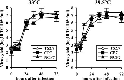
To further characterize viral growth properties, MDBK cells were infected in duplicate six-well plates with BVDV TS2.7, CP7, and the ncp virus NCP7 at an MOI of 1 and incubated at 33°C and 39.5°C for 48 h. As expected, there was no CPE detectable after infection of cells with NCP7 at both temperatures. Infection with TS2.7 at 33°C caused a strong CPE indistinguishable from that produced after infection with parent CP7 (Fig. 1C, top). In agreement with the results obtained from plaque assays, the ability of TS2.7 to cause cytopathogenicity was drastically inhibited at 39.5°C (Fig. 1C, bottom). In addition, the growth kinetics of TS2.7, CP7, and NCP7 at both temperatures were determined. Duplicate sets of MDBK cells were infected at an MOI of 0.1 and incubated in parallel at 33°C and 39.5°C. Virus released into the medium was sampled for a 3-day period, and the respective virus titers were determined by IF analysis. Each of the three viruses reached titers of >5 × 10⁶ TCID50/ml at 48 h after infection at both temperatures (Fig. 2). With regard to infections at 33°C, the peak titer of the mutant virus TS2.7 reached at 48 h p.i. was 2.3 × 10⁷ TCID50/ml and thus slightly higher than the peak titer of CP7 (5.9 × 10⁶ TCID50/ml); the peak titer of NCP7 was 1.8 × 10⁷ TCID50/ml. With respect to infections at 39.5°C, the titers of TS2.7 reached at 24 h and 36 h were about three- and four-fold lower, respectively, than the titers of the parental CP7 strain. Taken together, the growth kinetics of TS2.7, CP7, and NCP7 were very similar at both temperatures. Similar results, including the absence of significant (>4-fold) differences between the titers of T2.7 and CP7, were obtained by two additional comparative analyses of the growth kinetics of TS2.7 and CP7 (data not shown).
FIG. 2.
Growth kinetics of BVDV CP7, NCP7, and TS2.7 at 33°C (left) and 39.5°C (right). MDBK cells were infected with the indicated viruses at an MOI of 0.1. The titers of released virus were determined by IF analysis over a time period of 72 h. Error bars indicate the ± ranges from quadruplicate titrations.
To study possible effects of temperature shifts on the induction of cytopathogenicity, sets of MDBK cells were infected with TS2.7 at MOIs of 1.0, 10−1, 10−2, 10−3, and 10−4; incubated at various combinations of temperatures for 7 days; and examined daily by light microscope. Periods of 6 h and 24 h at 33°C before shifting to 39.5°C did not result in increased cytopathogenicity of TS2.7 at 39.5°C (data not shown). Vice versa, a period of 6 h at 39.5°C before shifting to 33°C did not attenuate the cytopathogenicity of TS2.7 at 33°C, while incubation for 24 h at 39.5°C before shifting to 33°C led to a temporal delay of the occurrence of CPEs (data not shown).
Genetic characterization of TS2.7.
To determine the genetic alteration(s) present in the genome of TS2.7, the entire genomic sequence of TS2.7 was determined and compared to the sequence of BVDV CP7; RNA from the second tissue culture passage of TS2.7 was used for the respective analysis. When differences from the wild-type BVDV CP7 sequence were detected, the sequencing procedure was repeated and only the confirmed changes were considered to be mutations present in the genome of TS2.7. A total of five nucleotide changes were found in the genomic regions encoding structural proteins C and E2 as well as NS proteins NS2 and NS5B. Four of these mutations resulted in changes of the deduced amino acid sequence. The identified mutations and corresponding deduced amino acid changes found in the genome of TS2.7 are detailed in Fig. 3A.
FIG. 3.
Genetic basis of the ts phenotype of BVDV TS2.7. (A) Genetic characterization of TS2.7. A schematic representation of the genome organization of the parental BVDV strain CP7 is shown on the top. Nucleotide sequencing of the entire genome of BVDV TS2.7 and subsequent comparison with the sequence of BVDV CP7 led to identification of five nucleotide changes at positions 1139, 3575, 4394, 10156, and 10542. The resulting four amino acid changes are indicated on the bottom. The mutation at position 4394 together with the resulting substitution of a tyrosine (Y) residue by a histidine (H) residue at position 1338 of the CP7 polyprotein is highlighted by a box. (B) The ts phenotype of BVDV mutant CP7-Y1338 carrying the point mutation at position 4394 in the CP7 genome. Plaque assays of cells infected with BVDV CP7-Y1338H at 33°C resulted in detection of plaques (top), while infection at 39.5°C led to production of foci (bottom). Plaques and foci were visualized by immunostaining 7 days after infection of cells.
A single nucleotide substitution is responsible for the ts phenotype of TS2.7.
To identify the mutation(s) responsible for the ts phenotype of TS2.7, each of the four individual substitutions causing amino acid changes was transferred into the infectious cDNA clone pCP7-388. After in vitro transcription and transfection of MDBK cells with each of the resulting RNAs carrying a single substitution and incubation at 33°C for 3 days, infectious cp viruses were recovered. In two independent experiments the specific infectivity of the four mutant RNAs ranged between 4.2 × 10⁵ and 2.0 × 10⁶ PFU/μg RNA and thus were very similar to that of parent CP7 (6.0 × 10⁵ and 2.0 × 10⁶ PFU/μg RNA). To examine whether one of these mutations was responsible for the ts phenotype of TS2.7, duplicate plaque assays were performed using cells infected with dilutions of the first passage of the respective viruses and incubated at 33°C and 39.5°C for 7 days. Like those of the parental CP7 strain, dilutions (up to 10−5) of the three mutants with single nucleotide substitutions in the genomic regions encoding C, E2, and NS5B (Fig. 3A) resulted in cell lysis and formation of large plaques at both temperatures (data not shown). Infection of cells with dilutions (up to 10−5) of the mutant CP7-Y1338H carrying the single mutation at nucleotide position 4394 located in the NS2 gene also resulted in cell lysis and plaque formation at 33°C. However, CP7-Y1338H was not able to produce plaques at 39.5°C when dilutions between 10−2 and 10−5 were used for infection of cells but resulted in the formation of infected foci (visualized by immunoperoxidase staining) without any signs of cytopathogenicity (Fig. 3B). Accordingly, the phenotype of CP7-Y1338H is very similar to the ts phenotype of TS2.7. Taken together, the results of these analyses demonstrated that a single mutation at position 4394 within the NS2 gene resulted in attenuation of cytopathogenicity of BVDV CP7 at high temperature.
Growth characteristics of BVDV CP7-Y1338H.
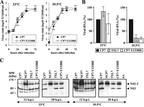
To compare the growth characteristics of the reconstructed ts mutant CP7-Y1338H and the parental virus strain CP7, growth rates and virus yields were determined using duplicate sets of MDBK cells infected at an MOI of 0.1 and incubated at 33°C and 39.5°C. Virus titers were determined for a 3-day period. As observed for the original TS2.7 mutant (Fig. 2), the growth kinetics of CP7-Y1338H and CP7 were similar and both viruses replicated at similar titers at 33°C and 39.5°C (Fig. 4A). Similar results were obtained by an additional comparative analysis of the growth kinetics of CP7-Y1338H and CP7 (data not shown). Taken together, the results show that the Y1338H mutation had no significant effect on virus yields and growth rates. Furthermore, the results of this analysis showed that attenuation of cytopathogenicity of CP7-Y1338H at 39.5°C does not correlate with reduced viral growth.
FIG. 4.
Characterization of BVDV CP7-Y1338H. (A) Growth kinetics of BVDV CP7 and the genetically engineered ts mutant CP7-Y1338H at 33°C (left) and 39.5°C (right). MDBK cells were infected at an MOI of 0.1 with supernatants of the second cell culture passages of the indicated viruses. The titers of released virus were determined by IF analysis over a time period of 72 h. Error bars indicate the ± ranges from quadruplicate titrations. (B) Relative amounts of accumulated viral genomic RNAs obtained after infection of MDBK cells with BVDV CP7, TS2.7, and CP7-Y1338H at 33°C (left) and 39.5°C (right). For infection an MOI of 1 was used. Total cellular RNAs were extracted at 10 h p.i. and subjected to quantitative BVDV-specific real-time RT-PCR. Results are indicated as percentages of the mean value obtained for reference strain CP7 (100%). Data show the means ± standard deviation ranges from two independent experiments, each analyzed by the measurement of triplicates. (C) Immunoblot. MDBK cells infected with CP7, NCP7, TS2.7, and CP7-Y1338H at 33°C (left side) and 39.5°C (right side) were lysed 12 and 20 h p.i. (h.p.i.). For infection an MOI of 2 was used. The samples were separated by sodium dodecyl sulfate-polyacrylamide gel electrophoresis (8% polyacrylamide) under reducing conditions, transferred to nitrocellulose, and incubated with the anti-NS3 monoclonal antibody 8.12.7 (15). Noninfected (n.i.) cells served as negative controls. The sizes (in kDa) of the molecular mass marker proteins (in thousands) are indicated on the left. The positions of NS2-3 and NS3 are indicated on the right.
Analysis of viral RNA synthesis and expression of NS2-3 and NS3.
It is well known that cp and ncp BVDV strains significantly differ in viral RNA production. For CP7 and other cp BVDV strains, amounts of viral RNA accumulated in infected cells were about 5- to 10-fold larger than the viral RNA amounts produced by their ncp counterparts (7, 32, 50). Another striking difference between cp and ncp BVDV concerns expression of viral NS3. While cp BVDV strains produce NS3 at early and late time points postinfection, for ncp BVDV, NS3 is usually no longer detectable after the first 12 h of infection (26). It was therefore interesting to investigate whether the observed attenuation of cytopathogenicity of TS2.7 and CP7-Y1338H correlates with changes of viral RNA synthesis and/or expression of NS3.
For analysis of viral RNA synthesis, cells were infected with BVDV CP7, TS2.7, and CP7-Y1338H at an MOI of 1. To minimize effects of potential differences in the levels of viral spread and replication efficiency of the individual viruses, total cellular RNAs were prepared 10 h p.i.; this time point precedes the end of the first viral replication cycle (7, 32, 50). The relative amounts of accumulated viral RNAs were determined by quantitative real-time RT-PCR analysis. Comparative analysis revealed that the amounts of viral RNA produced at 33°C were very similar for CP7, TS2.7, and the reconstructed ts mutant; significant differences were not observed (Fig. 4B). In contrast, the amounts of accumulated viral RNA after infection of cells with TS2.7 and CP7-Y1338H at 39.5°C were about fivefold lower than the amount produced after infection with parental BVDV CP7.
To monitor expression of NS3 and NS2-3, sets of cells were infected with BVDV CP7, NCP7, TS2.7, and CP7-Y1338H at an MOI of 2 and incubated at 33°C and 39.5°C. Cells were lysed and harvested at 12 h and 20 h p.i., and aliquots corresponding to 6.0 × 10⁴ cells (collected at 12 h p.i.) and 6.0 × 10³ cells (collected at 20 h p.i.) were used for immunoblot analysis (Fig. 4C). The results of this analysis revealed that CP7, TS2.7, and CP7-Y1338H express significant amounts of NS2-3 and NS3 throughout infection at both temperatures. Compared to parental CP7, infection with TS2-7 and CP7-Y1338H resulted in slightly decreased NS2-3 processing. This phenomenon was observed at both temperatures (Fig. 4C). In contrast, after infection with NCP7 at 33°C and 39.5°C, expression of detectable levels of NS3 was limited to the early phase of infection (12 h p.i.), while large amounts of NS2-3 were expressed at 12 h and 20 h p.i. Taken together, the results show that attenuation of cytopathogenicity of BVDV CP7 at 39.5°C caused by the Y1338H substitution correlated with reduced viral RNA synthesis but that production of NS3 was not affected in a temperature-dependent manner.
DISCUSSION
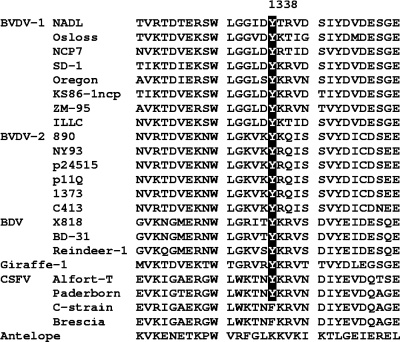
The existence of two biotypes represented by cp and ncp viruses is a particularly interesting feature of pestiviruses. In this study we describe isolation and characterization of a ts mutant of cp BVDV strain CP7 whose ability to cause cytopathogenicity at 39.5°C is strongly reduced despite efficient viral propagation. This is the first report on a BVDV mutant which differs from the parental virus by temperature-dependent attenuation of viral cytopathogenicity. Our analysis demonstrated that the genetic basis of this unique phenotype is represented by a single tyrosine-to-histidine substitution (Y1338H) at residue 193 of NS2. An alignment of partial NS2 protein sequences of a representative set of pestiviruses showed that Y1338 is highly conserved among pestivirus species BVDV-1, BVDV-2, BDV, and the tentative species “Giraffe” but not among CSFV and a pestivirus isolated from a pronghorn antelope (Fig. 5).
FIG. 5.
Y1338 of NS2 is highly conserved among pestivirus species BVDV-1, BVDV-2, BDV, and “Giraffe” but not among CSFV and a pestivirus from a pronghorn antelope. Fragments of polyproteins (of a representative set of 23 pestiviruses) that include amino acid Y1338 (highlighted by a black box and white letters) were aligned (see Materials and Methods). The virus strains (and their GenBank accession numbers) are BVDV-1 strains NADL (M31182), Osloss (M96687), NCP7 (U63513), SD-1 (M96751), Oregon (AF041040), KS86-1ncp (AB078950), ZM-95 (AF526381), and ILLC (U86599); BVDV-2 strains 890 (U18059), NY93 (AF502339), p24515 (AY149216), p11Q (AY149215), 1373 (AF145967), and C413 (AF002227); BDV strains X818 (AF037405), BD-31 (U70263), and Reindeer-1 (AF144618); pestivirus strain Giraffe-1 (NC_003678); CSFV strains Alfort-T (J04358), Paderborn (AY072924), C-strain (Z46258), and Brescia (P21530); and a pestivirus from pronghorn antelope (AAX12371).
A common characteristic of cp BVDV strains is the efficient production of NS3 throughout infection, which represents a major difference from ncp BVDV (15, 18, 35, 40). Cytopathogenic BVDV strains can use various strategies for expression of NS3, including processing of the precursor protein NS2-3 (1, 9, 24, 32-34, 36, 37, 42, 49). This strategy, which is used by BVDV CP7 and some other cp BVDV strains, depends on the autoproteolytic activity of NS2 (26). Studies of cp BVDV strain Oregon demonstrated that point mutations within NS2 can influence processing of NS2-3 (24, 25). Mutants of BVDV Oregon carrying point mutations in NS2 which significantly reduced NS2-3 processing were either nonviable or propagated much more slowly and rapidly reverted to viruses, allowing efficient NS2-3 cleavage. These viruses apparently caused cytopathogenicity only after reversion (25). The results of these studies significantly contributed to the conclusion that cytopathogenicity of BVDV correlates with increased NS3 expression and efficient viral replication. It was therefore expected that attenuation of cytopathogenicity of TS2.7 correlates with reduced processing of NS2-3 at high temperature. However, the results of our study showed that, irrespective of the incubation temperature used, infections with the original ts mutant TS2.7 and the reconstructed mutant CP7-Y1338H resulted in expression of substantial amounts of NS3 (Fig. 4C). This observation uncouples cytopathogenicity of BVDV from efficient production of NS3 at early and late phases of infection and shows that a single point mutation in NS2 can suppress cytopathogenicity of BVDV CP7 at 39.5°C without compromising NS2-3 processing in a temperature-dependent manner.
Another major difference between cp and ncp BVDV concerns the efficiency of viral RNA synthesis. Analyses of several BVDV pairs, including the isogenic pair consisting of CP7 and NCP7, have shown that the cp viruses produce amounts of viral RNA about 5- to 10-fold larger than their ncp counterparts (7, 32, 50). The ability of the ts mutants described here to produce large amounts of viral RNA at 39.5°C was significantly reduced (Fig. 4B). Accordingly, attenuation of viral cytopathogenicity correlated with reduced amounts of accumulated viral RNAs. This result supports the hypothesis that synthesis of large amounts of viral RNA, including double-stranded RNA, triggers cp BVDV-induced apoptosis (31, 54).
Apart from the work described here, there is one other report describing isolation and characterization of several ncp variants of a cp BVDV strain (NADL) (43). Similar to the ts mutant TS2.7, these ncp variants expressed substantial amounts of NS3 and produced slightly elevated accumulation of NS2-3 relative to that of NS3. However, for these ncp variants of NADL, attenuation of cytopathogenicity did not correlate with significantly reduced amounts of viral RNA, as was observed for TS2.7 and CP7-Y1338H. For all these ncp variants, a substitution of a tyrosine residue at position 15 of NS4B by a cysteine residue was responsible for the change of biotype (43). The results of this study with the ncp variants of BVDV NADL and our study demonstrate that efficient production of NS3 throughout infection does not inevitably result in a CPE and that at least two viral NS proteins, NS4B and NS2, can attenuate viral cytopathogenicity. While NS4B represents an essential component of the viral RNA replication complex, NS2 is dispensable for viral RNA replication. Furthermore, analyses of several naturally occurring and genetically engineered cp BVDV replicon RNAs lacking the NS2 coding region of the viral genome have shown that NS2 is also not required for induction of viral cytopathogenicity (7, 10, 49). This suggests that the Y1338H mutation identified in NS2 of TS2.7 causes induction of an antiapoptotic function of NS2 rather than suppression of a proapoptotic function residing in NS2. The mechanism by which NS2 of TS2.7 attenuates cytopathogenicity remains to be determined. It can be speculated that the Y1338H mutation results in a temperature-dependent alteration of the NS2 structure and that the respective structural change modulates the interaction of NS2 with viral or cellular proteins.
In the field of virology ts mutants have been exploited extensively to study biological processes. For many plus-strand RNA viruses, including members of the Togaviridae, Picornaviridae, Coronaviridae, and Flaviviridae, a number of ts mutants have been described (references 11, 14, 19, 23, 30, 38, 41, 45, and 47 and references therein). To our knowledge, only one other ts pestivirus (cp BVDV strain RIT 4350) has been reported so far (29). The genome of RIT 4350 harbors two cellular insertions and a large duplication of viral sequences (6). While these genomic alterations were shown to be responsible for expression of NS3 and induction of cytopathogenicity, the molecular basis of the ts phenotype remains unknown (6, 7). In contrast to the yield of the ts mutants described here, the virus yield obtained after infection of cells with RIT 4350 at high temperature was significantly reduced by at least two orders of magnitudes (29). Our own evaluation of the virus titers produced after infection with RIT 4350 confirmed that its ts phenotype is characterized by a significant reduction of virus propagation at nonpermissive temperature (data not shown). Irrespective of the incubation temperature used, the infectious virus titers of RIT 4350 determined by plaque assays were identical to the titers obtained by subsequent IF analysis. Thus, the phenotype of the ts mutants TS2.7 and CP7-Y1338H is clearly different from that of BVDV RIT 4350.
In this study a novel ts mutant of a cp BVDV strain exhibiting a unique phenotype was isolated and characterized. The results of our analyses showed that a single point mutation in NS2 interferes with viral RNA synthesis in a temperature-dependent manner and attenuates BVDV-induced cytopathogenicity despite efficient production of NS3 and high levels of infectious virus yield. Thus, NS2 can play an unexpected role in attenuation of BVDV cytopathogenicity. Accordingly, this study provided further insights into the role of NS proteins NS2 and NS3 in viral cytopathogenicity. Future experiments will concentrate on the mechanism by which NS2 of TS2.7 causes the ts attenuation of cytopathogenicity. Such studies will contribute to our understanding of the molecular basis of pestivirus-induced apoptosis.
Acknowledgments
We thank M. Orlich for excellent technical assistance in the initial stage of the project.
This study was supported by Sonderforschungsbereich 535, Invasion Mechanisms and Replication Strategies of Infectious Agents (project B8), and grant BE 2333/2-1 from the Deutsche Forschungsgemeinschaft (DFG). P.B. is supported by a Heisenberg professorship from the DFG (BE 2333/1-1).
Footnotes
Published ahead of print on 23 September 2009.
REFERENCES
- 1.Baroth, M., M. Orlich, H.-J. Thiel, and P. Becher. 2000. Insertion of cellular NEDD8 coding sequences in a pestivirus. Virology 278:456-466. [DOI] [PubMed] [Google Scholar]
- 2.Becher, P., M. Orlich, M. König, and H.-J. Thiel. 1999. Nonhomologous RNA recombination in bovine viral diarrhea virus: molecular characterization of a variety of subgenomic RNAs isolated during an outbreak of fatal mucosal disease. J. Virol. 73:5646-5653. [DOI] [PMC free article] [PubMed] [Google Scholar]
- 3.Becher, P., M. Orlich, A. D. Shannon, G. Horner, M. König, and H.-J. Thiel. 1997. Phylogenetic analysis of pestiviruses from domestic and wild ruminants. J. Gen. Virol. 78:1357-1366. [DOI] [PubMed] [Google Scholar]
- 4.Becher, P., M. Orlich, and H.-J. Thiel. 1998. Complete genomic sequence of border disease virus, a pestivirus from sheep. J. Virol. 72:5165-5173. [DOI] [PMC free article] [PubMed] [Google Scholar]
- 5.Becher, P., M. Orlich, and H.-J. Thiel. 2000. Mutations in the 5′ nontranslated region of bovine viral diarrhea virus result in altered growth characteristics. J. Virol. 74:7884-7894. [DOI] [PMC free article] [PubMed] [Google Scholar]
- 6.Becher, P., M. Orlich, and H.-J. Thiel. 1998. Ribosomal S27a-coding sequences upstream of ubiquitin-coding sequences in the genome of a pestivirus. J. Virol. 72:8697-8704. [DOI] [PMC free article] [PubMed] [Google Scholar]
- 7.Becher, P., M. Orlich, and H.-J. Thiel. 2001. RNA recombination between persisting pestivirus and a vaccine strain: generation of cytopathogenic virus and induction of lethal disease. J. Virol. 75:6256-6264. [DOI] [PMC free article] [PubMed] [Google Scholar]
- 8.Becher, P., and H.-J. Thiel. 2002. Genus Pestivirus (Flaviviridae), p. 327-331. In C. A. Tidona and G. Darai (ed.), The Springer index of viruses. Springer-Verlag, Heidelberg, Germany.
- 9.Becher, P., H.-J. Thiel, M. Collins, J. Brownlie, and M. Orlich. 2002. Cellular sequences in pestivirus genomes encoding gamma-aminobutyric acid (A) receptor-associated protein and Golgi-associated ATPase enhancer of 16 kilodaltons. J. Virol. 76:13069-13076. [DOI] [PMC free article] [PubMed] [Google Scholar]
- 10.Behrens, S. E., C. W. Grassmann, H.-J. Thiel, G. Meyers, and N. Tautz. 1998. Characterization of an autonomous subgenomic pestivirus RNA replicon. J. Virol. 72:2364-2372. [DOI] [PMC free article] [PubMed] [Google Scholar]
- 11.Blaney, J. E. J., D. H. Johnson, C. Y. Firestone, C. T. Hanson, B. R. Murphy, and S. S. Whitehead. 2001. Chemical mutagenesis of dengue virus type 4 yields mutant viruses which are temperature sensitive in Vero cells or human liver cells and attenuated in mice. J. Virol. 75:9731-9740. [DOI] [PMC free article] [PubMed] [Google Scholar]
- 12.Bolin, S. R., A. W. McClurkin, R. C. Cutlip, and M. F. Coria. 1985. Severe clinical disease induced in cattle persistently infected with noncytopathogenic bovine viral diarrhea virus by superinfection with cytopathogenic bovine viral diarrhea virus. Am. J. Vet. Res. 46:573-576. [PubMed] [Google Scholar]
- 13.Brownlie, J., M. C. Clarke, and C. J. Howard. 1984. Experimental production of fatal mucosal disease in cattle. Vet. Rec. 114:535-536. [DOI] [PubMed] [Google Scholar]
- 14.Burge, B. W., and E. R. Pfefferkorn. 1967. Temperature-sensitive mutants of Sindbis virus: biochemical correlates of complementation. J. Virol. 1:956-962. [DOI] [PMC free article] [PubMed] [Google Scholar]
- 15.Corapi, W. V., R. O. Donis, and E. J. Dubovi. 1988. Monoclonal antibody analyses of cytopathic and noncytopathic viruses from fatal bovine viral diarrhea infections. J. Virol. 62:2823-2827. [DOI] [PMC free article] [PubMed] [Google Scholar]
- 16.Devereux, J., P. Haeberli, and O. A. Smithies. 1984. A comprehensive set of sequence analysis programs for the VAX. Nucleic Acids Res. 12:387-395. [DOI] [PMC free article] [PubMed] [Google Scholar]
- 17.Gillespie, J. H., J. A. Baker, and K. McEntee. 1960. A cytopathogenic strain of virus diarrhea virus. Cornell Vet. 50:73-79. [PubMed] [Google Scholar]
- 18.Greiser-Wilke, I., K. E. Dittmar, B. Liess, and V. Moennig. 1992. Heterogeneous expression of the non-structural protein p80/p125 in cells infected with different pestiviruses. J. Gen. Virol. 73:47-52. [DOI] [PubMed] [Google Scholar]
- 19.Hahn, Y. S., E. G. Strauss, and J. H. Strauss. 1989. Mapping of RNA− temperature-sensitive mutants of Sindbis virus: assignment of complementation groups A, B, and G to nonstructural proteins. J. Virol. 63:3142-3150. [DOI] [PMC free article] [PubMed] [Google Scholar]
- 20.Heinz, F. X., M. S. Collett, R. H. Purcell, E. A. Gould, C. R. Howard, M. Houghton, R. J. M. Moormann, C. M. Rice, and H.-J. Thiel. 2000. Family Flaviviridae, p. 859-878. In C. M. Fauquet, M. H. V. van Regenmortel, D. H. L. Bishop, E. B. Carstens, M. K. Estes, S. M. Lemon, J. Maniloff, M. A. Mayo, D. J. McGeoch, C. R. Pringle, and R. B. Wickner (ed.), Virus taxonomy. 7th report of the International Committee on Taxonomy of Viruses. Academic Press, San Diego, CA.
- 21.Hoff, H. S., and R. O. Donis. 1997. Induction of apoptosis and cleavage of poly(ADP-ribose) polymerase by cytopathic bovine viral diarrhea virus infection. Virus Res. 49:101-113. [DOI] [PubMed] [Google Scholar]
- 22.Kao, C. C., A. M. Del Vecchio, and W. Zhong. 1999. De novo initiation of RNA synthesis by a recombinant flaviviridae RNA-dependent RNA polymerase. Virology 253:1-7. [DOI] [PubMed] [Google Scholar]
- 23.Koolen, M. J., S. Love, W. Wouda, J. Calafat, M. C. Horzinek, and B. A. van der Zeijst. 1987. Induction of demyelination by a temperature-sensitive mutant of the coronavirus MHV-A59 is associated with restriction of viral replication in the brain. J. Gen. Virol. 68:703-714. [DOI] [PubMed] [Google Scholar]
- 24.Kümmerer, B., D. Stoll, and G. Meyers. 1998. Bovine viral diarrhea virus strain Oregon: a novel mechanism for processing of NS2-3 based on point mutations. J. Virol. 72:4127-4138. [DOI] [PMC free article] [PubMed] [Google Scholar]
- 25.Kümmerer, B. M., and G. Meyers. 2000. Correlation between point mutations in NS2 and the viability and cytopathogenicity of bovine viral diarrhea virus strain Oregon analyzed with an infectious cDNA clone. J. Virol. 74:390-400. [DOI] [PMC free article] [PubMed] [Google Scholar]
- 26.Lackner, T., A. Müller, A. Pankraz, P. Becher, H.-J. Thiel, A. E. Gorbalenya, and N. Tautz. 2004. Temporal modulation of an autoprotease is crucial for replication and pathogenicity of an RNA virus. J. Virol. 78:10765-10775. [DOI] [PMC free article] [PubMed] [Google Scholar]
- 27.Lee, K. M., and J. H. Gillespie. 1957. Propagation of virus diarrhea virus of cattle in tissue culture. Am. J. Vet. Res. 18:953. [PubMed] [Google Scholar]
- 28.Lindenbach, B. D., H.-J. Thiel, and C. M. Rice. 2007. Flaviviridae: the viruses and their replication, p. 1101-1152. In D. M. Knipe and P. M. Howley (ed.), Fields virology, 5th ed. Lippincott-Raven, Philadelphia, PA.
- 29.Lobmann, M., P. Charlier, G. Florent, and N. Zygraich. 1984. Clinical evaluation of a temperature-sensitive bovine viral diarrhea vaccine strain. Am. J. Vet. Res. 45:2498-2503. [PubMed] [Google Scholar]
- 30.MacKenzie, J. S. 1975. Virulence of temperature-sensitive mutants of foot-and-mouth disease virus. Arch. Virol. 48:1-8. [DOI] [PubMed] [Google Scholar]
- 31.Mätzener, P., I. Magkouras, T. Rümenapf, E. Peterhans, and M. Schweizer. 2009. The viral RNase Erns prevents IFN type-I triggering by pestiviral single- and double-stranded RNAs. Virus Res. 140:15-23. [DOI] [PubMed] [Google Scholar]
- 32.Mendez, E., N. Ruggli, M. S. Collett, and C. M. Rice. 1998. Infectious bovine viral diarrhea virus (strain NADL) RNA from stable cDNA clones: a cellular insert determines NS3 production and viral cytopathogenicity. J. Virol. 72:4737-4745. [DOI] [PMC free article] [PubMed] [Google Scholar]
- 33.Meyers, G., D. Stoll, and M. Gunn. 1998. Insertion of a sequence encoding light chain 3 of microtubule-associated proteins 1A and 1B in a pestivirus genome: connection with virus cytopathogenicity and induction of lethal disease in cattle. J. Virol. 72:4139-4148. [DOI] [PMC free article] [PubMed] [Google Scholar]
- 34.Meyers, G., N. Tautz, P. Becher, H.-J. Thiel, and B. Kümmerer. 1996. Recovery of cytopathogenic and noncytopathogenic bovine viral diarrhea viruses from cDNA constructs. J. Virol. 70:8606-8613. [DOI] [PMC free article] [PubMed] [Google Scholar]
- 35.Meyers, G., N. Tautz, E. J. Dubovi, and H.-J. Thiel. 1991. Viral cytopathogenicity correlated with integration of ubiquitin-coding sequences. Virology 180:602-616. [DOI] [PMC free article] [PubMed] [Google Scholar]
- 36.Meyers, G., and H.-J. Thiel. 1996. Molecular characterization of pestiviruses. Adv. Virus Res. 47:53-118. [DOI] [PubMed] [Google Scholar]
- 37.Nagai, M., Y. Sakoda, M. Mori, M. Hayashi, H. Kida, and H. Akashi. 2003. Insertion of cellular sequence and RNA recombination in the structural protein coding region of cytopathogenic bovine viral diarrhoea virus. J. Gen. Virol. 84:447-452. [DOI] [PubMed] [Google Scholar]
- 38.Nichol, F. R., and D. R. Tershak. 1968. Rescue of temperature-sensitive poliovirus. J. Virol. 2:415-420. [DOI] [PMC free article] [PubMed] [Google Scholar]
- 39.Pankraz, A., H. J. Thiel, and P. Becher. 2005. Essential and nonessential elements in the 3′ nontranslated region of bovine viral diarrhea virus. J. Virol. 79:9119-9127. [DOI] [PMC free article] [PubMed] [Google Scholar]
- 40.Pocock, D. H., C. J. Howard, M. C. Clarke, and J. Brownlie. 1987. Variation in the intracellular polypeptide profiles from different isolates of bovine viral diarrhea virus. Arch. Virol. 94:43-53. [DOI] [PubMed] [Google Scholar]
- 41.Pringle, C. R. 1996. Temperature-sensitive mutant vaccines, p. 17-32. In A. Robinson, G. H. Graham, and C. N. Wiblin (ed.), Methods in molecular medicine: vaccine protocols. Humana, Totowa, NJ. [DOI] [PubMed]
- 42.Qi, F., J. F. Ridpath, and E. S. Berry. 1998. Insertion of a bovine SMT3B gene in NS4B and duplication of NS3 in a bovine viral diarrhea virus genome correlate with the cytopathogenicity of the virus. Virus Res. 57:1-9. [DOI] [PubMed] [Google Scholar]
- 43.Qu, L., L. K. McMullan, and C. M. Rice. 2001. Isolation and characterization of noncytopathogenic pestivirus mutants reveals a role for nonstructural protein NS4B in viral cytopathogenicity. J. Virol. 75:10651-10662. [DOI] [PMC free article] [PubMed] [Google Scholar]
- 44.Reed, K. E., A. E. Gorbalenya, and C. M. Rice. 1998. The NS5A/NS5 proteins of viruses from three genera of the family Flaviviridae are phosphorylated by associated serine/threonine kinases. J. Virol. 72:6199-6206. [DOI] [PMC free article] [PubMed] [Google Scholar]
- 45.Sawicki, S. G., D. L. Sawicki, D. Younker, Y. Meyer, V. Thiel, H. Stokes, and S. G. Siddell. 2005. Functional and genetic analysis of coronavirus replicase-transcriptase proteins. PLoS Pathog. 1:e39. [DOI] [PMC free article] [PubMed] [Google Scholar]
- 46.Tamura, J. K., P. Warrener, and M. S. Collett. 1993. RNA-stimulated NTPase activity associated with the p80 protein of the pestivirus bovine viral diarrhea virus. Virology 193:1-10. [DOI] [PubMed] [Google Scholar]
- 47.Tarr, G. C., and A. S. Lubiniecki. 1976. Chemically-induced temperature sensitive mutants of dengue virus type 2. I. Isolation and partial characterization. Arch. Virol. 50:223-235. [DOI] [PubMed] [Google Scholar]
- 48.Tautz, N., K. Elbers, D. Stoll, G. Meyers, and H.-J. Thiel. 1997. Serine protease of pestiviruses: determination of cleavage sites. J. Virol. 71:5415-5422. [DOI] [PMC free article] [PubMed] [Google Scholar]
- 49.Tautz, N., H.-J. Thiel, E. J. Dubovi, and G. Meyers. 1994. Pathogenesis of mucosal disease: a cytopathogenic pestivirus generated by internal deletion. J. Virol. 68:3289-3297. [DOI] [PMC free article] [PubMed] [Google Scholar]
- 50.Vassilev, V. B., and R. O. Donis. 2000. Bovine viral diarrhea virus induced apoptosis correlates with increased intracellular viral RNA accumulation. Virus Res. 69:95-107. [DOI] [PubMed] [Google Scholar]
- 51.Warrener, P., and M. S. Collett. 1995. Pestivirus NS3 (p80) protein possesses RNA helicase activity. J. Virol. 69:1720-1726. [DOI] [PMC free article] [PubMed] [Google Scholar]
- 52.Wiskerchen, M. A., and M. S. Collett. 1991. Pestivirus gene expression: protein p80 of bovine viral diarrhea virus is a proteinase involved in polyprotein processing. Virology 184:341-350. [DOI] [PubMed] [Google Scholar]
- 53.Xu, J., E. Mendez, P. R. Caron, C. Lin, M. A. Murcko, M. S. Collett, and C. M. Rice. 1997. Bovine viral diarrhea virus NS3 serine proteinase: polyprotein cleavage sites, cofactor requirements, and molecular model of an enzyme essential for pestivirus replication. J. Virol. 71:5312-5322. [DOI] [PMC free article] [PubMed] [Google Scholar]
- 54.Yamane, D., K. Kato, Y. Tohya, and H. Akashi. 2006. The double-stranded RNA-induced apoptosis pathway is involved in the cytopathogenicity of cytopathogenic Bovine viral diarrhea virus. J. Gen. Virol. 87:2961-2970. [DOI] [PubMed] [Google Scholar]
- 55.Zhang, G., A. M., M. C. Clarke, and J. W. McCauley. 1997. Cell death induced by cytopathic bovine viral diarrhea virus is mediated by apoptosis. J. Gen. Virol. 77:1677-1681. [DOI] [PubMed] [Google Scholar]
- 56.Zhong, W., L. L. Gutshall, and A. M. Del Vecchio. 1998. Identification and characterization of an RNA-dependent RNA polymerase activity within the nonstructural 5B region of bovine viral diarrhea virus. J. Virol. 72:9365-9369. [DOI] [PMC free article] [PubMed] [Google Scholar]


