Full text
PDF
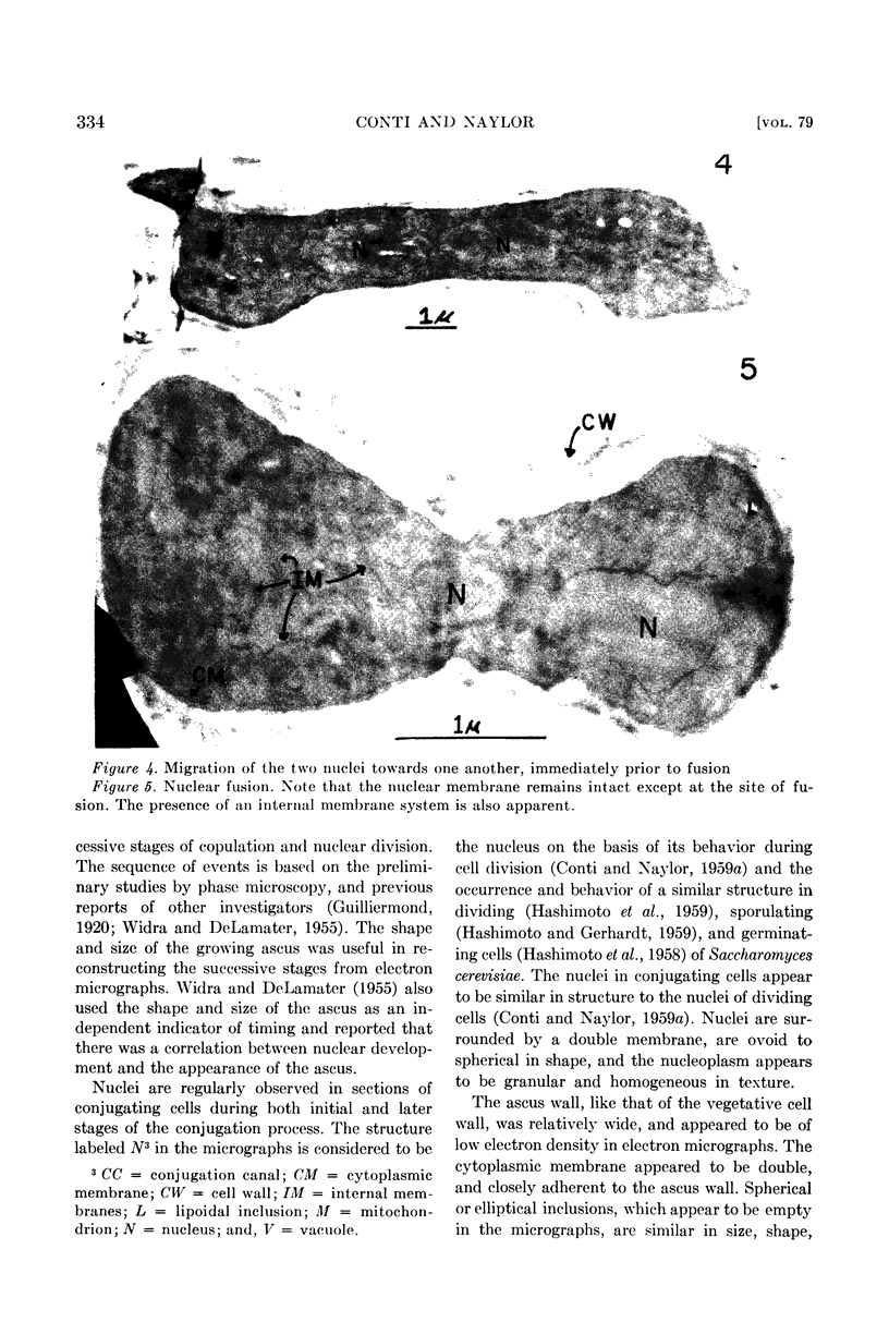
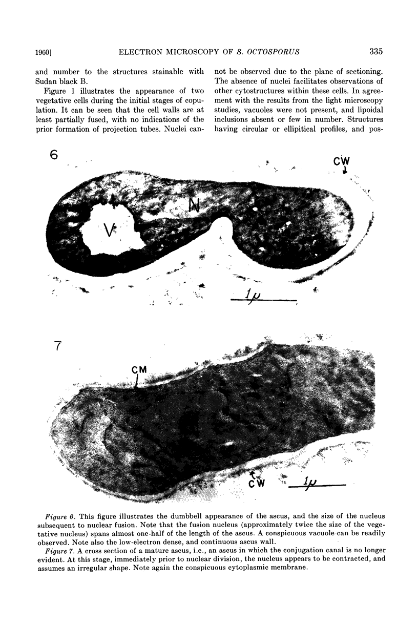
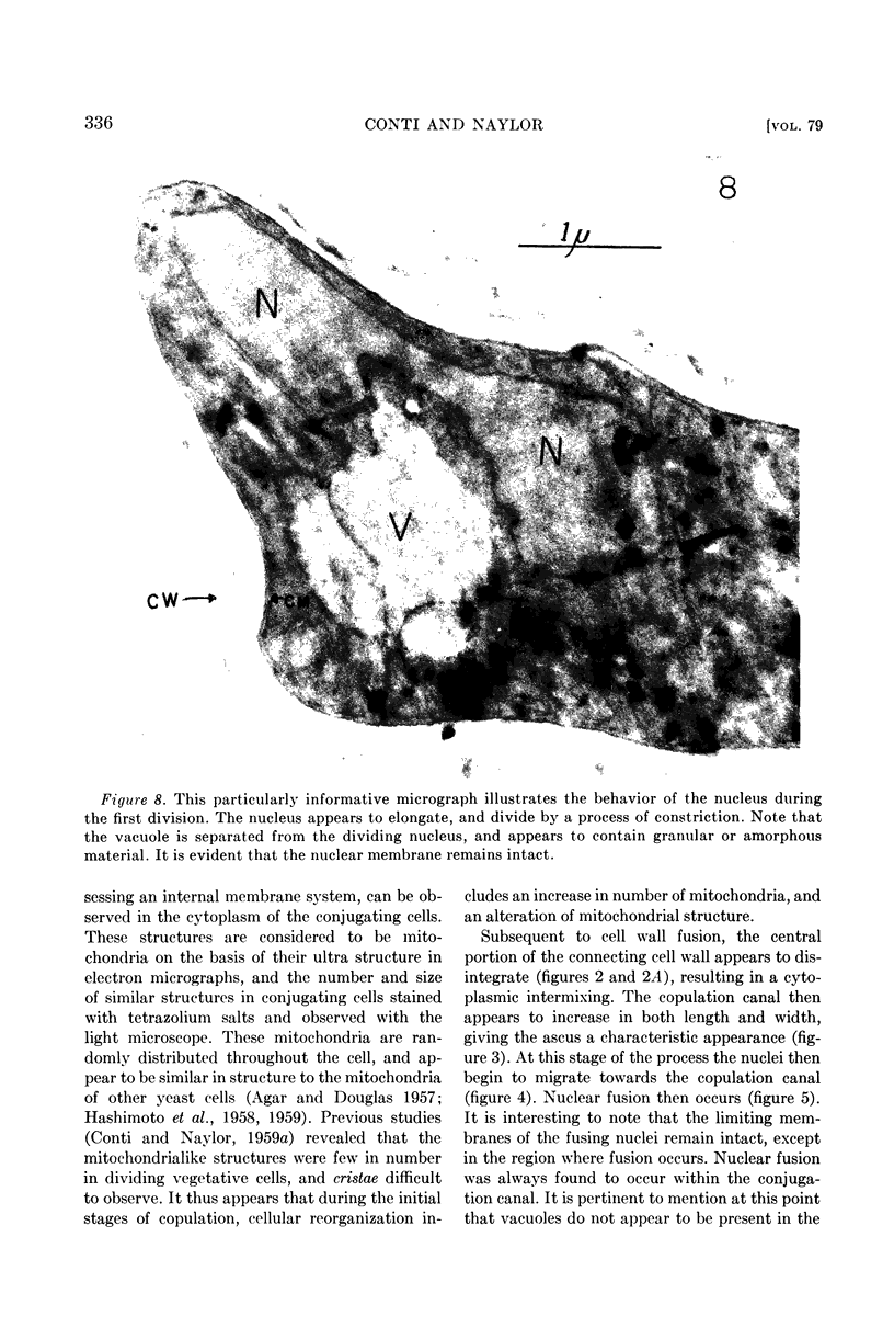
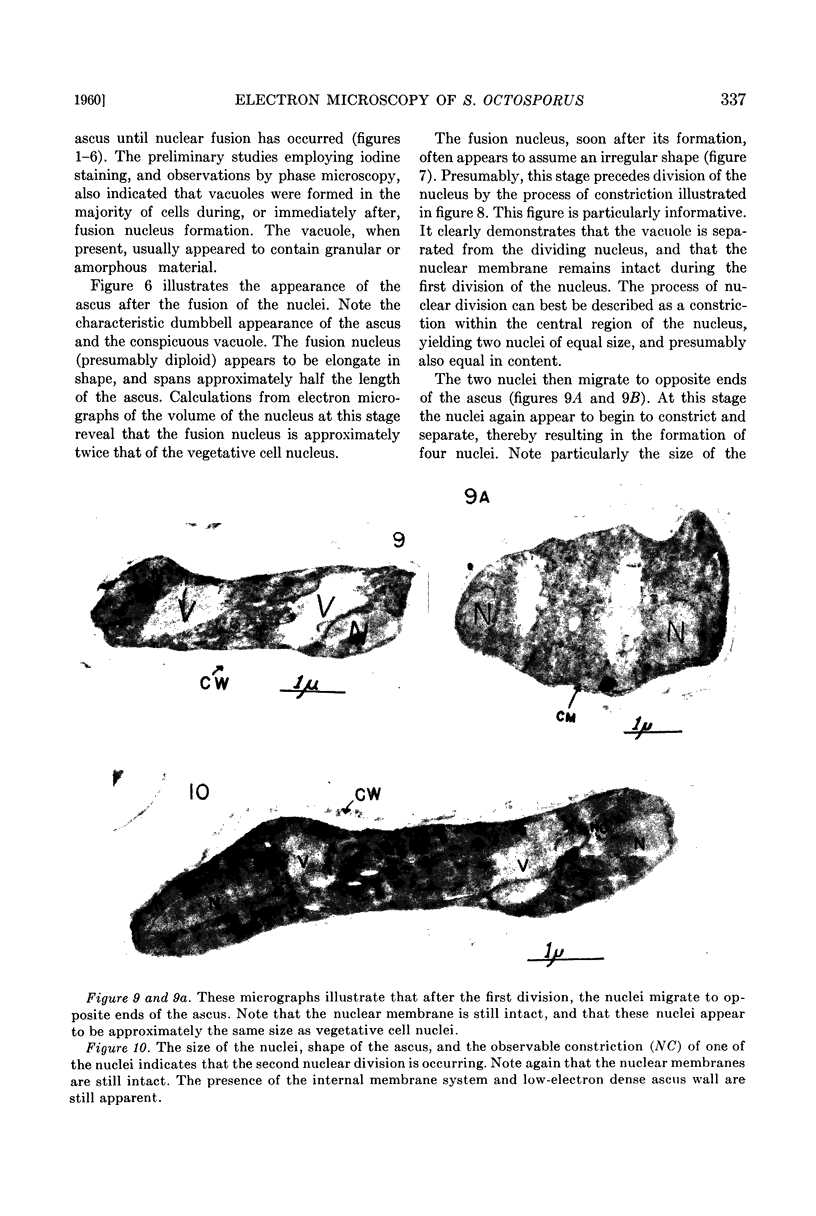
Images in this article
Selected References
These references are in PubMed. This may not be the complete list of references from this article.
- AGAR H. D., DOUGLAS H. C. Studies on the cytological structure of yeast: electron microscopy of thin sections. J Bacteriol. 1957 Mar;73(3):365–375. doi: 10.1128/jb.73.3.365-375.1957. [DOI] [PMC free article] [PubMed] [Google Scholar]
- CONTI S. F., NAYLOR H. B. Electron microscopy of ultrathin sections of Schizosaccharomyces octosporus. I. Cell division. J Bacteriol. 1959 Dec;78:868–877. doi: 10.1128/jb.78.6.868-877.1959. [DOI] [PMC free article] [PubMed] [Google Scholar]
- CUTTER V. M., Jr The cytology of the fungi. Annu Rev Microbiol. 1951;5:17–34. doi: 10.1146/annurev.mi.05.100151.000313. [DOI] [PubMed] [Google Scholar]
- GANESAN A. T., SWAMINATHAN M. S. Staining the nucleus in yeasts. Stain Technol. 1958 May;33(3):115–121. doi: 10.3109/10520295809111834. [DOI] [PubMed] [Google Scholar]
- HASHIMOTO T., CONTI S. F., NAYLOR H. B. Fine structure of microorganisms. III. Electron microscopy of resting and germinating ascospores of Saccharomyces cerevisiae. J Bacteriol. 1958 Oct;76(4):406–416. doi: 10.1128/jb.76.4.406-416.1958. [DOI] [PMC free article] [PubMed] [Google Scholar]
- HASHIMOTO T., CONTI S. F., NAYLOR H. B. Studies of the fine structure of microorganisms. IV. Observations on budding Saccharomyces cerevisiae by light and electron microscopy. J Bacteriol. 1959 Mar;77(3):344–354. doi: 10.1128/jb.77.3.344-354.1959. [DOI] [PMC free article] [PubMed] [Google Scholar]
- MOSES M. J. Studies on nuclei using correlated cytochemical, light, and electron microscope techniques. J Biophys Biochem Cytol. 1956 Jul 25;2(4 Suppl):397–406. doi: 10.1083/jcb.2.4.397. [DOI] [PMC free article] [PubMed] [Google Scholar]
- SATIR P. G., PEACHEY L. D. Thin sections. II. A simple method for reducing compression artifacts. J Biophys Biochem Cytol. 1958 May 25;4(3):345–348. doi: 10.1083/jcb.4.3.345. [DOI] [PMC free article] [PubMed] [Google Scholar]


