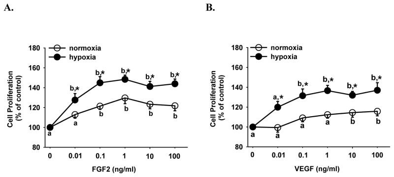
Figure 2.
Effects of hypoxia on FGF2- and VEGF-stimulated HPAE cell proliferation. Cells seeded in 96-well plates (5000 cells/well) were cultured in serum containing medium for 24 hr under normoxia and hypoxia. After 24 hr of serum starvation, cells were treated without or with FGF2 (A) or VEGF (B) for additional 72 hr. Cell numbers were then determined. Data are expressed as means ± SEM percent of the control (no growth factor). Numbers of cells per well in the controls after 72 hr were 5573 ± 633 and 4728 ± 327 under normoxia and hypoxia, respectively. a,b Within each growth factor treatment group, means with different letters differ (p < 0.05). *differs (p < 0.05) from the normoxia at the corresponding dose of growth factor.

