Full text
PDF
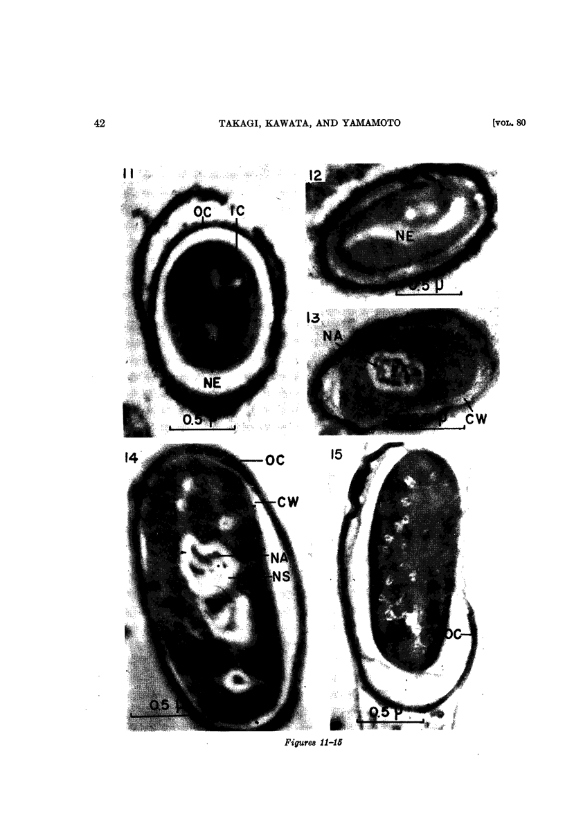
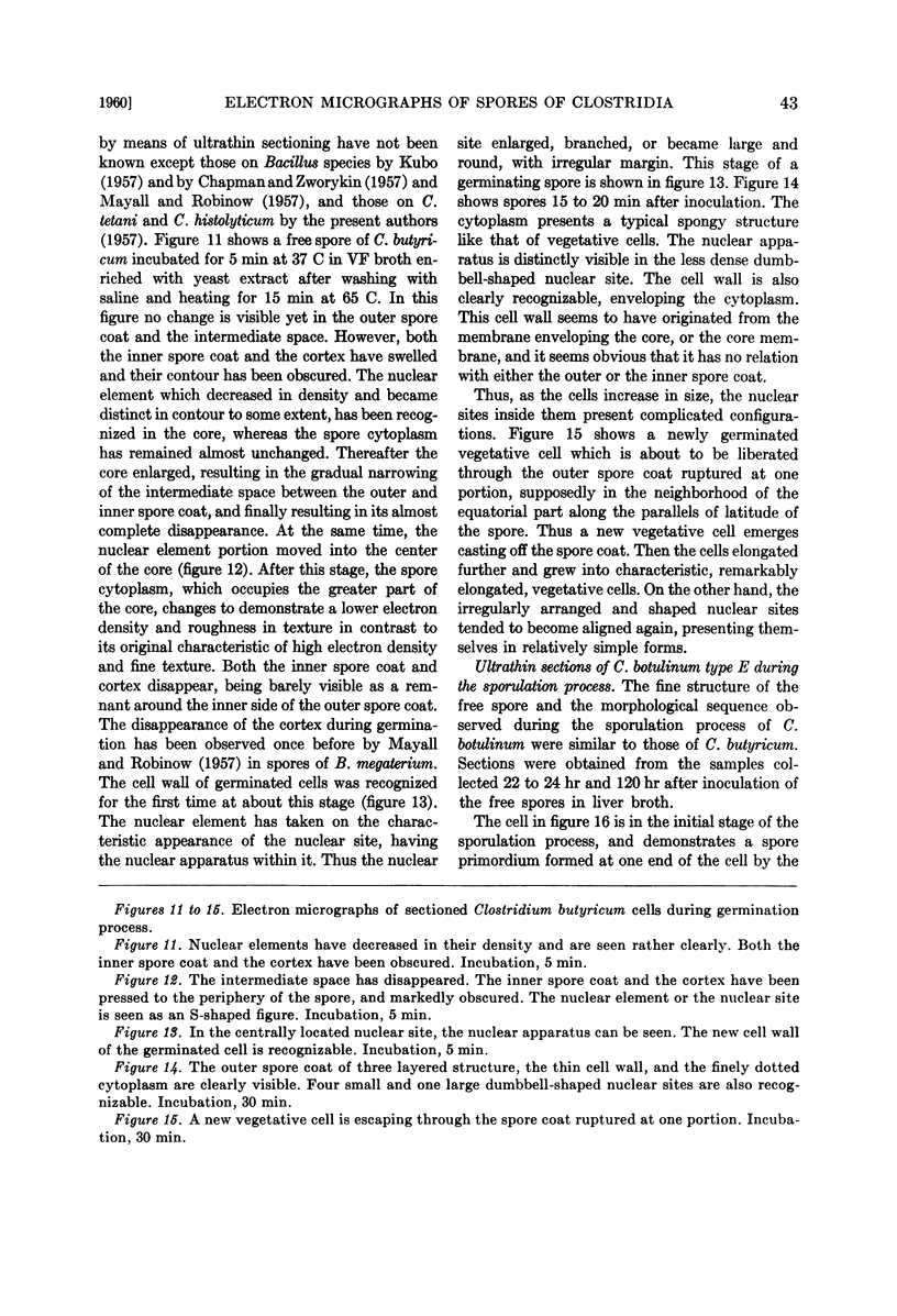
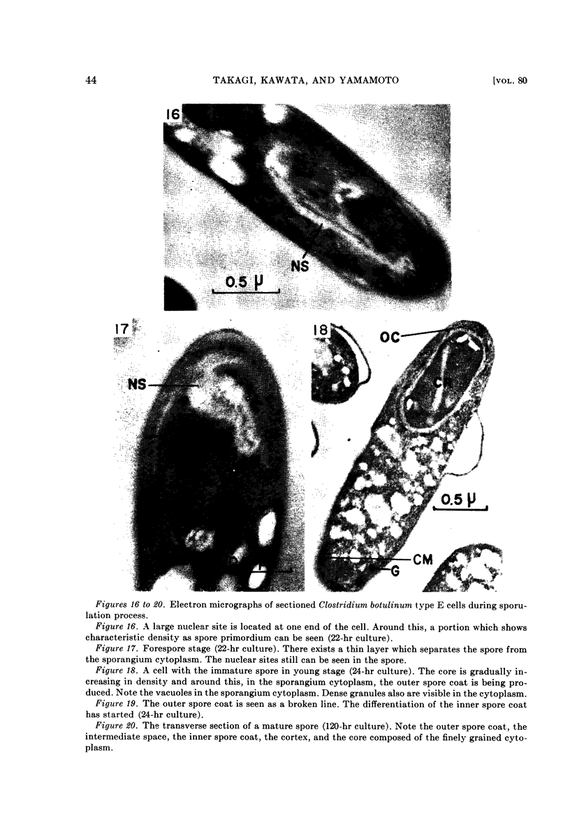
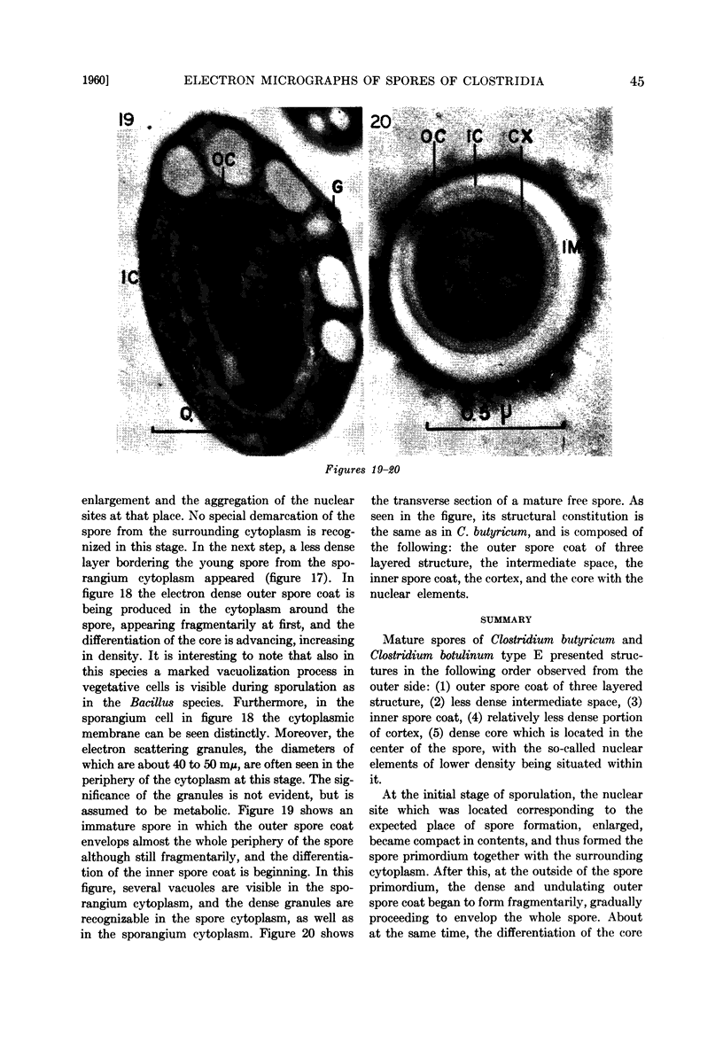
Images in this article
Selected References
These references are in PubMed. This may not be the complete list of references from this article.
- BISSET K. A. The sporulation of clostridium tetani. J Gen Microbiol. 1950 Jan;4(1):1–3. doi: 10.1099/00221287-4-1-1. [DOI] [PubMed] [Google Scholar]
- CHAPMAN G. B. Electron microscopy of ultra-thin sections of bacteria. II. Sporulation of Bacillus megaterium and Bacillus cereus. J Bacteriol. 1956 Mar;71(3):348–355. doi: 10.1128/jb.71.3.348-355.1956. [DOI] [PMC free article] [PubMed] [Google Scholar]
- CHAPMAN G. B., ZWORYKIN K. A. Study of germinating Bacillus cereus spores employing television microscopy of living cells and electron microscopy of ultrathin sections. J Bacteriol. 1957 Aug;74(2):126–132. doi: 10.1128/jb.74.2.126-132.1957. [DOI] [PMC free article] [PubMed] [Google Scholar]
- DAVIS J. C., MUDD S. Reducing sites in bacterial cells of the anaerobic genus Clostridium and their differentiation from other cell structures. J Histochem Cytochem. 1957 May;5(3):254–263. doi: 10.1177/5.3.254. [DOI] [PubMed] [Google Scholar]
- HASHIMOTO T., NAYLOR H. B. Studies of the fine structure of microorganisms. II. Electron microscopic studies on sporulation of Clostridium sporogenes. J Bacteriol. 1958 Jun;75(6):647–653. doi: 10.1128/jb.75.6.647-653.1958. [DOI] [PMC free article] [PubMed] [Google Scholar]
- KELLENBERGER E., RYTER A., SECHAUD J. Electron microscope study of DNA-containing plasms. II. Vegetative and mature phage DNA as compared with normal bacterial nucleoids in different physiological states. J Biophys Biochem Cytol. 1958 Nov 25;4(6):671–678. doi: 10.1083/jcb.4.6.671. [DOI] [PMC free article] [PubMed] [Google Scholar]
- ROBINOW C. F. Spore structure as revealed by thin sections. J Bacteriol. 1953 Sep;66(3):300–311. doi: 10.1128/jb.66.3.300-311.1953. [DOI] [PMC free article] [PubMed] [Google Scholar]
- RYTER A., KELLENBERGER E., BIRCHANDERSEN A., MAALOE O. Etude au microscope électronique de plasmas contenant de l'acide désoxyribonucliéique. I. Les nucléoides des bactéries en croissance active. Z Naturforsch B. 1958 Sep;13B(9):597–605. [PubMed] [Google Scholar]
- SMITH A. G., ELLNER P. D. Cytological observations on the sporulation process of Clostridium perfringens. J Bacteriol. 1957 Jan;73(1):1–7. doi: 10.1002/path.1700730102. [DOI] [PMC free article] [PubMed] [Google Scholar]
- TOKUYASU K., YAMADA E. Fine structure of Bacillus subtilis. II. Sporulation progress. J Biophys Biochem Cytol. 1959 Jan 25;5(1):129–134. doi: 10.1083/jcb.5.1.129. [DOI] [PMC free article] [PubMed] [Google Scholar]


