Abstract
Diphyllobothrium nihonkaiense was first described by Yamane in 1986 but the taxonomical features have been obscure due to lack of critical morphologic criteria in its larval and adult stages. In Korea, this tapeworm had long been known as Diphyllobothrium latum. In this study, we observed 62 specimens collected from Korean residents and analyzed them by morphological features and nucleotide sequences of mitochondrial cox1 gene as well as the ITS1 region. Adult tapeworms were examined after carmine or trichrome stain. Longitudinal sections of the gravid proglottids showed an obtuse angle of about 150 degree between the cirrus sac and seminal vesicle. This angle is known as a major differential point compared with that of D. latum. Nucleotide sequence differences between D. latum and the specimens from Koreans represented 17.3% in mitochondrial DNA cox1 gene. Sequence divergence of ITS1 among 4 Korean isolates was 0.3% and similarity was 99.7% with D. nihonkaiense and D. klebanovskii. All of the Korean specimens analyzed in this study were identified as being D. nihonkaiense (n = 62). We propose its Korean name as "Dong-hae-gin-chon-chung" which means 'long tapeworm of the East Sea' for this newly analyzed diphyllobothriid tapeworm in Korea.
Keywords: Diphyllobothrium nihonkaiense, Diphyllobothrium latum, genetic identification, distribution, Korea
INTRODUCTION
Cestodes of the genus Diphyllobothrium (Cobbold, 1858) are known to be widely distributed in northwestern Europe and Far-East Asia as a causative agent of diphyllobothriasis. Diphyllobothriasis is an intestinal parasitosis caused by the ingestion of raw freshwater fish containing infectious larvae of the genus Diphyllobothrium. Human diphyllobothriid cestodes have been reported so far as many as 18 species in the genus Diphyllobothrium (Yamane et al. 'Forum Cheju' in 1996, Japan). Among them, D. latum (Linnaeus, 1758), the broad fish tapeworm, is the most common human species whose life cycle is dependent on fish transport hosts. D. latum is mildly pathogenic in humans where it may cause pernicious anemia by absorbing large amounts of vitamin B12. Human diphyllobothriasis in Korea was first reported in 1919 by Kojima and Ko, and was morphologically identified by the adult worm as D. latum [1]; Diphyllobothrium yonagoense [2] and D. latum parvum type [3] were also reported.
D. nihonkaiense was first described by Yamane [4] in 1986 in Japan. He described morphologic differences of D. latum in Japan from that in Finland in the adult worms, eggs, and plerocercoids and proposed reconsideration about the taxonomic status of D. latum in Japan. Since Japan and Korea share the East Sea which locates between the 2 countries, and the salmons return to both countries from the Pacific Ocean, we speculate the possibility of diphyllobothriid tapeworms of identical species present in both countries. The morphologic identification of Diphyllobothrium in Korea has been obscure because of lack of detailed observations. Therefore, a reliable taxonomic criterion is needed based on morphologic features combined with molecular data. Molecular approaches to differential identification of D. latum and D. nihonkaiense have been done by restriction fragment length polymorphisms (RFLP) of ribosomal DNA [5], sequence differences of mitochondrial cytochrome c oxidase I (cox1) gene [6,7] and differentiating molecular makers [8].
The present study focused on differences in the morphology, sequence variations in the mitochondrial cox1, and rDNA internal transcribed spacer I (ITS1) PCR as markers for identifying diphyllobothriid tapeworms in Korea.
MATERIALS AND METHODS
Specimens
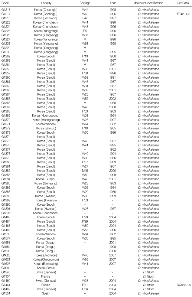
A total of 68 Diphyllobothrium tapeworms (1982-2007) were analyzed in this study (Table 1). Six isolates of D. latum were collected from France (n = 1), Russia (n = 1), Spain (n = 1), and Switzerland (n = 3). Sixty-two isolates were originated from Korea. Among them, 57 collections were given from other laboratories in Korea, and the rest 5 were collected from infected Korean patients who passed proglottids naturally in the stool or after treatment with niclosamide or praziquantel with purgation using MgSO4. These sample materials were divided each into 2 parts, which were then preserved in a -70℃ deep freezer and in 70% ethanol. Thirty-seven specimens were preserved in 10% formalin, 24 specimens were kept in 70% ethanol, and 7 specimens were kept between -20℃ and -70℃.
Table 1.
Diphyllobothriid specimens analyzed in this study collected between the years 1982 and 2007
-: unknown.
The rest 5 treated patients, all from Chungbuk Province in Korea, were addressed as follows. The code number G1213 was a 44-year-old man living in Cheongju City. He had eaten many unknown kinds of sea fishes raw and brought with an apolysed 1 m long strobila without scolex in 1998. G1215 was a 42 year-old woman living in Jochiwon, who had eaten sea fishes, brought an apolysed 130 cm long strobila without scolex, and a 5 m long strobila was collected after treatment in 1997. G1214 was a 42-year-old man living in Cheongju City, who he had eaten salmons and other sea fishes, brought an apolysed 2 m long strobila without scolex in 1996. G1623 was a 41-year-old man living in Eumseong. He had eaten 'masou salmon' and brought an apolysed 1 m long strobila, and a 5 m strobila was discharged after treatment in 2007. G1621 was a 65-year-old man living in Cheongwon. He also had eaten 'masou salmon' and a 6 m long strobila was collected after treatment in 2007.
Morphologic analysis
Ten or more gravid proglottids were longitudinally disrupted with a dissecting needle and fresh eggs were collected from terminal proglottids in saline and were prepared for scanning electron microscopy. After drying with critical point drier (CPD) and gold coating, specimens were observed by SEM (Hitachi S-570, Tokyo, Japan). The tapeworms were pressed and fixed in alcohol-formalin-acetic acid (AFA) for carmine stain. Some part of the specimen was used in hematoxylin and eosin (H-E) and trichrome stain after longitudinal sections for observation of the cirrus pouch, seminal vesicle, uterus, and uterine pore.
Genetic analysis
DNA extraction
Total genomic DNA was extracted from a single specimen that was chopped into small pieces and then the DNeasy Tissue Kit (Qiagen, Valencia, California, USA) was used according to manufacturer's instructions. The specimens were crushed in liquid nitrogen, soaked in TE buffer for 3-5 hr, and then digested for 30 min in DNA extraction buffer (100 mM NaCl, 50 mM EDTA [pH8.0], 50 mM Tris base/HCl [pH 8.0], 50 mM EDTA, 10% SDS, and 20 mg/ml proteinase K) at 56℃ After the incubation was continued in a hexadecyltrimethylammonium bromide (CTAB)/NaCl solution for 3 hr at 65℃, the cellular debris was removed and the genomic DNA was extracted using the phenol/ chloroform extraction protocol. DNA was then precipitated in 3 M sodium acetate (pH 5.2) and 2 volumes of 95% ethanol were added, after which the DNA was dried. The pellet was subsequently dissolved in 50 µl TE buffer.
PCR
18S small subunit nuclear ribosomal DNA (18S rDNA), ribosomal DNA ITS1, and mitochondrial cox1 gene were amplified by PCR. PCR were performed in a reaction mixture of 50 µl with 0.01 µg/µl of genomic DNA, 10 × PCR buffer (20 mM Mg+), 10 mM dNTP mixture, 10 pmoles of each primer, and 2.5 U/µl Taq DNA polymerase (High Fidelity PCR system, Roche, Mannheim, Germany). PCRs were performed in a GeneAmp PCR System 9700 (Applied Biosystems, Langen, Germany) and involved 1 cycle of initial denaturation at 94℃ for 3 min, followed by 35 cycles of denaturation (94℃ for 30 sec), annealing (52℃ for 30 sec for ITS1), and extension (72℃ for 1 min), with a final extension at 72℃ for 10 min. The PCR primers used were 5'-AACAAGGTTTCC GTAGGTG-3'and 5'-AGCAGTCTGCGATTCACATT-3'for ITS1 which yielded a 534 bp product including 18S rDNA and 5.8S rDNA, and 84C (5'-TGATTTTTTGGCCACCCCGAA AGTATA-3') and 85C (5'-TGACATTACATAGTGGAAGTGAGCTAC-3'), which yield a 348-bp product and designed from D. nihonkaiense [6]. These primers were used in PCR, which employed 35 cycles of 94℃ for 20 sec, 46℃ for 40 sec, 72℃ for 1 min, and incubation at 72℃ for 4 min. This resulted in 370 bp DNA fragments, which were isolated on a 1.0% agarose gel, excised under long-wave UV light, and extracted using a QIAquick PCR purification kit (Qiagen Co.).
DNA sequencing and analyses
The purified PCR-amplified fragments of ITS1 were then separately cloned. Ligation of the fragments was performed overnight at 15℃ using the pGEM-T easy vector kit (Promega, Madison, Wisconsin, USA). The ligates were transformed into the DH5α cell line. Plasmid DNA was then purified by using a QIA-prep spin miniprep kit (Qiagen). The primer walking method was employed to obtain overlapping sequences for each of the amplified fragments. Cyclic sequencing from both ends of the fragments was performed by using a Big-Dye Terminator sequencing kit (Applied Biosystems, Foster City, California, USA) and the reaction products were electrophoresed on an automated DNA sequencer (model 3730KL, Applied Biosystems).
The sequences were assembled and aligned by using CLUSTAL X multiple alignment program [9] and the Bioedit program version 5.0.6 (BIOSOFT, Ferguson, Missouri, USA). The sequencing regions were identified by comparing them using BLAST searches with those of Platyhelminthes that had been deposited in the GenBank database. The molecular identification of Diphyllobothrium tapeworm specimens was based on the similarity of nucleotide sequences of cox1 gene and ITS1 region, and phylogenetic relationships with those of D. nihonkaiense (Gen-Bank accession number EF420138), D. latum (Genbank accession number DQ985706). Phylogenetic analyses were determined by the neighbor-joining (NJ), maximum-parsimony (MP), and minimum-evolution (ME) methods using the Mega 3.1 program [11]. NJ analysis was performed using a distance matrix calculated using the Kimura 2 parameter method. Bootstrap analysis was performed with 3,000 replications.
RESULTS
Morphologic characteristics of D. nihonkaiense
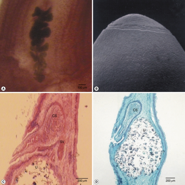
Morphologic observation was based on 5 specimens collected from the Korean people; G1213-5, G1261, and G1623. Whitish-yellow adult tapeworms were 1-5 m long with longitudinal central line of genital pores. The widest gravid proglottids measured 11 mm. The uterine structure showed typical diphyllobothriid tapeworm's feature showing rosette formation swirling 5-7 loops in carmine-stained specimens (Fig. 1A). Average size of eggs was 55.5 (± 1.0) × 40.5 (± 1.5) µm and the ratio of length and width was 1.37 (± 0.06) µm (n = 20). The eggshell showed shallow pits on the smooth surface in SEM (Fig. 1B). The ovary was renal shape and located at the posterior side. Testes were follicular and the ovaries were dumbbell shape. Longitudinal sections of the gravid proglottids showed an obtuse angle of about 150 degree between the cirrus sac and seminal vesicle, which looked different from D. latum after H-E or trichrome stain (Fig. 1C, D). The uterine and genital pores were separated on the midline with 150-300 µm. The uterine pore opened slightly posterior to the genital pore. The genital pore was located ventral on the middle at 1/3 of the proglottids. The cirrus sacs in sagittal sections were 430-480 µm in length and 275-310 µm in width. The seminal vesicles were round to elliptical and 300-420 in length by 170-300 µm in diameter.
Fig. 1.
Gravid proglottids and an egg of Diphyllobothrium nihonkaiense. (A) Whole mounted specimens of a proglottid showing the uterus and cirrus sac (× 25), (B) A SEM photo of the eggshell showing shallow pits and opercular structure (× 3,000), (C, D) Longitudinal sections of a mature proglottid showing the cirrus sac (CS), seminal vesicle (SV), and uterus (U) (C: H-E stain, D: trichrome stain).
Sequence divergence of mitochondrial cox1 and ITS1 of human diphyllobothriid tapeworms
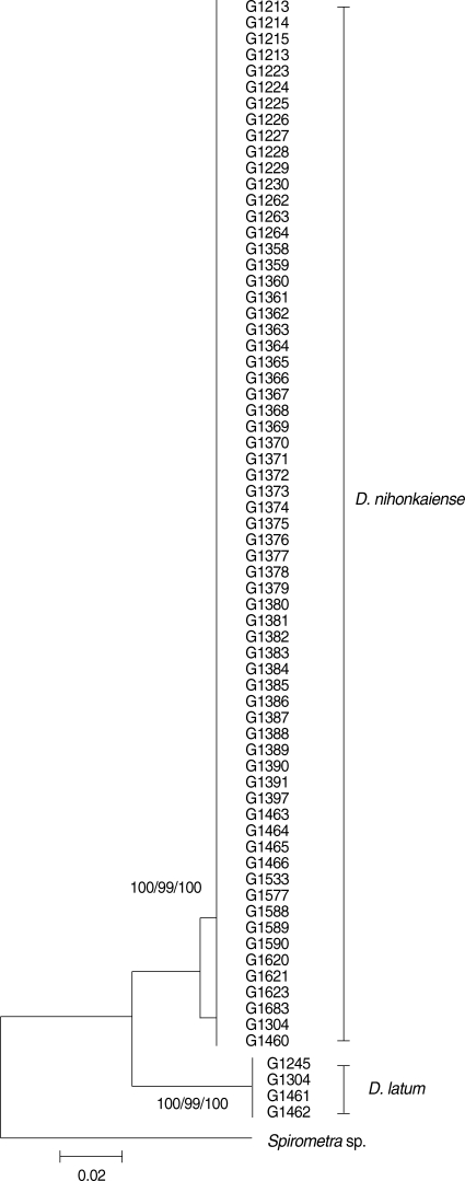
The cox1 sequences (335 bp) of 62 Korean isolates showed 99% similarity to reference sequences of the Japanese origin D. nihonkaiense (GenBank No. AB015755) and 83.7% similarity with reference sequences of the Russian origin D. latum (G1461; GenBank No. DQ985706). Phylogenetic analysis of the mitochondrial DNA cox1 (mtDNA cox1) sequences for a total 62 isolates identified Spirometra sp. as basal to the D. nihonkaiense-D. latum clade. The mtDNA cox1 sequences of D. nihonkaiense and D. latum differed by 17.3% based on Kimura's 2-parameter model. Trees topology using various analytical methods (NJ, MP, and ME) generated very high confidence values (bootstrap values of 100%, 99%, and 100% in NJ, MP and ME, respectively) for 2 major branches representing each of D. nihonkaiense and D. latum (Fig. 2). The ITS1 sequences of Spain (G1551) and Switzerland (G1460) presented 100% similarity (525 bp) to the Japanese reference sequences of D. nihonkaiense (GenBank No. AB375175), and other 4 Korean isolates (G1213-5 and G1621) also presented 100% identity to that of D. nihonkaiense. The analyses of cox1 and ITS1 sequences identified all 62 Korean specimens as D. nihonkaiense.
Fig. 2.
A phylogenetic tree of diphyllobothriid tapeworms based on partial cox1 sequences inferred from neighbor-joining (NJ) analysis. Numbers on branches indicate the bootstrap supporting values based on the 3,000 replicates. There were 335 bps corresponding to positions 735-1107 of the cox1 gene.
DISCUSSION
Diphyllobothriid tapeworm infections have been reported in 43 cases in South Korea since 1971, including D. latum parvum type and D. yonagoense [11]. The major symptoms were gastrointestinal discomfort, except in a case of a child reported in 1983 by Joo and colleagues who showed microcytic hypochromic anemia (25th annual meeting of The Korean Society for Parasitology in Seoul). The suspected sources of infection of diphyllobothriid tapeworms in Korea were salmon, mullet, perch, and trout, but there have been no reports on plerocercoid infections from these fish intermediate hosts. The main infective source of D. nihonkaiense is now known to be Oncorhynchus masou, O. keta, and Hucho perryi (Salmoniidae) in Japan [6]. These species migrate around the Okhotsk, Bering, and Pacific Ocean. The salmons return back to the East Sea which is located between Korea and Japan; that is the reason why we consider D. nihonkaiense is shared by the 2 countries. The prevalence of diphyllobothriasis among Koreans was considered to be caused mainly by infection with D. latum until the address of D. nihonkaiense by Eom and colleagues in 2001 (35th annual meeting of The Korean Society for Parasitology at Kwangju, Korea). Characterization of the complete mitochondrial genome of D. nihonkaiense was also reported by Kim et al. in 2007 [8]. More recently, revised identification of D. nihonkaiense Yamane et al., 1986 and D. klebanovskii Muratov and Posokhov, 1988 [12] has been studied, and they were considered as the same species according to Arizono et al. in 2009 [13].
D. nihonkaiense was first identified by Yamane et al. in 1986 [4] with establishment of distinct characteristics of this parasite, such as an angle of the axis of the cirrus sac and seminal vesicle, of which morphology was first addressed by Kamo in 1978 [14]. Morphologic differentiation of human-infecting diphyllobothriid tapeworms is based on the features like the pit shape of egg shells, genital atrium openings, the angle between the long axis of the cirrus sac and seminal vesicle. The average size of D. nihonkaiense eggs was 55.5 (± 1.0) × 40.5 (± 1.5) µm which is smaller than that of D. latum [7,15]. The egg shells of D. nihonkaiense exhibit shallower pits distributing on the smooth surface. The genital pore and uterine pore were closer in D. nihonkaiense (150-300 µm) than in D. latum (260-1,240 µm) [15]. The angle between the long axis of the cirrus sac and seminal vesicle was sharper in D. nihonkaiense than those of D. latum. Nevertheless, species differentiation between D. latum and D. nihonkaiense is not clear sometimes due to their morphological similarities.
Recently, the taxonomic status of diphyllobothriid tapeworm infections was questioned because only D. latum was reported in Korea. Consequently, it was necessary to clarify the distribution of these tapeworms in this country. Only 15 specimens could be examined on the morphological basis; the rest 47 specimens could not afford it. Most of them were preserved in 10% formalin or improper for morphologic examinations. The cox1 sequences of 62 specimens showed 2 polymorphic sites with 2 non-synonymous substitution in D. nihonkaiense Korean isolates. The overall sequence difference in the full mitochondrial cox1 gene between D. nihonkaiense and D. latum was 7.7%, whereas the full mitochondrial genome differed by 10.1% [8]. The sequence divergence of ITS1 among 4 Korean isolates was 0.3% and the similarity was 99.7% with D. nihonkaiense (GenBank No. AB375175) and D. klebanovskii (GenBank No. AB375657-AB375671). These results clearly indicate that D. nihonkaiense is a dominant species distributing in Korea without exception in all specimens examined (n = 62).
ACKNOWLEDGEMENTS
This work was supported by a research grant from Chungbuk National University in 2009. Parasite materials PRB50850001-050910001, PRB50990001, PRB051000001, PRB051090001-PRB051130001, PRB051500001, PRB051650001-PRB05167-0001, PRB060880826, PRB061180022, PRB061190020, PRB070061180, PRB070490002, PRB071140100, PRB071150100, PRB071170100, PRB071400002, PRB071410100, PRB071630001, PRB071640001, PRB071650001 used in this study were provided by the Parasite Resource Bank of Korea National Research Resource Center (R21-2005-000-10007-0), Republic of Korea. The authors thank Prof. Yamane for morphological identification of D. nihonkaiense based on the sectioned slide specimens as well as for providing references. Thanks are also due to Prof. T.S. Yong for suggesting its Korean name, Dong-hae-gin-chon-chung.
References
- 1.Cho SY, Seo BS, Ahn JH. One case report of Diphyllobothrium latum infection in Korea. Seoul J Med. 1971;12:157–163. [Google Scholar]
- 2.Lee SH, Chai JY, Hong ST, Sohn WM, Choi DI. A case of Diphyllobothrium yonagoense infection. Seoul J Med. 1988;29:391–395. [Google Scholar]
- 3.Lee SH, Chai JY, Seo M, Kook J, Huh S, Ryang YS, Ahn YK. Two rare cases of Diphyllobothrium latum parvum type infection in Korea. Korean J Parasitol. 1994;32:117–120. doi: 10.3347/kjp.1994.32.2.117. [DOI] [PubMed] [Google Scholar]
- 4.Yamane Y, Kamo H, Bylund G, Wikgren BJP. Diphyllobothrium nihonkaiense sp. nov. (Cestoda: Diphyllobothriidae)-revised identification of Japanese broad tapeworm. Shimane J Med Sci. 1986;10:29–48. [Google Scholar]
- 5.Matsuura T, Bylund G, Sugane K. Comparison of restriction fragment length polymorphisms of ribosomal DNA between Diphyllobothrium nihonkaiense and D. latum. J Helminthol. 1992;66:261–266. doi: 10.1017/s0022149x00014693. [DOI] [PubMed] [Google Scholar]
- 6.Ando K, Ishokura K, Nakakug T, Shimono Y, Tamai T, Sugawa M, Limviroj W, Chinzei Y. Five cases of Diphyllobothrium nihonkaiense infection with discovery of plerocercoids from an infective source, Oncorhynchus masou ishikawae. J Parasitol. 2000;87:96–100. doi: 10.1645/0022-3395(2001)087[0096:FCODNI]2.0.CO;2. [DOI] [PubMed] [Google Scholar]
- 7.Wicht B, Marval F, Peduzzi R. Diphyllobothrium nihonkaiense (Yamane et al., 1986) in Switzerland: first molecular evidence and case reports. Parasitol Int. 2007;56:195–199. doi: 10.1016/j.parint.2007.02.002. [DOI] [PubMed] [Google Scholar]
- 8.Kim KH, Jeon HK, Kang S, Sultana T, Kim GJ, Eom KS, Park JK. Characterization of the complete mitochondrial genome of Diphyllobothrium nihonkaiense (Diphyllobothriidae: Cestoda), and development of molecular markers for differentiating fish tapeworms. Mol Cell. 2007;23:379–390. [PubMed] [Google Scholar]
- 9.Thompson JD, Gibson TJ, Plewniak F, Higgins DG. The Clustal X windows interface: flexible strategies for multiple sequence alignment aided by quality analysis tools. Nucleic Acids Res. 1997;25:4876–4882. doi: 10.1093/nar/25.24.4876. [DOI] [PMC free article] [PubMed] [Google Scholar]
- 10.Kumar S, Tamura K, Nei M. MEGA3: integrated software for molecular evolutionary genetics analysis and sequencing alignment. Brief Bioinformat. 2004;5:150–163. doi: 10.1093/bib/5.2.150. [DOI] [PubMed] [Google Scholar]
- 11.Lee EB, Song JH, Park NS, Kang BK, Lee HS, Han YJ, Kim HJ, Shin EH, Chai JY. A case of Diphyllobothrium latum infection with a brief review of diphyllobothriasis in the Republic of Korea. Korean J Parasitol. 2007;3:219–223. doi: 10.3347/kjp.2007.45.3.219. [DOI] [PMC free article] [PubMed] [Google Scholar]
- 12.Muratov IV, Posokhov PS. Causative agent of human diphyllobothriasis-Diphyllobothrium klebanovskii sp. Parazitologiia. 1988;22:165–170. [PubMed] [Google Scholar]
- 13.Arizono N, Shedko M, Yamada M, Uchikawa R, Tegoshi T, Takeda K, Hashimoto K. Mitochondrial DNA divergence in populations of tapeworm Diphyllobothrium nihonkaiense and its phylogenetic relationship with Diphyllobothrium klebanovskii. Parasitol Int. 2009;58:22–28. doi: 10.1016/j.parint.2008.09.001. [DOI] [PubMed] [Google Scholar]
- 14.Kamo H. Reconsideration of taxonomic status of Diphyllobothrium latum (Linnaeus, 1758) in Japan with special regard to species specific characters. Jpn J Parasitol. 1978;27:135–142. [Google Scholar]
- 15.Raush RL, Hillard DK. Studies on the helminth fauna of Alaska. XLIX. The occurrence of Diphyllobothrium latum (Linnaeus, 1758) (Cestoda: Diphyllobothriidae) in Alaska, with notes on other species. Can J Zool. 1970;48:1201–1219. doi: 10.1139/z70-210. [DOI] [PubMed] [Google Scholar]


