

Abstract
Imaging with labeled monoclonal antibodies may be useful in detecting, staging and monitoring tumors. Despite their high affinity and specificity, a critical limitation of antibody imaging is the high background signal due to prolonged clearance from the blood which reduces the tumor-to-background ratio (TBR). To address this problem, we developed an molecular imaging probe consisting of multiple self-quenching, fluorophores (Cy5.5 or Alexa680) conjugated to a monoclonal antibody (trastuzumab-Tra) to synthesize Tra-Cy5.5(SQ) or Tra-Alexa680(SQ) respectively. This agent only becomes fluorescently “active” after cellular internalization but is quenched in the unbound state leading to high TBRs. The in vitro quenching capacity for both conjugates was approximately 9-fold. In vivo imaging experiments were performed in mice bearing both 3T3/HER2+ and Balb/3T3/ZsGreen/HER2- xenografts. Tra-Alexa680(SQ) produced specific enhancement in the 3T3/HER2+ tumors but not in the HER2- control tumors. However, Tra-Cy5.5(SQ) produced non-specific enhancement in both 3T3/HER2+ and control tumors. In conclusion, while Cy5.5 produced non-specific results as well as rapid liver accumulation, conjugating multiple Alexa680 molecules to a single monoclonal antibody resulted in a near-infrared (NIR) optical agent that activated within specific target tumors with high TBR with considerable potential for clinical translation.
Keywords: molecular imaging, activatable, cancer, near infrared, humanized antibody
Introduction
Molecular imaging with antibodies has the potential not only to improve the detection of tumors but also to characterize them by their cell surface expression profiles (1,2). However, antibody delivery to a tumor relies on the high binding affinity and the low off-rate of antibodies to their cell surface antigens as well as their abundant blood supply (3,4) with leaky tumor vasculature leading to enhanced permeability and retention (EPR) (5,6) thus increasing antibody accumulation. Since the EPR effect depends only on the physical characteristics of the macromolecules injected and not on their binding characteristics it often leads to non-specific tumor uptake. In order to achieve specific antibody imaging, sufficient time for clearance of the unbound antibody is needed to reduce background signal resulting in favorable target-to-background ratios (TBR). Meanwhile, the long clearance times of antibodies make delayed imaging a necessity raising practical issues with regard to patient and physician acceptance. Therefore, in vivo antibody-based target-specific molecular imaging is limited by the EPR effect and prolonged clearance times leading to reduced TBR which lowers both sensitivity and specificity.
Humanized antibodies, which are antigen specific CDR-grafted human IgG molecules, have been used for clinical cancer therapy because they produce antigen-dependent cellular cytotoxicity with minimal toxicity due to low immunogenicity. Therefore, the humanized antibody is a realistic choice as a targeting moiety for molecular imaging probes. However, imaging with humanized antibodies has achieved limited success. Despite their highly specific accumulation in target tumors, a critical limitation of humanized antibody imaging is the high background signal due to prolonged blood clearance which reduces the tumor-to-background ratio (TBR). Of the clinically available imaging techniques for labeling antibodies only positron emission tomography (PET) and single-photon emission computed tomography (SPECT) have been widely used in vivo and then only with long lived isotopes. However, because PET or SPECT probes constantly emit signal (decreasing as a function of half life of the radioisotope), EPR related signal and background signal are quite high, especially when humanized antibodies are used. Therefore, in order to optimize the pharmacokinetics and clearance, genetic or enzymatic modifications of antibodies have been investigated, however, these alterations may reduce the therapeutic value of the antibody (2). Optically labeled antibodies, in theory, suffer from the same limitations as radioisotopes, however, optical probes differ because they can be activated or switched on only at the target cancer cells in response to specific intracellular environmental stimuli. By activating the fluorescence signal only within the target cells, non specific accumulation due to EPR and in the blood pool is minimized.
Several activatable optical probes have recently been reported (7-11). These are largely based on self-quenching mechanisms whereby enzymatic cleavage of flurophores held in close steric alignment results in fluorescent activation as the fluorophores move away from each other. Among the various choices for imaging fluorophores, near-infrared (NIR) probes have the advantage of better depth penetration within tissue and are amenable to self-quenching (12). For instance, when two or more Cy5.5 dyes are conjugated to generation-6 polyamidoamine dendrimers, which are similar in hydrodynamic diameter to antibody molecules, self-quenching occurs and the degree of self quenching increases as the number of Cy5.5 dyes increases (13). However, conjugation of multiple fluorophores to the same macromolecule risks altering the pharmacokinetics of the conjugate. Relatively few reports focus on activatable optical probes conjugated to antibodies.
In this study, we synthesized and tested a self-quenching activatable probe conjugated to a monoclonal antibody using cyanine-based NIR fluorophores, AlexaFluor 680 (Alexa680) and Cy5.5 as described in Supplemental figure 1. In this study we employ trastuzumab, a humanized monoclonal IgG1 antibody, which binds to human epidermal growth factor receptor type 2 (HER2). After binding to HER2, trastuzumab is gradually internalized within the target cells and then undergoes degradation in the lysosome (14). Within the lysosome the conjugate is dequenched and light is emitted. Thus, the signal is quenched while the antibody-conjugate is outside the cell but is activated after it is internalized intracellularly.
Materials and Methods
Reagents
Trastuzumab (Tra), an FDA-approved humanized anti-HER-2 antibody, which has a complimentary determination region (CDR) against HER-2 grafted on a human IgG1 framework, was purchased from Genentech Inc. (South San Francisco, CA). Cy5.5-NHS ester was purchased from GE Healthcare (Piscataway, NJ). Alexa680-NHS ester was purchased from Invitrogen Corporation (Carlsbad, CA). ZsGreen plasmid was purchased from Clontech Laboratories, Inc. (Mountain View, CA). All other chemicals used were of reagent grade.
Structural analysis of Cy5.5 and Alexa680
The chemical structure of Cy5.5 has been widely published (24). The structure of Alexa680 has been determined with mass spectroscopic analysis and NMR as shown in the Supplemental data and figure 3,4.
Figure 3.
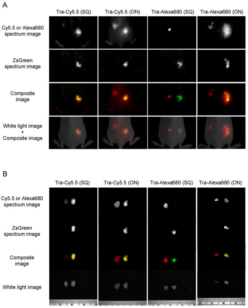
In vivo (A) and ex vivo fluorescence images with tumor bearing mice two days after injection. Trastuzumab-Cy5.5 or Alexa680 conjugates were injected intravenously into mice bearing 3T3/HER2+ (left) and Balb/3T3/ZsGreen (right) tumors. Cy5.5 or Alexa680 spectrum images shows target specific image with Tra-Alexa680(SQ). In contrast, the Cy5.5 signal was low in the target tumor (3T3/HER2+) for Tra-Cy5.5(SQ).
Figure 4.
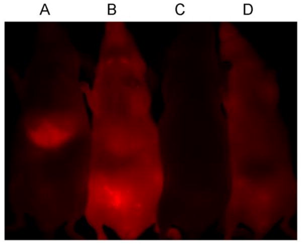
Supine Cy5.5 or Alexa680 spectrum images of trastuzumab-Cy5.5 or Alexa680 conjugate injected tumor bearing mice two days after the injection. A; Tra-Cy5.5(SQ), B; Tra-Cy5.5(ON), C; Tra-Alexa680(SQ), D; Tra-Alexa680(ON). Tra-Cy5.5(SQ) showed high liver uptake. The background was fairly low for Tra-Alexa680(SQ).
Briefly, mass spectroscopic analyses (MS and MS/MS) were performed with LCMS-IT-TOF systems (Shimadzu America Co., Columbia, MD). H- and C-NMR analyses were performed with Gemini or a Mercury 300 MHz spectrometer (Varian, Palo Alto, CA). By summarizing both results with the reference from a patent document (15), the structure of Alexa680 was suggested as shown in Supplemental figure 1.
Synthesis of Cy5.5 or Alexa680 conjugated antibodies
To synthesize the “always-on” control conjugate, trastuzumab (1 mg, 6.8 nmol) was incubated with Cy5.5-NHS (11 μg, 10 nmol, 5mM in DMSO) or Alexa680-NHS (16 μg, 14 nmol, 5mM in DMSO) in 0.1M Na2HPO4 (pH 8.5) at room temperature for 30 min. The mixture was purified with a Sephadex G50 column (PD-10; GE Healthcare). The protein concentration was determined with Coomassie Plus protein assay kit (Pierce Biotechnology, Rockford, IL) by measuring the absorption at 595 nm with a UV-Vis system (8453 Value UV-Visible Value System; Agilent Technologies, Santa Clara, CA). The concentration of Cy5.5 or Alexa680 was measured by absorption with the UV-Vis system to confirm the number of fluorophore molecules conjugated to each trastuzumab molecule. The number of Cy5.5 or Alexa680 per antibody was approximately 1 in these control probes. The resulting compounds Tra-Cy5.5(ON) and Tra-Alexa680(ON) (the designation “ON” indicates that these probes are always active) were kept at 4°C in the refrigerator as a stock solution.
For the self-quenched conjugate, trastuzumab (1 mg, 6.8 nmol) was incubated with Cy5.5-NHS (77 μg, 68 nmol, 5mM in DMSO) or Alexa680-NHS (158 μg, 137 nmol, 5mM in DMSO) in 0.1M Na2HPO4 (pH 8.5) at room temperature for 30 min. Then, the mixture was purified and protein and Cy5.5 and Alexa680 concentrations were determined as described above. The number of Cy5.5 or Alexa680 per antibody was approximately 7. The resulting conjugates Tra-Cy5.5(SQ) and Tra-Alexa680(SQ) (the designation “SQ” indicates that these compounds are self quenched) were kept at 4°C in the refrigerator as a stock solution.
Determination of quenching ability in vitro
The quenching abilities of each conjugate were investigated by denaturalizing with 5% SDS and 2-mercaptoethanol (2-ME). Briefly, the conjugates were incubated with 5% SDS and 1% 2-ME in PBS at 95 °C for 2 min. As a control, the samples were incubated in PBS. The fluorescence signal intensity of each sample was measured with a fluorescence spectrometer (Perkin-Elmer LS55, Perkin-Elmer, Shelton, CT, USA).
Measurement of the lipophilicity of Cy5.5 and Alexa680
To evaluate the lipophilicity of Cy5.5 and Alexa680,the partition coefficient (logD) was determined. Fifty nmol of Cy5.5-NHS or Alexa680-NHS was mixed with 1 mL each of 1-octanol and 0.1 M phosphate buffer (pH 5.2, 7.3 and 8.5) in a test tube. Three tubes were used for each condition. The tubes were shaken intensely (3 × 1 min) and incubated for 20 min at room temperature, and then the whole process was repeated twice to ensure that the reaction had reached equilibrium. Then, 1 mL aliquots of each phase were collected, and the concentration of Cy5.5-NHS or Alexa680-NHS was measured by absorption with the UV-Vis system. The distribution ratios were determined as the logarithm value of the octanol-to-buffer ratio (logD).
Cell culture
For HER2 targeting studies, the HER2 gene transfected NIH3T3 (3T3/HER2+) cell line was used. As a negative control, green fluorescence protein transfected Balb/3T3 cell line (Balb/3T3/ZsGreen, HER2 negative) was employed. The Balb/3T3/ZsGreen cell line does not express HER2 receptor. The cell lines were grown in RPMI 1640 (Life Technologies, Gaithersburg, MD) containing 10% fetal bovine serum (Life Technologies), 0.03% L-glutamine, 100 units/mL penicillin, and 100 μg/mL streptomycin in 5% CO2 at 37°C.
Fluorescence microscopy studies
3T3/HER2+ (1 × 10⁴) were plated on a cover glass–bottomed culture well and incubated for 16 h. Then Tra-Cy5.5 or Tra-Alexa680 was added to the medium (30 μg/mL), and the cells were incubated for either 1 or 8 hr. Cells were washed once with PBS, and fluorescence microscopy was performed using an Olympus BX51 microscope (Olympus America, Inc., Melville, NY) equipped with the following filters: excitation wavelength 590 to 650nm, emission wavelength 662.5 to 747.5 nm. Transmitted light differential interference contrast images were also acquired. To investigate the receptor specificity, the conjugates were also incubated with Balb/3T3 (HER2 negative) cells.
Tumor model
All procedures were carried out in compliance with the Guide for the Care and Use of Laboratory Animal Resources (1996), National Research Council, and approved by the local Animal Care and Use Committee. Both receptor positive and negative tumor cell lines were implanted in the mice. For HER2 targeting studies, 3T3/HER2+ cells (2 × 10⁶ cells in PBS) were injected subcutaneously in the left dorsum of the mice, and Balb/3T3/ZsGreen cells (2 × 10⁶ cells in PBS) were injected subcutaneously into the right dorsum. For orthotopic tumor models, 3T3/HER2+ cells (1 × 10⁶ cells in PBS) and Balb/3T3/ZsGreen cells (1 × 10⁶ cells in PBS) were injected into the right and left mouse mammary pads, respectively. The experiments were performed at 5 - 8 days after cell injection.
In vivo spectral imaging studies
Tra-Cy5.5(ON), Tra-Cy5.5(SQ), Tra-Alexa680(ON) or Tra-Alexa680(SQ) (50 μg / 100 μL PBS) were injected via the tail vein into tumor bearing (3T3/HER2+ and Balb/3T3/ZsGreen HER2-) mice. The mice were anesthetized with intraperitoneally administered 10% sodium pentobarbital with 0.1 % scopolamine butylbromide, then spectral fluorescence images were obtained using the Maestro In Vivo Imaging System (CRi Inc., Woburn, MA) using two filter sets two days after the conjugate injection. The deep red filter sets were used to image Cy5.5 or Alexa680 fluorescence and the blue filter sets were used for ZsGreen fluorescence. The deep red filter sets use a band-pass filter from 642 to 680 nm (excitation) and a long-pass filter over 702 nm (emission), the blue filter sets use a band-pass filter from 437 to 476 nm (excitation) and a long-pass filter over 493 nm (emission). The tunable emission filter was automatically stepped in 10-nm increments from 650 to 950 nm for the deep red filter sets and from 500 to 800 nm for the blue filter sets while the camera captured images at each wavelength interval with constant exposure. The spectral fluorescence images consisting of autofluorescence spectra and the spectra from Cy5.5, Alexa680 and ZsGreen which were then unmixed based on their spectral patterns using commercial software (Maestro software, CRi, Inc., Woburn, MA). Mice were sacrificed with carbon dioxide immediately after completion of imaging. Then the tumors were resected and ex vivo imaging was done using the same Maestro settings.
Results
Chemical structures of Cy5.5 and Alexa680 indicates differences in non-specific protein binding
The chemical structures of Alexa680 and Cy5.5 based on previously published results (15) and our own analysis (see Supportive data and method for NMR and Mass spectroscopy as shown in Supplemental figures 2-4) are shown in Supplemental figure 1. In summary, Alexa680 is smaller with fewer aromatic rings than Cy5.5. Additionally, although Cy5.5 is negatively charged, Alexa680 has the neutral charge. From these chemical structures, it is predicted that Alexa680 is less lipophilic and charged with lower non-specific protein binding than Cy5.5.
Lipophilicity of Cy5.5 is higher than that of Alexa680
The partition coefficients of Cy5.5 and Alexa680 were obtained to determine their lipophilicity which might alter the pharmacokinetics of the conjugated antibody. The resulting logD values (higher values indicating higher lipophilicity), are shown in Table 1. As predicted from the chemical structures, the lipophilicity of Cy5.5 was higher than Alexa680 for all values of pH and was pH independent whereas the lipophilicity of Alexa680 was pH dependent. Therefore, Cy5.5 would be predicted to more strongly alter pharmacokinetics by non-specific protein binding.
Table 1.
Partition coefficient of Cy5.5 and Alexa680 (logD) in various pH.
| pH 5.2 | pH 7.3 | pH 8.5 | |
|---|---|---|---|
| Cy5.5 | -1.07 ± 0.06 | -0.98 ± 0.14 | -1.06 ± 0.02 |
| Alexa680 | -1.91 ± 0.03 | -1.72 ± 0.04 | -1.42 ± 0.14 |
Each value represents the mean ± S.D. of three experiments.
Quenching capacity of both antibody-fluorophore conjugates is high
The quenching capacity was measured by adding 5% SDS and 2-ME. The molecular interaction can be dissociated with SDS and the heavy and light chains of IgG can be separated from each other with 2-ME treatment. The quenching capacities were 9-fold, 2-fold, 8-fold and 2-fold for Tra-Cy5.5(SQ), Tra-Cy5.5(ON), Tra-Alexa680(SQ) and Tra-Alexa680(ON), respectively.
Fluorescence microscopy demonstrates fluorescent activation in target cells
Fluorescence microscopy studies were carried out to visualize the cellular binding location and behavior of fluorescently-labeled antibody in vitro. All the investigated trastuzumab conjugates showed florescent signal on the surface of 3T3/HER2+ cells 1hr after incubation (Figure 2A). The fluorescence was observed inside the cell after 8hr incubation for all conjugates, and the fluorescent dots were brighter for the self-quenched conjugates than always-on conjugates which were not self quenching (Figure 2A). The microscopic images were also obtained using HER2 negative Balb/3T3 cells (Figure 2B). Tra-Alexa680(ON), Tra-Alexa680(SQ) and Tra-Cy5.5(ON) showed no fluorescent signal thus demonstrating their specificity for HER2 expressing tumors. However, Tra-Cy5.5(SQ) demonstrated non-specific fluorescence inside HER2 negative Balb/3T3 cells after 8h incubation probably because of nonspecific protein binding. These findings are well correlated and supported by the FACS analysis shown in the Supplemental figure 5.
Figure 2.

Fluorescence microscopy studies. A, 3T3/HER2+ cells were incubated with trastuzumab-Cy5.5 or Alexa680 conjugates for 1hr or 8hr. The fluorescent signal was on the surface of the cell after 1hr incubation. The higher fluorescent signal was detected by self-quenched conjugates after internalization into the cells after 8hr incubation. B, The conjugates were incubated with Balb/3T3 (HER2 negative) cells. Only Tra-Cy5.5(SQ) showed fluorescent signal inside the cell after an 8hr incubation.
The self-quenched antibody-Alexa680 conjugate showed high tumor-to-background ratio during in vivo fluorescence imaging whereas Cy5.5 conjugates did not
The results of imaging with trastuzumab conjugates in both 3T3/HER2+ (HER2 positive) and Balb/3T3/ZsGreen (HER2 negative) tumor bearing mice are summarized in Figure 3. In the whole body image (Figure 3A), the targeted tumor (3T3/HER2+) was greatly enhanced with Tra-Alexa680(SQ) and whereas the control tumor (Balb/3T3/ZsGreen) was minimally enhanced. With Tra-Alexa680(ON), the NIR signal could be detected not only in the targeted tumor but also in the control tumor, although the signal was higher in the targeted tumor. The NIR signal was also shown around the control tumor with Tra-Alexa680(ON). On the other hand, the targeted tumor could not be clearly imaged with Tra-Cy5.5(SQ), and the NIR signal was higher for the control tumor including the peripheral regions indicating non-specific binding. High NIR signal was observed both in the target and control tumor with Tra-Cy5.5(ON). The resected tumors also showed the advantage of Tra-Alexa680(SQ), i.e., the NIR from the labeled antibody in the HER2+ tumors did not overlap with the ZsGreen fluorescence signal arising from the HER2-tumors (Figure 3B). With Cy5.5 conjugated trastuzumab, high NIR signal could be seen in non-target tumor. In addition, the whole body abdominal images demonstrated reduced background for Tra-Alexa680(SQ) but high liver uptake with Tra-Cy5.5(ON) likely related to the latter's lipophilicity (Figure 4). Similar results were obtained with the orthotropic mammary pad tumor model (Figure 5).
Figure 5.

In vivo fluorescence imaging with orthotopic (breast) tumor bearing mice. The images were obtained two days after the trastuzumab-Cy5.5 or Alexa680 conjugates injection. The target tumor (3T3/HER2+) was specifically imaged with low background with Tra-Alexa680(SQ).
Discussion
The fundamental barriers to optical imaging in tissue are high light scattering, autofluorescence, high absorption by hemoglobin and other macromolecules, and reduced depth penetration with decreasing wave length. One solution for these problems is to use a very bright fluorescence source such as quantum dots (16-18) or high efficiency fluorescence proteins (19). In reality, even single cell labeled with fluorescence proteins could reportedly be tracked even in vivo (20,21). However, the physical sizes of such high efficiency fluorescence sources strongly influence the biodistribution of the agent. As a result, it is difficult to use such agents as injectable fluorescent molecular imaging probes especially in concert with antibodies, which are themselves large molecules. Therefore, smaller organic fluorophores remain the preferred choice as labels for intravenous optical molecular imaging probes. Among the many organic fluorophores available, NIR dyes are often selected because of the advantages of reduced autofluorescence and improved tissue penetration compared to the visible dyes (12).
In this study, we investigated two kinds of NIR fluorophores attached to the monoclonal antibody, trastuzumb: Cy5.5 and Alexa680. Trastuzumab is a complementary determining region (CDR)-grafted humanized monoclonal antibody (IgG1 subclass), which binds to human epidermal growth factor receptor type 2 (HER2) expressed on the cell surface of some tumors and the antibody has been highly successful in treating patients with breast cancers that have amplified expression of HER2. Since the humanized antibody is made by using the human IgG1 framework and grafting 12 CDR sequences, the advantage of using the same subclass of humanized antibody is that >98% of protein sequences have homology, even when the target antigen of the antibody is changed from HER2 to other cancer specific target molecules. Therefore, one would expect the optical features of the antibody conjugate to be the same regardless of the antigen used. This was demonstrated when the self-quenching efficiency of the conjugate was identical, when we switched the antibody from trastuzumab to daclizumab (Dac), which is also a CDR-grafted humanized monoclonal antibody (IgG1 subclass) targeting CD25 (IL-2 receptor α subunit). Indeed, the identical chemistry was used underscoring the flexibility of the concept. The quenching capacities were 12-fold, 2-fold, 10-fold and 1-fold for Dac-Cy5.5(SQ), Dac-Cy5.5(ON), Dac-Alexa680(SQ) and Dac-Alexa680(ON), respectively.
The pharmacokinetic characteristics of humanized antibodies, with clearance times typically measured in weeks, are well matched to the self-quenching system described here (22). Prolonged circulating humanized antibody is advantageous from a therapeutic perspective because of the continuous delivery of drug to the target tumor, however, this becomes a disadvantage when trying to image with the same antibody labeled with radioisotopes or “always on” fluorophores. By employing a self-quenching activatable strategy, the unbound circulating agent yields minimal signal, while the bound agent activates after internalization leading to high signal from target tumors.
Quenching was achieved by conjugating a large number (∼7) of Cy5.5 and Alexa680 to each antibody. In vitro microscopy revealed that fluorescence markedly increased after 8hr of incubation due to internalization (Figure 2). The quenching effect and the fluorescence increase after the internalization was also shown for Tra-Alexa680(SQ) with FACS flow cytometry studies (Supplemental figure 5). Among these conjugates the most successful for in vivo imaging was Tra-Alexa680(SQ) (Figure 3). The background fluorescence from the blood pool with this agent was low compared to Tra-Alexa680(ON) (Figure 4). In contrast, Tra-Alexa680(ON) resulted in signal from non-target tumor as well as target tumor. Although the rich blood supply and EPR effect might cause this non-specific fluorescence with an agent, which is always on (i.e. always fluoresces) the self-quenched conjugates were able to activate only within tumor cells, thus, taking advantage of the EPR effect without suffering the consequences of possible non-specific uptake. The identical findings were observed, when the daclizumab-CD25 targeting tumor system was employed (Supplemental Figure 2).
Tra-Cy5.5(SQ) was less successful as a tumor specific imaging agent. The multiplicity of Cy5.5 molecules altered the biodistribution and pharmacokinetics of the antibody. For instance, as shown in Figure 4, fluorescence was detected in liver immediately after i.v. injection of Tra-Cy5.5(SQ). In comparison the fluorescence in the liver was substantially lower for Tra-Alexa680(SQ) and even Tra-Cy5.5(ON). The rapid uptake by the liver could sequester Tra-Cy5.5(SQ) and prevent it from binding to the HER2 receptor in the tumor. The lipophilicity and charge of Cy5.5 likely results in its uptake and metabolism in the liver. Since the lipophilicity was higher for Cy5.5 than Alexa680 (Table 1), the pharmacokinetics could be largely affected by the conjugated fluorophore itself because of the large number of fluorophore molecules on a single antibody. The minimal liver accumulation with Tra-Cy5.5(ON) supports this hypothesis.
In addition, it is known that non-specific binding to plasma protein increases with lipophilicity (23). In microscopic studies, Tra-Cy5.5(SQ) showed non-specific uptake for HER2 negative Balb/3T3 cells. The non-specific protein binding of Cy5.5 may be related to its lipophilicity, although other factors (e.g. charge, molecular weight) might also affect binding.
In conclusion, we have successfully demonstrated an antibody targeted, activatable NIR molecular imaging probe based on self-quenching. The high quenching ability of Tra-Alexa680(SQ) and low non-specific binding derived from its more hydrophilic character leads to a high TBR in both the xenograft and orthotopic models of cancer. Humanized antibodies conjugated with self-quenched Alexa680 can image HER2 and CD25 in vivo without the disadvantages typical of antibody imaging. This technology could be universally applied to humanized antibodies of a similar class (IgG1).
Supplementary Material
Figure 1.

The scheme of a self-quenching activation system. A, the “activatable” probe, Tra-Cy5.5(SQ) or Tra-Alexa680(SQ) is self-quenched outside of the cell and has no fluorescence. When it binds to HER2 and is internalized, it is catabolized within the endosome/lysosome and dequenched. As a result, fluorescence signal appears within the cell. B, the “always-on” probe, Tra-Cy5.5(ON) or Tra-Alexa680(ON) has fluorescence outside as well as inside of the cell.
Acknowledgments
This research was supported by the Intramural Research Program of the NIH, National Cancer Institute, Center for Cancer Research. We thank to Drs. Masatoshi Takahashi and Masayuki Nishimura in Shimadzu Scientific Instruments, Inc., Columbia, MD for their great assistance for the various mass spectroscopy analyses of fluorescence dyes.
Footnotes
Significance of the work:
Imaging with labeled monoclonal antibodies is expected to be useful in detecting and staging tumors and monitoring therapy, however, to date this strategy has achieved limited success. Humanized antibodies, which are antigen specific CDR-grafted human IgG molecules, have been used for clinical cancer therapy because they induce antigen-dependent cellular cytotoxicity with minimal immunogenic toxicity. Although, humanized monoclonal antibodies would seem ideal candidates as targeting moieties for molecular imaging probes, little success has been achieved to date. Despite their high specificity and affinity, a critical limitation of humanized antibody imaging is the high background signal due to prolonged clearance from the blood which reduces the tumor-to-background ratio (TBR). To address this problem, we developed a self-quenching, fluorescently activatable monoclonal antibody, based on the humanized anti-HER2 antibody, trastuzumab, which produces little signal when unbound but activates after cellular internalization and dequenching leading to a high TBR. We validated this system by comparing it with a similarly labeled humanized anti-CD25 antibody, daclizumab to demonstrate that this method is widely applicable to a broad range of humanized antibodies. This target specific, NIR, self-quenching antibody-based molecular imaging system employing multiple Alexa680 molecules is a potential platform technology for detecting, diagnosing, and characterizing tumors in vivo by defining their surface antigen expression with high TBR.
References
- 1.Barrett T, Koyama Y, Hama Y, et al. In vivo diagnosis of epidermal growth factor receptor expression using molecular imaging with a cocktail of optically labeled monoclonal antibodies. Clin Cancer Res. 2007;13:6639–48. doi: 10.1158/1078-0432.CCR-07-1119. [DOI] [PubMed] [Google Scholar]
- 2.Wu AM, Senter PD. Arming antibodies: prospects and challenges for immunoconjugates. Nat Biotechnol. 2005;23:1137–46. doi: 10.1038/nbt1141. [DOI] [PubMed] [Google Scholar]
- 3.Shchors K, Evan G. Tumor angiogenesis: cause or consequence of cancer? Cancer Res. 2007;67:7059–61. doi: 10.1158/0008-5472.CAN-07-2053. [DOI] [PubMed] [Google Scholar]
- 4.McDonald DM, Choyke PL. Imaging of angiogenesis: from microscope to clinic. Nat Med. 2003;9:713–25. doi: 10.1038/nm0603-713. [DOI] [PubMed] [Google Scholar]
- 5.Torchilin VP. Drug targeting. Eur J Pharm Sci. 2000;11 2:S81–91. doi: 10.1016/s0928-0987(00)00166-4. [DOI] [PubMed] [Google Scholar]
- 6.Baban DF, Seymour LW. Control of tumour vascular permeability. Adv Drug Deliv Rev. 1998;34:109–19. doi: 10.1016/s0169-409x(98)00003-9. [DOI] [PubMed] [Google Scholar]
- 7.Alencar H, Funovics MA, Figueiredo J, Sawaya H, Weissleder R, Mahmood U. Colonic adenocarcinomas: near-infrared microcatheter imaging of smart probes for early detection--study in mice. Radiology. 2007;244:232–8. doi: 10.1148/radiol.2441052114. [DOI] [PubMed] [Google Scholar]
- 8.Hama Y, Urano Y, Koyama Y, Gunn AJ, Choyke PL, Kobayashi H. A self-quenched galactosamine-serum albumin-rhodamineX conjugate: a “smart” fluorescent molecular imaging probe synthesized with clinically applicable material for detecting peritoneal ovarian cancer metastases. Clin Cancer Res. 2007;13:6335–43. doi: 10.1158/1078-0432.CCR-07-1004. [DOI] [PubMed] [Google Scholar]
- 9.Hama Y, Urano Y, Koyama Y, et al. A target cell-specific activatable fluorescence probe for in vivo molecular imaging of cancer based on a self-quenched avidin-rhodamine conjugate. Cancer Res. 2007;67:2791–9. doi: 10.1158/0008-5472.CAN-06-3315. [DOI] [PubMed] [Google Scholar]
- 10.Law B, Weissleder R, Tung CH. Protease-sensitive fluorescent nanofibers. Bioconjug Chem. 2007;18:1701–4. doi: 10.1021/bc070054z. [DOI] [PMC free article] [PubMed] [Google Scholar]
- 11.Weissleder R, Tung CH, Mahmood U, Bogdanov A., Jr In vivo imaging of tumors with protease-activated near-infrared fluorescent probes. Nat Biotechnol. 1999;17:375–8. doi: 10.1038/7933. [DOI] [PubMed] [Google Scholar]
- 12.Ntziachristos V, Bremer C, Weissleder R. Fluorescence imaging with near-infrared light: new technological advances that enable in vivo molecular imaging. Eur Radiol. 2003;13:195–208. doi: 10.1007/s00330-002-1524-x. [DOI] [PubMed] [Google Scholar]
- 13.Koyama Y, Talanov VS, Bernardo M, et al. A dendrimer-based nanosized contrast agent dual-labeled for magnetic resonance and optical fluorescence imaging to localize the sentinel lymph node in mice. J Magn Reson Imaging. 2007;25:866–71. doi: 10.1002/jmri.20852. [DOI] [PubMed] [Google Scholar]
- 14.Harari D, Yarden Y. Molecular mechanisms underlying ErbB2/HER2 action in breast cancer. Oncogene. 2000;19:6102–14. doi: 10.1038/sj.onc.1203973. [DOI] [PubMed] [Google Scholar]
- 15.Leung W, Cheung C, Yue S, inventors. Molecular Probes, Inc.; Modified carbocyanine dyes and their conjugates. United States patent 6974873. 2005 assignee.
- 16.Cai W, Chen X. Preparation of peptide-conjugated quantum dots for tumor vasculature-targeted imaging. Nat Protoc. 2008;3:89–96. doi: 10.1038/nprot.2007.478. [DOI] [PubMed] [Google Scholar]
- 17.Kobayashi H, Hama Y, Koyama Y, et al. Simultaneous multicolor imaging of five different lymphatic basins using quantum dots. Nano Lett. 2007;7:1711–6. doi: 10.1021/nl0707003. [DOI] [PubMed] [Google Scholar]
- 18.Lee KH. Quantum dots: a quantum jump for molecular imaging? J Nucl Med. 2007;48:1408–10. doi: 10.2967/jnumed.107.042069. [DOI] [PubMed] [Google Scholar]
- 19.Hoffman RM. The multiple uses of fluorescent proteins to visualize cancer in vivo. Nat Rev Cancer. 2005;5:796–806. doi: 10.1038/nrc1717. [DOI] [PubMed] [Google Scholar]
- 20.Hoffman RM. Advantages of multi-color fluorescent proteins for whole-body and in vivo cellular imaging. J Biomed Opt. 2005;10:41202. doi: 10.1117/1.1992485. [DOI] [PubMed] [Google Scholar]
- 21.Chishima T, Miyagi Y, Wang X, et al. Cancer invasion and micrometastasis visualized in live tissue by green fluorescent protein expression. Cancer Res. 1997;57:2042–7. [PubMed] [Google Scholar]
- 22.Morell A, Terry WD, Waldmann TA. Metabolic properties of IgG subclasses in man. J Clin Invest. 1970;49:673–80. doi: 10.1172/JCI106279. [DOI] [PMC free article] [PubMed] [Google Scholar]
- 23.Chien YW, Hofmann LM, Lambert HJ, Tao LC. Binding of spirolactones to human plasma proteins. J Pharm Sci. 1976;65:1337–40. doi: 10.1002/jps.2600650919. [DOI] [PubMed] [Google Scholar]
- 24.Ballou B, Ernst LA, Waggoner AS. Fluorescence imaging of tumors in vivo. Curr Med Chem. 2005;12:795–805. doi: 10.2174/0929867053507324. [DOI] [PubMed] [Google Scholar]
Associated Data
This section collects any data citations, data availability statements, or supplementary materials included in this article.