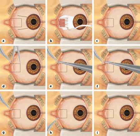
Fig. 1.
Schematic representation of Harms’ conjunctival opening and the new transposition MISS technique for rectus muscle posterior fixation. The eye is represented from above as seen by the surgeon. So far, rectus muscle posterior fixation sutures have been performed using the large Harms’ limbal opening (a, b). With the new MISS technique, after applying a limbal traction suture, two small L-shaped cuts are performed slightly anterior to the site where the scleromuscular sutures will be placed (c). The episcleral tissue is separated from the muscle sheath and the sclera with blunt Wescott scissors. Then, a curved ruler is used to determine the exact placement of the scleromuscular sutures (d). The posterior fixation is performed by first passing a nonresorbable suture through the sclera (e), followed by the muscle suture, which will include one third of the muscle (f). g Final appearance of the scleromuscular suture. Then, using the same technique, a posterior fixation suture is placed at the other border of the muscle (h). The surgical procedure is completed by applying resorbable single sutures to the two small cuts (i). For better visualization of the operating site, the cuts can be enlarged or joined at the limbus in order to obtain the usual limbus approach (a).

