Abstract
Episodes of high temperature at anthesis, which in rice is the most sensitive stage to temperature, are expected to occur more frequently in future climates. The morphology of the reproductive organs and pollen number, and changes in anther protein expression, were studied in response to high temperature at anthesis in three rice (Oryza sativa L.) genotypes. Plants were exposed to 6 h of high (38 °C) and control (29 °C) temperature at anthesis and spikelets collected for morphological and proteomic analysis. Moroberekan was the most heat-sensitive genotype (18% spikelet fertility at 38 °C), while IR64 (48%) and N22 (71%) were moderately and highly heat tolerant, respectively. There were significant differences among the genotypes in anther length and width, apical and basal pore lengths, apical pore area, and stigma and pistil length. Temperature also affected some of these traits, increasing anther pore size and reducing stigma length. Nonetheless, variation in the number of pollen on the stigma could not be related to measured morphological traits. Variation in spikelet fertility was highly correlated (r=0.97, n=6) with the proportion of spikelets with ≥20 germinated pollen grains on the stigma. A 2D-gel electrophoresis showed 46 protein spots changing in abundance, of which 13 differentially expressed protein spots were analysed by MS/MALDI-TOF. A cold and a heat shock protein were found significantly up-regulated in N22, and this may have contributed to the greater heat tolerance of N22. The role of differentially expressed proteins and morphology during anther dehiscence and pollination in shaping heat tolerance and susceptibility is discussed.
Keywords: Anther, high temperature, pollen, proteomics, rice, spikelet fertility
Introduction
Nearly half the worlds population depends on rice, and an increase in rice production by 0.6–0.9% annually until 2050 is needed to meet the demand (Carriger and Vallee, 2007). As a result, rice (Oryza sativa L.) is increasingly cultivated in more marginal environments that experience warmer temperatures where day/night temperatures average 28/22 °C (Prasad et al., 2006). In these environments, day temperatures frequently exceed the critical temperature of 33 °C for seed set, resulting in spikelet sterility and reduced yield (Nakagawa et al., 2002). The vulnerability of the crop will be increased with a projected global average surface temperature increase of 2.0–4.5 °C and the possibility of increased variability about this mean by the end of this century (IPCC, 2007). Hence, in the future, rice will be grown in much warmer environments (Battisti and Naylor, 2009) with a greater likelihood of high temperatures coinciding with heat-sensitive processes during the reproductive stage.
Seed set under high ambient air temperature primarily depends on successful pollination and fertilization. As was shown in reciprocal studies with pollen from control plants on heat-stressed pistils and vice versa, the male gametophyte and not the pistil, is responsible for spikelet sterility under high temperature in rice (Yoshida et al., 1981). Morphologically, large anthers (Hashimoto, 1961; Suzuki, 1981) and longer stigmas (Suzuki, 1982) contribute to increased tolerance to cold stress during the booting stage, and the same may be true for high temperature tolerance at flowering (Matsui and Omasa, 2002). Among physiological processes occurring at anthesis, anther dehiscence is perceived to be the most critical stage affected by high temperature (Matsui et al., 1997a, b, 2001). Spikelet opening triggers rapid pollen swelling, leading to anther dehiscence and pollen shedding from the anthers’ apical and basal pores (Matsui et al., 1999). Increased basal pore length in a dehisced anther was found to contribute significantly to successful pollination (Matsui and Kagata, 2003), probably because of its proximity to the stigmatic surface. Longer stigmas may also be important for the same reason. After pollination, it takes about 30 min for the pollen tube to reach the embryo sac (Cho, 1956). Genotypic differences in pollen number and germinating pollen on the stigma (Matsui et al., 1997a) and spikelet fertility (Matsui and Omasa, 2002; Prasad et al., 2006) under high temperatures in rice have been well described. Similarly, in several other crops, pollen germination and pollen tube growth are shown to be sensitive to high temperatures (Kakani et al., 2002, 2005; Salem et al., 2007).
Attempts have also been made to study molecular mechanisms conferring heat tolerance through the analysis of pollen gene expression in Arabidopsis (Arabidopsis thaliana) (Haralampidis et al., 2002). However, the data revealed a poor correlation between transcript level and protein expression (Becker et al., 2003; Honys and Twell, 2003), possibly due to alternative splicing and/or post-translational modifications (Lockhart and Winzeler, 2000). Therefore, 2D gel electrophoresis has been used to study differential protein expression under varying conditions in different crops with diverse objectives (Tsugita et al., 1996; Koller et al., 2002; Salekdeh et al., 2002; Lin et al., 2005; Yan et al., 2005). To understand the molecular basis of male gametophyte development, proteomic analysis at different stages of pollen development and mature pollen (Imin et al., 2001; Kerim et al., 2003; Dai et al., 2006) were conducted. The effect of cold stress on young microspores was studied at the anther protein level (Imin et al., 2004) while the effect of heat stress on anther (male reproductive organ) protein expression has not been studied.
Addressing the physiological and molecular mechanisms conferring heat tolerance during anthesis will help to develop rice germplasm capable of adapting to changing climates. Experiments were therefore carried out under control (29 °C) and high temperatures (38 °C): (i) to evaluate the effect of high temperature during anthesis on the morphology of the reproductive organs and to identify heat-sensitive physiological processes; (ii) to identify and compare high-temperature-responsive anther proteins in rice genotypes at anthesis; and (iii) to determine genotypic differences in reproductive organ morphology, and physiological processes to spikelet fertility of rice.
Materials and methods
Greenhouse
Plants were grown in a temperature controlled greenhouse maintained at 29/21 °C day/night temperature [Actual: 27.7 oC (SD=0.80)/19.9 oC (SD=0.11)] and RH of 75% [Actual 82.7% (SD=2.83)] under natural sunlight conditions at the International Rice Research Institute (IRRI), Philippines. Plants were placed on a bench spaced at 30 cm intervals to avoid shading effects. Ambient air temperature and RH were measured using thermocouples every 10 s and averaged over 10 min (Chessell 392, USA).
Crop husbandry
Three rice varieties, japonica type Moroberekan (highly susceptible to high temperature during anthesis), indica type IR64 (moderately tolerant), and an Aus type N22 (highly tolerant) were chosen for this study based on data from previous work (Jagadish et al., 2008). Pregerminated seeds were sown into 2l trays containing natural clay loam soil with 2.5 g ammonium sulphate (NH4)2SO4, 0.5 g muriate of potash (KCl), and 0.5 g single superphosphate (SSP) incorporated into the soil before sowing. After 15 d, three seedlings each were transplanted into 10 l pots with a solid bottom containing 7.4 kg of the same clay loam soil with 7.5 g (NH4)2SO4, 1.5 g KCl, and 1.5 g SSP. An additional 2.5 g of (NH4)2SO4 was added 25–30 d after transplanting. For each of the three genotypes, 34 pots were sown. The plants were grown under flooded conditions throughout the crop cycle. Cypermethrin (Cymbush) 0.42 g l−1 was sprayed in 15 d intervals, starting 30 d after transplanting, to control white flies (Bemisia spp). There were no other pest or disease problems.
Growth chambers and heat treatment
On the first day of anthesis (i.e. the appearance of anthers), plants were transferred at 08.00 h into growth chambers (Thermoline, Australia) with temperatures gradually increasing from 29 °C to 38 °C by 09.00 h (2.5 h after dawn) and maintained at 38 °C (SD=0.13) until 15.00 h, with an RH of 75% (SD=1.10). Immediately after the heat treatment, plants were moved back to the control conditions (29/21 °C) before being returned to the growth cabinets and exposed to the same conditions the following morning at 09.00 h, i.e. plants were exposed to 2 d of high temperature.
A thermocouple placed above the canopy in the growth chamber measured the ambient air temperature and RH every 10 s and averaged over 10 min (Chessell 392, USA). Photosynthetic photon flux density was maintained at 640 μmol m−2 s−1. Temperature of the ambient air cycled from outside into the cabinets was warmed with the help of heaters. CO2 concentration was not measured.
Sampling
Seventeen pots of each genotype were exposed to high (38 °C) or ambient (29 °C) temperature for 6 h. Two pots (six plants) were used for scoring spikelet fertility both at high and control temperatures while the remaining 15 pots were used to sample spikelets for the morphological and proteomic analyses.
Spikelet fertility was measured on spikelets opening between 09.00 h and 15.00 h on the first day of anthesis and marked with acrylic paint for identification (Jagadish et al., 2007, 2008). Ten to 12 days later, the marked spikelets were scored for fertility by pressing the marked spikelets individually. 121 to 184 and 110 to 174 spikelets were scored under control and high temperature treatments, respectively.
Ten unopened spikelets, each positioned third on the top most rachis branch of the panicle predicted to open the next day (Matsui and Kagata, 2003) were sampled at the end of the treatment period (at 15.00 h) to measure anther length and width. Following 1 h of high temperature exposure on the first day of high temperature treatment, spikelets just beginning to open were carefully marked and collected in order to record: the pistil length (20 spikelets); total number of pollen, number of germinated pollen on the stigma, and stigma length (15 spikelets); and pollen tube length (10 spikelets). All spikelets were transferred immediately into fixative containing 1:3 glacial acetic acid: absolute alcohol (v/v) in glass vials.
Photographs to measure anther pore opening and size were taken in situ three times a day, taking care to ensure chambers were only open for a short period of about 3 min. Photographs were taken from panicles not used for any other observations to prevent manual influence on the anther pore area.
Samples for proteomic analysis were collected from the top four rachis branches simultaneously from both high temperature and control treatments and stored in falcon tubes suspended in liquid N at –80 °C for further use (Ishimaru et al., 2003). Samples for proteomic analyses were stored.
Microscopic observations
The dissection of spikelets was done using a stereomicroscope (Olympus SZX7, Olympus Corp, Japan) and images were taken with DP70 digital camera attached to an Axioplane 2 microscope (Carl Zeiss, Germany) at ×50 for all morphological characters. Numbers of pollen and germinated pollen on the stigma were monitored at ×100 magnification. All measurements were done using Image Pro Plus 5.1 software after calibration. The length of all four anther locules from 30 anthers, excised from five randomly chosen spikelets was analysed. The width of the same 30 anthers was measured at the top and bottom quarter and in the middle to compute the mean.
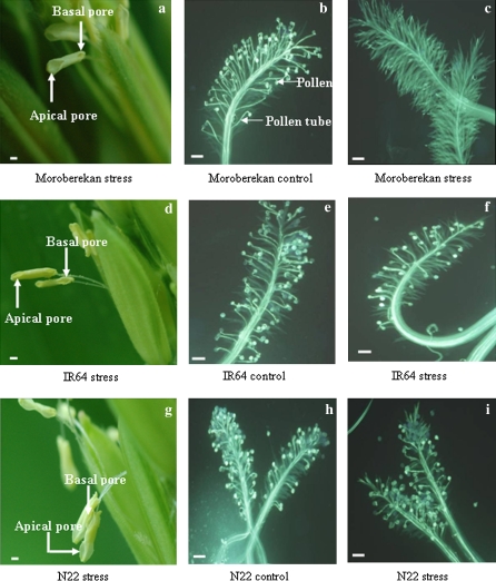
Photographs were taken with a Nikon D70 camera (Nikon Corp., Japan) when the anther pores were completely open (Fig. 1). All the photographs were taken at 1.2 magnification to maintain uniformity. Thirty anthers were used for measuring apical and basal pore areas and the longitudinal lengths of the same pores were recorded.
Fig. 1.
Images of Moroberekan (a–c), IR64 (d–f), and N22 (g–i) showing the apical and basal pore sizes under heat stress, and stigmas with germinated pollen under control and high temperature conditions. Bars=100 μm. (This figure is available in colour at JXB online.)
Spikelets collected with minimum disturbance were used to record pollen count and number of pollen germinated on the stigma. The stigmas were cleared using 8 N NaOH for 3–5 h and subsequently stained with 2% aniline blue. A pollen was scored as germinated when the pollen tube length was either equal to or greater than the diameter of the pollen (Luza et al., 1987).
The length of the stigma was measured from the tip of the stigma to the base of both stigma branches and the mean was taken for analysis. For measuring pollen tube length, two to three images were taken at ×50 and the total length was meticulously determined from the three images. The pollen tube was measured from the base of the germ pore to the tip of the germinating pollen tube. Pistil length was measured from the tip of the stigma to the base of the ovary as described for pollen tube length measurement.
Protein extraction and 2D gel electrophoresis
Proteins from the anthers of three different genotypes collected under control and heat-stressed conditions were extracted by trichloroacetic acid (TCA) precipitation with minor modifications (Salekdeh et al., 2002). Three biological replicate anther tissue samples of 1 g each were ground in liquid nitrogen and suspended in 10% w/v TCA in acetone with 0.07% w/v DTT at –20 °C for 1 h, followed by centrifugation for 25 min at 10 000 rpm. The pellets were washed with ice-cold acetone containing 0.07% DTT, incubated at –20 °C for 1 h, and centrifuged again at 4 °C. The above step was repeated three times and the pellets were lyophilized. The lyophilized sample was solubilized in lysis buffer (9 M urea, 4% w/v CHAPS, 2.5% w/v Pharmalyte pH 3–10, 1% w/v DTT, 35mM TRIS) and the protein concentration was determined using a Bradford assay with BSA (bovine serum albumin) as the standard (Salekdeh et al., 2002).
2D-PAGE separation of proteins was carried out with minor modifications according to Gorg et al. (1988, 1998). Equal amount of proteins were rehydrated into 17 cm IPG strips (pH 4–7) for analytical (100 μg) and preparative gels (500 μg). IEF was carried out using a Pharmacia Multiphore II kit (Amersham Pharmacia Biotech) at 20 °C under high voltage (500 V for 1 h, 1000 V for 1 h, and finally 2950 V for 14 h). Second dimension protein separation was performed using 12% SDS-PAGE gels. The protein spots in analytical gels were visualized by staining with silver nitrate according to Blum et al. (1987), with some modifications as published at http://www.weihenstephan.de/bim/deg. Preparative gels were stained with colloidal Coomassie Brilliant Blue G-250 (Smith et al., 1995; Neuhoff et al., 1998).
Image acquisition and data analysis
GS-800 densitometer (Bio-Rad) was used to scan silver-stained 2D gels with a resolution of 600 dots and 12 bits per inch. Image visualization, spot detection, and protein quantification was carried out using the Melanie 3 software (GeneBio, Geneva, Switzerland). Twelve bit images were used for detecting the spots using optimized parameters as follows: Number of smooths 1; Laplacian threshold, 5; Partial threshold, 5; Saturation, 90; Peakness increase, 100; Minimum perimeter, 30. Comparison between the treatments across genotypes was done by calculating the abundance ratio of spots (% volume of spot in treated samples/% volume of spot in control samples) (Yan et al., 2005). The % volume was determined based on the area occupied and the intensity of the protein spot using the above mentioned optimized parameters. Experimental molecular weight of the protein spots was determined by co-electrophoresis of standard protein markers (Bio-Rad) as internal markers, accounting for any inconsistencies in gel compositions. The Isoelectric point (pI) was determined by migration of the spot along the 17 cm IPG strip (4–7 pH).
Protein identification
Proteins were initially separated over pH ranges of 3–10 and 4–7. Since proteins clumped at 3–10 pH, IPG strips of 4–7 pH were used for further analyses. Protein spots of interest showing significant quantitative changes during heat stress were excised from the CBB-stained gels and used for trpysin digestion. Digested proteins were further analysed using a MALDI-TOF MS 4700 proteomics analyser at the Australian Proteome Analysis Facility (http://www.proteome.org.au/). The identities of the proteins were determined using MASCOT (Matrix Science, London, UK) software (peptide mass tolerance of ±100 ppm, the maximum number of missed tryptic cleavages 1, allowing for iodoacetamide modifications including oxidation of methionine). The physical position of proteins on the rice genome was identified using the NCBI (www.ncbi.nlm.nih.gov) and TIGR (http://www.tigr.org/tdb/e2k1/osa1/index.shtml) databases.
Statistical analysis
All morphological and physiological data were analysed as a completely randomized design of two treatments (control and high temperature), three genotypes and 30, 20, 15, and 10 replicates for anther length and width, anther apical and basal pore area and length; pistil length; pollen count, number of germinated pollen on stigma, stigma length, and pollen tube length, respectively using Genstat ver. 8.1 (Rothamsted Experimental Station). Spikelet fertility (231–356 observations per genotype) was treated as binomial data taking into account each individual marked spikelet as a data point from all the treatments and analysed using the Generalised Linear Mixed Model in Genstat ver. 8.1 (Jagadish et al., 2007, 2008). Linear regressions and comparison of linear regressions were also carried out using Genstat.
Protein volume (%) obtained from Melanie 3 software (GeneBio, Geneva, Switzerland) was analysed as a randomized design using Genstat 8.1. Three replications were used to determine the significance of differences between the control and heat-stressed samples within the genotypes and difference in % volume between genotypes under high temperature.
Results
Spikelet fertility
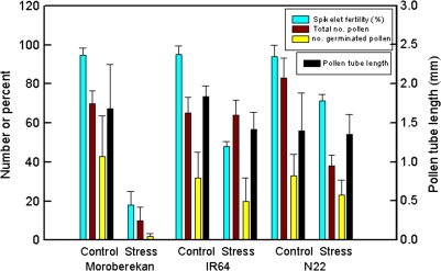
The three contrasting rice genotypes selected were exposed to temperatures of 38 °C and 29 °C at anthesis. Spikelet fertility was between 95% and 96% under control (29 °C) conditions (Fig. 2). The 6 h high temperature treatment (38 °C) during anthesis had a significant impact on spikelet fertility. Moroberekan was highly sensitive (18% fertility), IR64 intermediate (48%), and N22 tolerant (71%) to high temperature.
Fig. 2.
Spikelet fertility (%Odds Ratio), total and germinated pollen number on the stigma, and pollen tube length (mm) under control and high temperature stress in the three rice genotypes Bars indicate ±SE. (This figure is available in colour at JXB online.)
Anther and pore size
Anther size (length and width) varied significantly between genotypes (P <0.001) but was not affected by temperature. There was no temperature×genotype interaction for pore area, length or width (P >0.05). IR64 had the longest and widest anthers, resulting in a cross-sectional area of 2.44 mm2, nearly double that of Morobekan and N22 which had similar sized anthers. However, larger anther size did not contribute to larger apical (r >–0.78; n=6) or basal (r=0.07 to –0.3) pore size or length, which were greater in N22 than other genotypes (Table 1). Apical pore size and length was greater than basal pore size or length, particularly in N22 and Moroberekan. High temperature increased apical and basal pore areas (P <0.05) and lengths (P <0.05). Apical and basal pore areas and lengths were not correlated in any genotype.
Table 1.
Effect of genotype and temperature during anthesis on anther dehiscence characteristics, anther length and width, stigma length and pistil length
| Anther |
Basal pore |
Apical pore |
Stigma | Pistil | ||||
| Length | Width | Area | Length | Area | Length | Length | Length | |
| (mm) | (mm) | (μm2) | (μm) | (μm2) | (μm) | (mm) | (mm) | |
| Genotype | ||||||||
| Moroberekan | 1.91 | 0.65 | 4700 | 471 | 10418 | 626 | 1.07 | 2.13 |
| IR64 | 2.91 | 0.84 | 5594 | 528 | 7629 | 517 | 1.07 | 2.34 |
| N22 | 1.79 | 0.59 | 6086 | 552 | 15626 | 685 | 0.72 | 1.85 |
| SEDa | 0.11*** | 0.03*** | 591.2ns | 27.1** | 949.2*** | 27.3*** | 0.02*** | 0.04*** |
| Temperature | ||||||||
| Control | 2.19 | 0.68 | 4929 | 495 | 10318 | 581 | 0.96 | 2.0 |
| High temperature | 2.22 | 0.70 | 5992 | 539 | 12132 | 638 | 0.94 | 2.0 |
| SEDa | 0.09ns | 0.03ns | 482.7* | 22.1* | 775.0* | 22.3* | 0.014ns | 0.04ns |
*P <0.05, **P <0.01, ***P <0.001; ns, non significant.
Stigma and pistil length
Genotypic differences in stigma and pistil length (P <0.001) were observed, but these differences were unaffected by temperature. Stigmas and pistils were significantly shorter in N22 than IR64 or Moroberekan (Table 1). Stigma length, and more so pistil length, was positively correlated with anther size (r=0.60 and 0.86, n=6, respectively) and negatively correlated with apical pore size (r >–0.90).
Number of germinated pollen and pollen tube length
The number of pollen on the stigma was affected by genotype (P <0.001), temperature (P <0.001), and their interaction (P <0.001). Under control conditions, pollen number on the stigma (Fig. 2) varied from 65 (IR64) to 83 (N22), with higher pollen counts being associated with larger apical and total pore sizes, and smaller anthers, stigmas, and pistils (Table 1). Clumping of pollen was also observed on N22 (see Supplementary Fig. S1 at JXB online), as Yoshida et al. (1981) also found. High temperature significantly reduced the number of pollen on the stigma in N22 (by 55%) and Moroberekan (by 86%), but not in IR64 (Fig. 2). Pollen number under stress was not associated strongly with any morphological traits.
There was also a significant effect of temperature on the number of germinated pollen in the three genotypes (Fig. 2), and germinated pollen number was highly correlated with spikelet fertility (r=0.94; n=6). In all three genotypes about 60% of pollen on the stigma germinated under control conditions (see Supplementary Fig. S2 at JXB online). However, at high temperature compared to the control, this proportion was significantly reduced in Moroberekan (17% of pollen grains germinated; P <0.01) and IR64 (40% pollen germination; P <0.05). However, high temperature had no significant (P >0.95) effect on the proportion of germinated pollen in N22.
Although mean spikelet fertility was correlated with the mean number of germinated pollen across genotypes and temperature treatments, Satake and Yoshida (1978) and Matsui et al. (2000) have shown that individual spikelets require a critical minimum number of between 10 and 20 germinated pollen grains per spikelet to be fertile. Overall, spikelet fertility was also strongly correlated (r=0.97, n=6) with the proportion of spikelets with >20 germinated pollen grains.
The rate of pollen tube growth was also significantly affected by genotype, high temperature and their interaction (all at P <0.001), with almost no pollen germination in Moroberekan at high temperature (Fig. 2). Under control conditions pollen tube length in all three genotypes 1 h after anthesis was between 1.4 mm and 1.84 mm, and this was equivalent to 0.75 to 0.79 of pistil length. At high temperature pollen tube length in IR64 was reduced from 1.84 to 1.42 mm and proportionately pollen tubes were shorter (0.61) relative to the length of the pistil. By contrast, in N22 pollen tube length (1.35 mm) and length relative to the pistil (0.73) was not significantly affected at high temperature. This difference in relative pollen tube length may contribute to greater spikelet fertility in N22.
Proteomic analysis of anthers exposed to heat stress
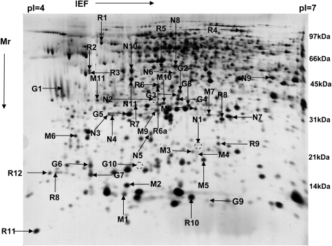
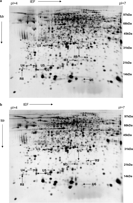
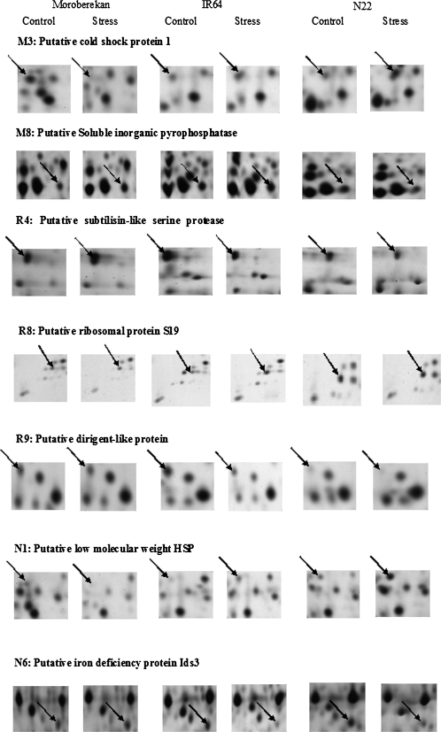
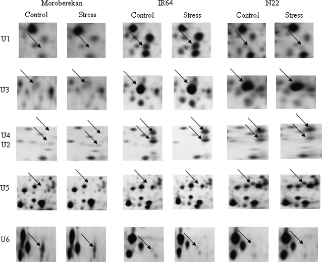
Following 2D gel electrophoresis, 46 protein spots were found to differ in abundance under heat stress compared with the control over a pH range of 4–7 and molecular weight (Mr) range of 10–102 kDa. A representative master gel that was used for a cross-comparison of protein expression patterns between treatments and genotypes is shown in Fig. 3 and a global protein expression map of IR64 under control and high temperature in Fig. 4. The variation in expression for: (i) genotype-dependent heat-responsive proteins and (ii) genotype-dependent, constitutively expressed proteins under control and high temperatures are presented in Table 2. Three replicate abundance ratios under both control and high temperature were taken. Spots shown to be differentially accumulated at the 0.05 level of significance with ≥2-fold changes were excised from 2D gels and considered for further analysis. Hence, 13 out of 46 protein spots fitting these criteria were analysed by mass spectrometry. Proteins were annotated based on the NCBI and TIGR databases using BLASTp analyses (Table 3; Figs 5, 6). For seven proteins, sequence similarities to annotated proteins were found (Fig. 5), coding for: a putative cold shock protein (CSP), an inorganic pyrophosphatase, a serine protease (AIR3), a dirigent-like protein, a ribosomal protein (S19), a small heat shock protein (sHSP), and an iron deficiency protein (ids3). Of the six remaining proteins, five proteins were classified as proteins with unknown functions following Luhua et al. (2008), and a hypothetical protein (Fig. 6).
Fig. 3.
A master gel from a 2D electrophoresis using 4–7 pH IGP strips showing all 46 proteins differentially expressed either within or across the genotypes under control and high temperature, as well as genotype-specific proteins. Arrow names starting with M, R, N, and G indicate that the differential expression was in Moroberekan, IR64, N22, and constitutive expression within the genotypes irrespective of conditions. Three protein spots G10, N1, and N2 indicated with a dashed circle has less visible protein spot on the master gel but there was a significant higher expression in the N22 gels (see Table 2).
Fig. 4.
2D gel electrophoresis of anther proteins. Representative 2D global protein expression map of IR64 mature anther under control conditions (top, 29 °C) and high temperature (bottom, 38 °C). Significantly changing protein spots are highlighted using arrows. Spots were excised from 2D gels of Moroberekan (M), IR64 (R), N22 (N), and U (unknown proteins); U1–U4 were excised from IR64 gels, U5 from N22, and U6 from Moroberekan. First-dimensional focusing was done by using 17 cm IPG strips with a pH gradient of 4–7 loaded with 100 μg of total anther protein. In the second dimensional SDS PAGE, 12% gels were used. Experimental molecular weight is indicated with Mr.
Table 2.
Differentially expressed proteins within each genotype under control and high temperature stress given in bold (M, Moroberekan; R, IR64; N, N22) and the expression pattern of these spots across the other two genotypes (normal font)
| Genotype | Comment | Moroberekan |
IR64 |
N22 |
|||
| Control | Heat | Control | Heat | Control | Heat | ||
| Heat responsive proteins within and across genotypes | |||||||
| Moro | M1 | 0.287±0.017 | 0.248±0.023 | 0.235±0.014 | 0.214±0.012 | 0.253±0.001 | 0.281±0.023 |
| Moro | M2 | 0.591±0.016 | 0.541±0.036 | 0.484±0.027 | 0.566±0.034 | 0.486±0.056 | 0.695±0.029 |
| Moro | M3 | 0.223±0.018 | 0.038±0.030 | 0.090±0.000 | 0.168±0.017 | 0.102±0.007 | 0.391±0.019 |
| Moro | M4 | 0.248±0.015 | 0.046±0.013 | 0.001±0.000 | 0.001±0.000 | 0.001±0.000 | 0.001±0.000 |
| Moro | M5 | 0.323±0.019 | 0.335±0.016 | 0.288±0.010 | 0.251±0.013 | 0.298±0.027 | 0.387±0.014 |
| Moro | M6 | 0.186±0.017 | 0.151±0.005 | 0.168±0.015 | 0.134±0.022 | 0.140±0.038 | 0.182±0.024 |
| Moro | M7 | 0.310±0.014 | 0.263±0.038 | 0.173±0.007 | 0.203±0.038 | 0.233±0.010 | 0.313±0.012 |
| Moro | M8 | 0.169±0.025 | 0.289±0.007 | 0.207±0.030 | 0.206±0.010 | 0.212±0.040 | 0.155±0.027 |
| Moro | M9 | 0.237±0.010 | 0.368±0.058 | 0.300±0.059 | 0.244±0.014 | 0.373±0.010 | 0.371±0.030 |
| Moro | M10 | 0.223±0.009 | 0.198±0.006 | 0.182±0.016 | 0.187±0.015 | 0.260±0.046 | 0.452±0.092 |
| Moro | M11 | 0.170±0.007 | 0.175±0.015 | 0.139±0.014 | 0.152±0.010 | 0.181±0.021 | 0.175±0.021 |
| IR64 | R1 | 0.308±0.037 | 0.332±0.061 | 0.230±0.023 | 0.372±0.046 | 0.173±0.023 | 0.203±0.041 |
| IR64 | R2 | 0.140±0.004 | 0.171±0.034 | 0.177±0.044 | 0.197±0.023 | 0.074±0.019 | 0.069±0.023 |
| IR64 | R3 | 0.177±0.024 | 0.177±0.014 | 0.155±0.052 | 0.271±0.024 | 0.179±0.063 | 0.130±0.021 |
| IR64 | R4 | 0.115±0.043 | 0.162±0.004 | 0.176±0.014 | 0.115±0.015 | 0.136±0.004 | 0.148±0.013 |
| IR64 | R5 | 0.241±0.076 | 0.186±0.019 | 0.152±0.006 | 0.108±0.017 | 0.130±0.005 | 0.166±0.012 |
| IR64 | R6 | 0.289±0.022 | 0.290±0.016 | 0.122±0.007 | 0.097±0.013 | 0.291±0.014 | 0.341±0.026 |
| IR64 | R6a | 0.001±0.000 | 0.001±0.000 | 0.138±0.012 | 0.102±0.002 | 0.001±0.000 | 0.001±0.000 |
| IR64 | R7 | 0.096±0.023 | 0.114±0.014 | 0.160±0.040 | 0.198±0.029 | 0.013±0.007 | 0.003±0.001 |
| IR64 | R8 | 0.078±0.023 | 0.003±0.001 | 0.097±0.016 | 0.198±0.023 | 0.223±0.032 | 0.238±0.008 |
| IR64 | R9 | 0.121±0.003 | 0.164±0.022 | 0.148±0.012 | 0.091±0.011 | 0.013±0.007 | 0.391±0.020 |
| IR64 | R10 | 1.226±0.062 | 1.584±0.131 | 0.814±0.132 | 0.644±0.083 | 1.086±0.133 | 0.923±0.110 |
| IR64 | R11 | 0.528±0.082 | 0.561±0.138 | 0.516±0.048 | 0.400±0.057 | 0.683±0.098 | 0.636±0.190 |
| IR64 | R12 | 0.156±0.033 | 0.281±0.072 | 0.109±0.014 | 0.023±0.004 | 0.157±0.026 | 0.072±0.014 |
| IR64 | R13 | 0.347±0.084 | 0.267±0.026 | 0.151±0.033 | 0.001±0.000 | 0.504±0.133 | 0.268±0.021 |
| N22 | N1 | 0.090±0.020 | 0.024±0.011 | 0.007±0.003 | 0.075±0.015 | 0.056±0.002 | 0.226±0.029 |
| N22 | N2 | 0.179±0.014 | 0.166±0.008 | 0.066±0.023 | 0.068±0.002 | 0.170±0.008 | 0.170±0.011 |
| N22 | N3 | 0.001±0.000 | 0.208±0.206 | 0.130±0.019 | 0.208±0.015 | 0.233±0.019 | 0.270±0.002 |
| N22 | N4 | 0.118±0.010 | 0.172±0.027 | 0.098±0.018 | 0.148±0.009 | 0.219±0.019 | 0.186±0.016 |
| N22 | N5 | 0.166±0.016 | 0.143±0.075 | 0.167±0.006 | 0.195±0.026 | 0.063±0.034 | 0.180±0.022 |
| N22 | N6 | 0.081±0.008 | 0.122±0.012 | 0.088±0.004 | 0.042±0.003 | 0.125±0.015 | 0.064±0.006 |
| N22 | N7 | 0.440±0.027 | 0.358±0.122 | 0.325±0.015 | 0.357±0.024 | 0.411±0.019 | 0.470±0.051 |
| N22 | N8 | 0.258±0.026 | 0.362±0.063 | 0.313±0.029 | 0.303±0.015 | 0.372±0.028 | 0.361±0.017 |
| N22 | N9 | 0.259±0.025 | 0.231±0.036 | 0.316±0.021 | 0.318±0.015 | 0.250±0.048 | 0.338±0.026 |
| N22 | N10 | 0.394±0.013 | 0.442±0.045 | 0.415±0.026 | 0.395±0.017 | 0.417±0.064 | 0.376±0.042 |
| N22 | N11 | 0.434±0.030 | 0.318±0.091 | 0.392±0.041 | 0.420±0.025 | 0.444±0.068 | 0.452±0.065 |
| Genotype dependent proteins | |||||||
| IR64 | G1 | 0.084±0.041 | 0.142±0.009 | 0.150±0.072 | 0.176±0.040 | 0.013±0.007 | 0.002±0.000 |
| IR64 | G2 | 0.032±0.030 | 0.002±0.000 | 0.339±0.031 | 0.367±0.021 | 0.161±0.015 | 0.142±0.019 |
| IR64 | G3 (U1) | 0.044±0.018 | 0.065±0.009 | 0.140±0.022 | 0.195±0.015 | 0.019±0.017 | 0.012±0.010 |
| IR64 | G4 | 0.027±0.014 | 0.037±0.004 | 0.252±0.022 | 0.221±0.009 | 0.253±0.020 | 0.263±0.007 |
| IR64 | G5 (U3) | 0.003±0.002 | 0.001±0.000 | 0.584±0.051 | 0.674±0.050 | 0.679±0.090 | 0.774±0.038 |
| IR64 | G6 (U4) | 0.001±0.000 | 0.001±0.000 | 0.279±0.010 | 0.339±0.008 | 0.345±0.027 | 0.380±0.057 |
| IR64 | G7 (U2) | 0.011±0.009 | 0.009±0.007 | 0.347±0.023 | 0.269±0.005 | 0.168±0.010 | 0.110±0.052 |
| N22 | G8 | 0.019±0.005 | 0.011±0.005 | 0.246±0.032 | 0.207±0.030 | 0.012±0.004 | 0.013±0.005 |
| N22 | G9 (U6) | 0.340±0.026 | 0.321±0.074 | 0.001±0.000 | 0.002±0.000 | 0.001±0.000 | 0.002±0.000 |
| N22 | G10 (U5) | 0.001±0.000 | 0.002±0.001 | 0.001±0.000 | 0.001±0.000 | 0.309±0.030 | 0.385±0.006 |
Genotype-specific proteins (G) in bold indicate proteins constitutively expressed under control and high temperatures. Figures in normal font show the same proteins nearly absent. Variation in protein expression level is given as ±SE (n=3).
Table 3.
Annotation of high temperature responsive proteins identified in rice anthers
| Spot | pIa | Mrb | Coverage (%) | Peptides matched | pIb | Mrb | Accession numberc |
| Putative cold shock protein – 1 (M3) | 5.81 | 21 | 85 | 168 | 6.28 | 19 | XP_479920 |
| Soluble inorganic pyrophosphatase (M8) | 5.83 | 36 | 89 | 183 | 5.90 | 23 | ABB47572 |
| Putative subtilisin-like serine protease AIR3 (R4) | 6.29 | 102 | 47 | 361 | 5.92 | 81 | XP_481633 |
| Putative ribosomal protein S19 (R8) | 4.38 | 17 | 78 | 108 | 10.04 | 16 | XP_468752 |
| Dirigent-like protein (R9) | 6.08 | 24 | 31 | 59 | 6.18 | 20 | ABF99471 |
| Putative low molecular weight hsp (N1) | 5.86 | 23 | 57 | 127 | 6.88 | 24 | XP_467890 |
| Putative iron deficiency protein Ids3 (N6) | 5.61 | 57 | 66 | 225 | 5.33 | 38 | XP_476744 |
| Hypothetical protein LOC_Os11g08820 (U1) | 5.64 | 42 | 38 | 87 | 5.56 | 24 | ABA91854 |
| Unnamed protein product (U2) | 4.71 | 16 | 66 | 104 | 4.78 | 16 | NP_913551 |
| Unknown protein (U3) | 5.14 | 35 | |||||
| Unknown protein (U4) | 4.71 | 18 | |||||
| Unknown protein (U5) | 5.27 | 18 | |||||
| Unknown protein (U6) | 6.08 | 14 |
Spots were excised from 2D gels of Moroberekan (M), IR64 (R), N22 (N) and proteins with unknown functions (U) spots; U1–U4 were excised from IR64 gels, U5 from N22, and U6 from Moroberekan. The spots were excised from gels where they were prominent.
Experimental pI, Mr.
Theoretical pI and Mr.
Accession number in NCBI.
dAll matches to rice.
Fig. 5.
Annotated proteins identified by 2D gel electrophoresis and the sequence similarity to the annotated proteins was obtained from the TIGR database using protein mass fingerprinting data from mass spectrometry.
Fig. 6.
Protein spots identified by 2D gel electrophoresis which had no significant matches with the annotated protein sequences (proteins of unknown functions following Luhua et al., 2008) from the database search using protein mass fingerprinting data from mass spectrometry.
In N22, the heat-tolerant genotype, the cold and heat shock proteins were highly up-regulated and the Fe-deficiency protein was down-regulated at high temperature compared with the control temperature. The level of the remaining four identified proteins (pyrophosphatase, serine protease, ribosomal, and dirigent proteins) did not change significantly (Table 4). By contrast, in the susceptible genotype Moroberekan, the heat and cold shock proteins were significantly down-regulated while there was a greater expression of the Fe-deficiency protein and soluble inorganic pyrophosphatase. Levels of serine protease, ribosomal, and dirigent proteins also did not change significantly in Moroberekan. In the moderately tolerant genotype IR64 there was significant up-regulation of both the shock proteins and the ribosomal protein, and a down-regulation of serine protease, dirigent, and Fe-deficiency proteins.
Table 4.
Mean abundance ratio (ratio of the % volume of the protein under heat stress over control) for the analysed 13 proteins among the three genotypes
| Protein (Spot) | Mean abundance ratioa |
||
| Moroberekan | IR64 | N22 | |
| (n=3) | (n=3) | (n=3) | |
| Putative cold shock protein – 1 (M3) | 0.172** | 1.864** | 3.843*** |
| Soluble inorganic pyrophosphatase (M8) | 1.710** | 0.992ns | 0.730 ns |
| Putative subtilisin-like serine protease AIR3 (R4) | 1.456 ns | 0.654** | 0.927 ns |
| Putative ribosomal protein S19 (R8) | 1.363 ns | 2.043** | 1.069 ns |
| Dirigent-like protein (R9) | 1.495 ns | 0.617** | 0.209 ns |
| Putative low molecular weight hsp (N1) | 0.262** | 10.789** | 4.031** |
| Putative iron deficiency protein Ids3 (N6) | 1.515** | 0.476*** | 0.513** |
| Hypothetical protein LOC_Os11g08820 (U1) | 1.48ns | 1.40ns | 0.64ns |
| Unnamed protein product (U2) | 0.80ns | 0.78** | 0.65ns |
| Unknown protein (U3) | 0.29ns | 1.16ns | 1.14ns |
| Unknown protein (U4) | 1.22ns | 1.22** | 1.10ns |
| Unknown protein (U5) | 1.64ns | 0.95ns | 1.25ns |
| Unknown protein (U6) | 0.94ns | 1.09ns | 1.67ns |
The mean abundance ratios were either significantly up or down-regulated.
** significant at 5%; *** significant at 1%; (ns) non-significant. Significantly up-regulated proteins are highlighted in bold.
A number of unidentified anther proteins also varied in expression between the three genotypes. At high temperature five of these proteins were not expressed in the susceptible genotype Moroberekan while four of them were strongly expressed in the tolerant N22 and the moderately tolerant IR64 (Fig. 6). By contrast, the hypothetical protein was highly expressed only in IR64 while the unknown protein U5 had a similarly high constitutive expression only in N22.
Discussion
The genotype N22 was clearly the most heat tolerant of the three genotypes, with 71% of spikelets setting seed at 38 °C compared with 48% and 18% in IR64 and Moroberekan, respectively. This value of seed-set at 38 °C in rice compares favourably with heat tolerance observed in other species, for example, peanut (Craufurd et al., 2003). The variation in spikelet fertility observed in this paper was closely associated with variation in the proportion of spikelets with ≥20 germinated pollen grains on the stigma and with pollen tube length. The most important heat-sensitive process determining variation in pollen number on the stigma is anther dehiscence (Matsui et al., 1997a, b, 2001) and basal pore length of the anther has been documented as an important morphological trait influencing pollen number on the stigma (Matsui and Omasa, 2002). Although high temperature increased both basal and apical pore size/length, there was no relationship between basal pore size and pollen number at 29 °C or 38 °C, although at 29 °C apical pore area/length was correlated with pollen number on the stigma. Large anthers have been associated with spikelet fertility at cool temperatures (Hashimoto, 1961; Suzuki, 1981), but although there were significant genotypic differences in anther size, there was no effect of temperature and no correlation with pollen number among the three gentoypes. Stigma and pistil lengths were also not affected by high temperature. It is therefore concluded that, among these three genotypes subjected to high temperature at anthesis, variation in anther and gynoecium morphology did not contribute to higher pollen number on the stigma. Moroberekan, nonetheless, had significantly fewer pollen on the stigma than the other two genotypes. Other factors that might influence pollen count on the stigma include: increased pollen stickiness which prevents pollen shedding even when the pores were open (Liu et al., 2006), the position of the style relative to the anther, and the timing of dehiscence relative to spikelet opening. The latter, a form of developmental escape (i.e. pollination before the spikelets open and expose the anthers and stigma to the ambient environment), may be particularly important.
Successful anther dehiscence depends on rupturing of the septa, expansion of locule walls, pollen swelling, and rupturing of the stomium (Liu et al., 2006). Among the significantly changing proteins in anthers under heat stress, the dirigent-like protein and subtilisin-like serine protease could influence anther dehiscence. Dirigent proteins are involved in lignin biosynthesis to strengthen/repair damaged cell walls acting as physical barriers (Johansson et al., 2000; Ralph et al., 2006). In anthers, an increased or persisting rigidity under heat stress due to increased lignification might delay the disintegration of tissues and thereby obstruct the normal process of anther dehiscence. A serine protease LIM9, expressed during microsporogenesis in lily (Lilium longiflorum), is thought to be responsible for the degradation of cell wall matrices and the release of microspores from tetrads (Kobayashi et al., 1994; Taylor et al., 1997). There was no significant difference in the expression of these two proteins at high temperature compared with their respective controls in Moroberekan and N22. However, a higher expression of dirigent protein in Moroberekan explicitly under heat stress may be responsible for a lower pollen count on the stigma.
Spikelet fertility was highly correlated with the number of germinated pollen grains on the stigma at 29 °C and 38 °C, and pollen germination was therefore an important trait. Pollen germination is known to be influenced by high temperature immediately prior to shedding as well as at dehiscence (Matsui et al., 1997a). Moroberekan, the most susceptible genotype, had very poor pollen germination at 38 °C while N22, the most tolerant genotype, had levels of pollen germination close to those at 29 °C. Iron is known to be an essential element for microspores to germinate embryonic structures (Babbar and Gupta, 1986; Kasha et al., 1990). The Fe requirement for proper microspore development or pollen germination could be met in N22 and IR64 through a chelating (Mihasi and Mori, 1989; Ogo et al., 2006) and transportation mechanism across the plasma membrane (Yeh et al., 1995) seen in barley roots under higher temperature. In Moroberekan, the efficiency of absorption could be reduced with higher Fe-deficiency protein expression, similar to barley roots under Fe deficiency, hindering pollen viability.
The high percentage of pollen germination at high temperature in N22 was also associated with the maintenance of pollen tube growth (relative to pistil length), initiating the changes needed in the transmitting tissue in the style (Lan et al., 2004). Similarly, in peanut, genotypic variation in percentage pollen germination in response to temperature, and significant variation in the optimum and lethal temperature for the rate of pollen tube growth, has been found (Kakani et al., 2002). Moreover, the male reproductive unit, pollen, has presynthesized mRNAs and protein (Dai et al., 2006) essential for pollen germination and pollen tube growth (Mascarenhas, 1993). Since ribosomal proteins are major components of cellular protein synthesis, any reduction in the level of one ribosomal protein limits the ribosome assembly rates and protein synthesis (Matsson et al., 2004). Accordingly, a significant decline in S19 expression under heat stress per se in Moroberekan could be an additional reason for its poor pollen germination.
The greater heat tolerance of N22 could be due to the accumulation of stress-responsive cold (sCSP) and heat (sHSP) shock proteins in the anthers. The chaperonic activity of sHSPs, which function as a reservoir of intermediates of denatured proteins preventing protein aggregation due to heat, has been shown by biochemical and structural studies (van Montfort et al., 2001). Therefore, increased accumulation of sHSPs could play an important role in protecting metabolic activities of the cell and is a key factor for organisms adapting to heat (Jinn et al., 1993; Yeh et al., 1995). This cold shock protein, with an experimental Mr of 21 kDa and theoretical Mr of 19 kDa, is a first report of a sCSP expressed differentially due to high temperature exposure in plants, particularly rice anthers. The sCSP has a RNA binding domain that functions as a RNA chaperone in bacteria and in eukaryotes as a Rho transcription termination factor. Moreover, Rikhsky et al. (2002) identified the activation of wound and pathogen responsive pathways when tobacco (Nicotiana tabacum) was exposed to drought and heat stress, illustrating the possible overlap of abiotic stress pathways with some unique gene clusters for stress responses. Since the expressed sCSP is known to have a chaperonic activity in bacteria, as shown for sHSP, it may well act as a protective mechanism. The beneficial effects of these proteins would be reflected in the high spikelet fertility of N22 under heat stress.
Apart from the proteins for which a similar sequence is present in the Nipponbare reference genome, four unknown proteins were constitutively expressed at a high level in N22 compared to Moroberekan. Identifying and validating the functions of such novel proteins might reveal insight into so far undisclosed stress responsive pathways and hence should not be neglected (Luhua et al., 2008; Gollery et al., 2006, 2007). The short-listed candidate genes are currently being cloned from the tolerant cultivar N22 for comparative sequence analyses. Subsequent transformation of candidate genes will reveal if functional polymorphisms exist that might lead to differences in heat tolerance levels in tolerant and susceptible rice varieties. Such further analysis is essential to avoid artefacts as conclusions drawn from differential expression using only 2D gels is complex and has to be interpreted with caution.
In conclusion, this study has demonstrated, both at the physiological and molecular level, that N22 is a heat-tolerant genotype exhibiting high levels of spikelet fertility at high temperature during anthesis. Across all genotypes and temperature treatments spikelet fertility was strongly related to the number of germinated pollen grains. Thirteen proteins were differentially expressed in response to temperature with clear differences between genotypes. Of particular interest are the cold and heat shock proteins that have been identified, which may contribute to heat tolerance at anthesis in the tolerant cv. N22.
Supplementary data
Supplementary data are available at JXB online.
Supplementary Fig. 1. Pollen clumping was seen only on the stigmas of N22 exclusively under control conditions.
Supplementary Fig. 2. Relation between number of germinated pollen and total pollen number in spikelets of Moroberekan, IR64, and N22 exposed to 29 °C and 38 °C.
Supplementary Material
Acknowledgments
We thank the Felix Scholarship for funding the PhD of SVK Jagadish. The University of Reading and The International Rice Research Institute are thanked for providing facilities to support this research. BMZ is thanked for further supporting the ongoing work in validating the functions of the candidate genes through allelic sequencing and transformation. This research has been facilitated by access to the Australian Proteome Analysis Facility established under the Australian Government's Major National Research Facilities Program. PB Malabanan, NS Estenor, FV Gulay, and BA Enriquez are thanked for theirtechnical assistance during the experiment.
References
- Babbar SB, Gupta SC. Obligatory and period specific requirement of iron for microspore embryogenesis in Datura metel anther culture. Botanical Magazine Tokyo. 1986;99:225–232. [Google Scholar]
- Battisti1 DS, Naylor RL. Historical warnings of future food insecurity with unprecedented seasonal heat. Science. 2009;323:240–244. doi: 10.1126/science.1164363. [DOI] [PubMed] [Google Scholar]
- Becker JD, Boavida LC, Carneiro J, Haury M, Feijó JA. Transcriptional profiling of arabidopsis tissues reveals the unique characteristics of the pollen transcriptome. Plant Physiology. 2003;133:713–725. doi: 10.1104/pp.103.028241. [DOI] [PMC free article] [PubMed] [Google Scholar]
- Blum H, Beier H, Gross HJ. Improved silver staining of proteins, RNA and DNA. Electrophoresis. 1987;8:93–99. [Google Scholar]
- Carriger S, Vallee D. More crop per drop. Rice Today. 2007;6:10–13. [Google Scholar]
- Cho J. Double fertilization in Oryza sativa L. and development of the endosperm with special reference to the aleurone layer. Bulletin of the National Institute of Agriculture Science. 1956;6:61–101. [Google Scholar]
- Craufurd PQ, Prasad PVV, Kakani VG, Wheeler TR, Nigam SN. Heat tolerance in groundnut. Field Crops Research. 2003;80:63–77. [Google Scholar]
- Dai S, Li L, Chen T, Chong K, Xue Y, Wang T. Proteomic analyses of Oryza sativa mature pollen reveal novel proteins associated with pollen germination and tube growth. Proteomics. 2006;6:2504–2529. doi: 10.1002/pmic.200401351. [DOI] [PubMed] [Google Scholar]
- Gollery M, Harper J, Cushman J, Mittler T, Girke T, Zhu JK, Bailey-Serres J, Mittler R. What makes species unique? The contribution of proteins with obscure features. Genome Biology. 2006;7:757. doi: 10.1186/gb-2006-7-7-r57. [DOI] [PMC free article] [PubMed] [Google Scholar]
- Gollery M, Harper J, Cushman J, Mittler T, Mittler R. POFs: what we don't know can hurt us. Trends in Plant Science. 2007;12:492–496. doi: 10.1016/j.tplants.2007.08.018. [DOI] [PubMed] [Google Scholar]
- Gorg A, Boguth G, Obermaier C, Weiss W. Two-dimensional electrophoresis of proteins in an immobilized pH 4–12 gradient. Electrophoresis. 1998;19:1516–1519. doi: 10.1002/elps.1150190850. [DOI] [PubMed] [Google Scholar]
- Gorg A, Postel W, Domscheit A, Gunther S. Two-dimensional electrophoresis with immobilized pH gradients of leaf proteins from barley (Hordeum vulgare): method, reproducibility and genetic aspects. Electorphoresis. 1988;9:681–692. doi: 10.1002/elps.1150091103. [DOI] [PubMed] [Google Scholar]
- Haralampidis K, Milioni D, Rigas S, Hatzopoulos P. Combinatorial interaction of cis elements specifies the expression of the Arabidopsis AtHsp90-1 gene. Plant Physiology. 2002;129:1138–1149. doi: 10.1104/pp.004044. [DOI] [PMC free article] [PubMed] [Google Scholar]
- Hashimoto K. On size of anther rice plant varieties. Bulletin of Hokkaido Study Meeting of Breeding and Crop Science. 1961;2:11. [Google Scholar]
- Honys D, Twell D. Comparative analysis of the arabidopsis pollen transcriptome. Plant Physiology. 2003;132:640–652. doi: 10.1104/pp.103.020925. [DOI] [PMC free article] [PubMed] [Google Scholar]
- Imin N, Kerim T, Rolfe BG, Weinman JJ. Effect of early cold stress on the maturation of rice anthers. Proteomics. 2004;4:1873–1882. doi: 10.1002/pmic.200300738. [DOI] [PubMed] [Google Scholar]
- Imin N, Kerim T, Weinman JJ, Rolfe BG. Characterisation of rice anther proteins expressed at the young microspore stage. Proteomics. 2001;1:1149–1161. doi: 10.1002/1615-9861(200109)1:9<1149::AID-PROT1149>3.0.CO;2-R. [DOI] [PubMed] [Google Scholar]
- IPCC. Summary for policy makers. In: Climate change 2007: the physical science basis, 9. 2007 [Google Scholar]
- Ishimaru T, Matsuda T, Ohsugi R, Yamagishi T. Morphological development of rice caryopses located at the different positions in a panicle from early to middle stage of grain filling. Functional Plant Biology. 2003;30:1139–1149. doi: 10.1071/FP03122. [DOI] [PubMed] [Google Scholar]
- Jagadish SVK, Craufurd PQ, Wheeler TR. High temperature stress and spikelet fertility in rice (Oryza sativa L.) Journal of Experimental Botany. 2007;58:1627–1635. doi: 10.1093/jxb/erm003. [DOI] [PubMed] [Google Scholar]
- Jagadish SVK, Craufurd PQ, Wheeler TR. Phenotyping parents of mapping populations of rice (Oryza sativa L.) for heat tolerance during anthesis. Crop Science. 2008;48:1140–1146. [Google Scholar]
- Jinn TL, Wu SH, Yeh KW, Hsieh MH, Yeh YC, Chen YM, Lin CY. Immunological kinship of class I low molecular weight heat shock proteins and thermo-stabilization of soluble proteins in vitro among plants. Plant and Cell Physiology. 1993;34:1055–1062. [Google Scholar]
- Johansson CI, Saddler JN, Beatson RP. Characterization of the polyphenolics related to the colour of western red cedar (Thuja plicata Donn) heartwood. Holzforschung. 2000;54:246–254. [Google Scholar]
- Kakani VG, Prasad PVV, Craufurd PQ, Wheeler TR. Response of in vitro pollen germination and pollen tube growth of groundnut (Arachis hypogaea L.) genotypes to temperature. Plant, Cell and Environment. 2002;25:1651–1661. [Google Scholar]
- Kakani VG, Reddy KR, Koti S, Wallace TP, Prasad PVV, Reddy RV, Zhao D. Differences in in vitro pollen germination and pollen tube growth of cotton cultivars in response to high temperatures. Annals of Botany. 2005;96:59–67. doi: 10.1093/aob/mci149. [DOI] [PMC free article] [PubMed] [Google Scholar]
- Kasha KJ, Ziauddin A, Cho UH. Haploids in cereal improvement: anther and microspore culture. In: Gustafson JP, editor. Gene manipulation in plant improvement, II. New York: Plenum Press; 1990. pp. 213–235. [Google Scholar]
- Kerim T, Imin N, Weinman JJ, Rolfe BG. Proteome analysis of male gametophyte development in rice anthers. Proteomics. 2003;3:738–751. doi: 10.1002/pmic.200300424. [DOI] [PubMed] [Google Scholar]
- Kobayashi T, Kobayashi E, Sato S, Hotta Y, Miyajima N, Tanaka A, Tabata S. Characterization of cDNAs induced in meiotic prophase in lily microsporocytes. DNA Research. 1994;1:15–26. doi: 10.1093/dnares/1.1.15. [DOI] [PubMed] [Google Scholar]
- Koller A, Washburn MP, Lange BM, et al. Proteomic survey of metabolic pathways in rice. Proceedings of the National Academy of Sciences, USA. 2002;99:11969–11974. doi: 10.1073/pnas.172183199. [DOI] [PMC free article] [PubMed] [Google Scholar]
- Lan L, ChenW Lai Y, et al. Monitoring of gene expression profiles and isolation of candidate genes involved in pollination and fertilization in rice (Oryza sativa L.) with a 10 K cDNA microarray. Plant Molecular Biology. 2004;54:471–487. doi: 10.1023/B:PLAN.0000038254.58491.c7. [DOI] [PubMed] [Google Scholar]
- Lin SK, Chang MC, Tsai YG, Lur HS. Proteomic analysis of the expression of proteins related to rice quality during caryopsis development and the effect of high temperature on expression. Proteomics. 2005;5:2140–2156. doi: 10.1002/pmic.200401105. [DOI] [PubMed] [Google Scholar]
- Liu JX, Liao DQ, Oane R, Estenor L, Yang XE, Li ZC, Bennett J. Genetic variation in the sensitivity of anther dehiscence to drought stress in rice. Field Crops Research. 2006;97:87–100. [Google Scholar]
- Lockhart DJ, Winzeler EA. Genomics, gene expression and DNA arrays. Nature. 2000;405:827–836. doi: 10.1038/35015701. [DOI] [PubMed] [Google Scholar]
- Luhua S, Ciftci-Yilmaz S, Harper J, Cushman J, Mittler R. Enhanced tolerance to oxidative stress in transgenic arabidopsis plants expressing proteins of unknown function. Plant Physiology. 2008;148:280–292. doi: 10.1104/pp.108.124875. [DOI] [PMC free article] [PubMed] [Google Scholar]
- Luza JG, Polito VS, Weinbaum SA. Staminate bloom date and temperature responses to pollen germination and tube growth in two walnut (Juglans) species. American Journal of Botany. 1987;74:1898–1903. [Google Scholar]
- Mascarenhas JP. Molecular mechanisms of pollen tube growth and differentiation. The Plant Cell. 1993;5:1303–1314. doi: 10.1105/tpc.5.10.1303. [DOI] [PMC free article] [PubMed] [Google Scholar]
- Matsson H, Davey EJ, Draptchinskaia N, Hamaguchi I, Ooka A, Leveen P, Forsberg E, Karlsson S, Dahl N. Targeted disruption of the ribosomal protein S19 gene is lethal prior to implantation. Molecular and Cellular Biology. 2004;24:4032–4037. doi: 10.1128/MCB.24.9.4032-4037.2004. [DOI] [PMC free article] [PubMed] [Google Scholar]
- Matsui T, Kagata H. Characteristics of floral organs related to reliable self pollination in rice (Oryza sativa L.) Annals of Botany. 2003;91:473–477. doi: 10.1093/aob/mcg045. [DOI] [PMC free article] [PubMed] [Google Scholar]
- Matsui T, Namuco OS, Ziska LH, Horie T. Effects of high temperature and CO2 concentration on spikelet sterility in indica rice. Field Crops Research. 1997b;51:213–219. [Google Scholar]
- Matsui T, Omasa K. Rice (Oryza sativa L.) cultivars tolerant to high temperature at flowering: anther characteristics. Annals of Botany. 2002;89:683–687. doi: 10.1093/aob/mcf112. [DOI] [PMC free article] [PubMed] [Google Scholar]
- Matsui T, Omasa K, Horie T. High temperature induced spikelet sterility of japonica rice at flowering in relation to air temperature, humidity, and wind velocity condition. Japanese Journal of Crop Science. 1997a;66:449–455. [Google Scholar]
- Matsui T, Omasa K, Horie T. Rapid swelling of pollen grains in response to floret opening unfolds anther locules in rice (Oryza sativa L.) Plant Production Science. 1999;2:196–199. [Google Scholar]
- Matsui T, Omasa K, Horie T. The difference in sterility due to high temperatures during the flowering period among japonica rice varieties. Plant Production Science. 2001;4:90–93. [Google Scholar]
- Matsui T, Omasa K, Horie T. High temperature at flowering inhibit swelling of pollen grains, a driving force for anther dehiscence in rice (Oryza sativa L.) Plant Production Science. 2000;3:430–434. [Google Scholar]
- Mihasi S, Mori S. Characterization of mugienic acid Fe transporter in Fe-deficient barley roots using the multi-compartment transport box method. BioMetals. 1989;2:146–154. [Google Scholar]
- Nakagawa H, Horie T, Matsui T. Effects of climate change on rice production and adaptive technologies. In: Mew TW, Brar DS, Peng S, Dawe D, Hardy B, editors. Rice science: innovations and impact for livelihood. China: International Rice Research Institute; 2002. pp. 635–657. [Google Scholar]
- Neuhoff V, Arnold N, Taube D, Ehrhardt W. Gel staining: colloidal coomassie staining. Electrophoresis. 1988;9:255–262. doi: 10.1002/elps.1150090603. [DOI] [PubMed] [Google Scholar]
- Ogo Y, Itai RN, Nakanishi H, Inoue H, Kobayashi T, Suzuki M, Takahashi M, Mori S, Nishizawa NK. Isolation and characterization of IRO2, a novel iron-regulated bHLH transcription factor in graminaceous plants. Journal of Experimental Botany. 2006;57:2867–2878. doi: 10.1093/jxb/erl054. [DOI] [PubMed] [Google Scholar]
- Prasad PVV, Boote KJ, Allen LH, Sheehy JE, Thomas JMG. Species, ecotype and cultivar differences in spikelet fertility and harvest index of rice in response to high temperature stress. Field Crops Research. 2006;95:398–411. [Google Scholar]
- Ralph S, Park JY, Bohlmann J, Shawn D. Dirigent proteins in conifer defense: gene discovery, phylogeny and differential wound and insect-induced expression of a family of DIR and DIR-like genes in spruce (Picea spp.) Plant Molecular Biology. 2006;60:21–40. doi: 10.1007/s11103-005-2226-y. [DOI] [PubMed] [Google Scholar]
- Rizhsky L, Hongjian L, Mittler R. The combined effect of drought stress and heat shock on gene expression in tobacco. Plant Physiology. 2002;130:1143–1151. doi: 10.1104/pp.006858. [DOI] [PMC free article] [PubMed] [Google Scholar]
- Salekdeh GH, Siopongco J, Wade LJ, Ghareyazie B, Bennett J. Proteomic analysis of rice leaves during drought stress and recovery. Field Crops Research. 2002;76:199–219. doi: 10.1002/1615-9861(200209)2:9<1131::AID-PROT1131>3.0.CO;2-1. [DOI] [PubMed] [Google Scholar]
- Salem MA, Kakani VG, Koti S, Reddy KR. Pollen based screening of soybean genotypes for high temperatures. Crop Science. 2007;47:219–231. [Google Scholar]
- Satake T, Yoshida S. High temperature induced sterility in indica rice at flowering. Japanese Journal of Crop Sceince. 1978;47:6–10. [Google Scholar]
- Smith DM, Tran HM, Epstein LB. Two-dimensional gel electrophoresis for purification, sequencing and identification of cytokine-induced proteins in normal and malignant cells. In: Balkwill FR, editor. Cytokines: a practical approach. Oxford: IRL Press; 1995. pp. 111–128. [Google Scholar]
- Suzuki S. Cold tolerance in rice plants with special reference to the floral characters. I. Varietal differences in anther and stigma lengths and effects of planting densities on these characters. Japanese Journal of Breeding. 1981;31:57–64. (in Japanese, with English abstract) [Google Scholar]
- Suzuki S. Cold tolerance with rice plants with special reference to the floral characters. II. Relations between floral characters and the degree of cold tolerance in segregating generations. Japanese Journal of Breeding. 1982;32:9–16. (in Japanese, with English abstract) [Google Scholar]
- Taylor AA, Horsch A, Rzepczyk A, Hasenkampf CA, Riggs CD. Maturation and secretion of a serine proteinase is associated with events of late microsporogenesis. The Plant Journal. 1997;12:1261–1271. doi: 10.1046/j.1365-313x.1997.12061261.x. [DOI] [PubMed] [Google Scholar]
- Tsugita A, Kamo M, Kawakami T, Ohki Y. Two dimensional electrophoresis of plant proteins and standardization of gel patterns. Electrophoresis. 1996;17:855–865. doi: 10.1002/elps.1150170507. [DOI] [PubMed] [Google Scholar]
- van Montfort RLM, Basha E, Friedrich KL, Slingsby C, Vierling E. Crystal structure and assembly of a eukaryotic small heat shock protein. Nature Structural Biology. 2001;8:1025–1030. doi: 10.1038/nsb722. [DOI] [PubMed] [Google Scholar]
- Yan S, Tang Z, Su W, Sun W. Proteomic analysis of salt stress-responsive proteins in rice root. Proteomics. 2005;5:235–244. doi: 10.1002/pmic.200400853. [DOI] [PubMed] [Google Scholar]
- Yeh CH, Yeh KW, Wu SH, Chang PFL, Chen YM, Lin CY. A recombinant rice 16.9 kDa heat shock protein can provide thermo-protection in vitro. Plant and Cell Physiology. 1995;36:1341–1348. [PubMed] [Google Scholar]
- Yoshida S, Satake T, Mackill DS. High temperature stress in rice. IRRI Research Paper Series. 1981;67 [Google Scholar]
Associated Data
This section collects any data citations, data availability statements, or supplementary materials included in this article.


