Abstract
Background
Associations between a functional polymorphism in the serotonin transporter gene and amygdala activation have been found in healthy, depressed, and anxious adults. This study explored these gene–brain associations in adolescents by examining predictive effects of serotonin transporter gene variants (S and LG allele carriers vs. LA allele homozygotes) and their interaction with diagnosis (healthy vs. patients) on amygdala responses to emotional faces.
Methods
Functional magnetic resonance data were collected from 33 healthy adolescents (mean age: 13.71, 55% female) and 31 medication-free adolescents with current anxiety or depressive disorders (or both; mean age: 13.58, 56% female) while viewing fearful, angry, happy, and neutral facial expressions under varying attention states.
Results
A significant three-way genotype-by-diagnosis-by-face-emotion interaction characterized right amygdala activity while subjects monitored internal fear levels. This interaction was decomposed to map differential gene–brain associations in healthy and affected adolescents. First, consistent with healthy adult data, healthy adolescents with at least one copy of the S or LG allele showed stronger amygdala responses to fearful faces than healthy adolescents without these alleles. Second, patients with two copies of the LA allele exhibited greater amygdala responses to fearful faces relative to patients with S or LG alleles. Third, although weaker, genotype differences on amygdala responses in patients extended to happy faces. All effects were restricted to the fear-monitoring attention state.
Conclusions
S/LG alleles in healthy adolescents, as in healthy adults, predict enhanced amygdala activation to fearful faces. Contrary findings of increased activation in patients with LALA relative to the S or LG alleles require further exploration.
Keywords: Adolescence, amygdala, anxiety, depression, emotional faces, serotonin transporter gene polymorphism
Adolescent anxiety and mood disorders strongly predict adult anxiety and mood disorders (1–2), possibly through genetic influences on brain circuitry development (3). Although relationships between genetic variation and brain function characterize healthy and disordered adults (4), these have not been studied in adolescents. Assessing gene–brain relationships in youth may elucidate early risk mechanisms for these disorders.
Similar to adults, anxious and depressed adolescents exhibit signs of enhanced amygdala responsivity (5–10). These anomalies emerge when attention is focused on internal fear evaluation (7) to fearful faces (5–7), occasionally extending to angry or happy faces as well (9–10).
A variable repeat sequence polymorphism in the promoter region of the serotonin transporter (5-HTT) gene (SLC6A4) has been implicated in anxiety and depression (11). This variant involves short (S) and long (L) alleles with a recently discovered single nucleotide polymorphism (A-G substitution) within the L allele generating LA and LG alleles (12). Adult carriers of LG and S alleles show lower levels of 5-HTT than LA-allele homozygotes (12), findings attributed to differential 5-HTT expression among allelic variants, but with mixed support (13). Nevertheless, with varying consistency, adult S-(and LG-)allele carriers report greater anxiety, depression, neuroticism, and harm avoidance (14,15). Conflicting results characterize younger samples. Although two studies found greater emotionality and shyness among S-allele carriers (16,17), others show these effects for L-allele carriers (18,19). Still others report associations only under certain environmental contexts (20–23).
Inconsistent gene–behavior associations reinforce the need to identify intermediate phenotypes, such as brain function. Among healthy and affected adults, S-(and LG-)allele carriers manifest greater amygdala activation to emotional stimuli than L-allele homozygotes (4,24–28). Here, we extend this work to adolescents by exploring effects of 5-HTT genotypes, diagnosis, and their interaction on amygdala responses to fearful faces during internal fear evaluation.
Methods and Materials
Participants
Thirty-one unmedicated adolescents with a current anxiety disorder, or major depressive disorder (MDD), or both and 33 psychiatrically healthy adolescents were recruited through community health practitioners and advertisements (Table 1). Data from 6 patients and 18 healthy adolescents have been presented previously (7,29). Patients with anxiety or MDD were combined based on evidence implicating 5-HTT allelic variants in risk for both (11). Excluding MDD-only patients showed no overall change in results.
Table 1.
Demographic, Diagnostic and Genotypic Characteristics of Healthy Subjects and Patients
| Healthy (n = 33) |
Patient (n = 31) |
|
|---|---|---|
| Demographics | ||
| Age, Mean (SD) | 13.71 (2.73) | 13.52 (2.32) |
| Males, n (%) | 15 (46) | 13 (42) |
| IQ, Mean (SD) | 111.00 (14.62) | 110.97 (17.06) |
| SES, Mean (SD) | 52.58 (21.17) | 43.42 (20.31) |
| Ethnic Ancestry Factor Scores | ||
| Europe | .60 (.38) | .60 (.36) |
| Middle East | .11 (.18) | .11 (.16) |
| Africa | .09 (.20) | .13 (.26) |
| Central Asia | .10 (.16) | .07 (.18) |
| America | .06 (.16) | .02 (.04) |
| Far East Asia | .03 (.09) | .07 (.20) |
| Oceania | .01 (.01) | .01 (.01) |
| Current DSM-IV Diagnoses, n (%) | ||
| Anxiety Disorder | 25 (81) | |
| Generalized Anxiety Disorder | 15 (48) | |
| Social Phobia | 14 (45) | |
| Separation Anxiety Disorder | 5 (16) | |
| Generalized Anxiety Disorder only | 6 (19) | |
| Social Phobia Only | 7 (23) | |
| Separation Anxiety Disorder only | 2 (6) | |
| Major Depressive Disorder | 13 (42) | |
| Major Depressive Disorder only | 6 (19) | |
| Genotype, n (Mean age, % males) | ||
| LALA | 9 (14.03, 33%) | 5 (14.72, 20%) |
| LALG | 3 (13.25, 67%) | 3 (13.97, 33%) |
| SLA | 16 (13.51, 56%) | 14 (13.44, 57%) |
| SS | 4 (14.83, 25%) | 8 (12.99, 38%) |
| SLG | 1 (9.83, 0%) | 1 (11.50, 0%) |
| LGLG | 0 | 0 |
| Final Genotype Groups, n (%) | ||
| LALA | 9 (27) | 5 (16) |
| LALG/SLA | 19 (58) | 17 (55) |
| SLG/SS | 5 (15) | 9 (28) |
L, long allele; S, short allele; SES, socioeconomic status.
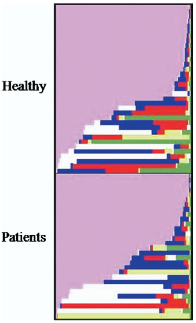
Patients and healthy subjects did not differ on age [t(62) = .25, p = .80], sex [χ2 = .08, p = .77], IQ [t(60) = .15, p = .88], or SES [t(55) = 1.66, p = .10]. Nor were there differences in ethnic ancestry factor scores between groups [ts < 1.42, ps = ns] or between genotypes within groups [ts < 1.67, ps = ns]. These scores were produced from a seven-factor solution of 186 ancestry-informative markers that differentiate continental and certain subcontinental populations (30). Ancestry distributions of individuals in each group are presented in Figure 1.
Figure 1.
Ancestry distributions across individuals in healthy and patient groups. Pink, Europe; blue, Middle East; white, Africa; red, Central Asia; green, America; yellow, Far East Asia; purple, Oceania. See journal Web site for full-color version of Figure 1.
The Kiddie Schizophrenia and Affective Disorders Schedule—Present and Lifetime Version (31) psychiatric interview was used to assign diagnoses. Of 18 anxiety-only patients, 12, 5, and 1 individuals met full criteria for one, two, and three current anxiety diagnoses, respectively; 5 patients received comorbid attention-deficit/hyperactivity disorder (ADHD) or oppositional defiant disorder diagnoses. Four patients met criteria for a past anxiety disorder, and two met criteria for prior alcohol abuse and ADHD. Other inclusion criteria comprised clinically significant symptoms for patients indexed by scores on the Pediatric Anxiety Rating Scale (≥ 10), the Children’s Depression Rating Scale (≥ 13), and the Child Global Assessment Scale (< 60). Exclusion criteria were current Tourette’s syndrome, obsessive-compulsive disorder, or conduct disorder; recent exposure to trauma;1 current use of any psychoactive substance;2 suicidal ideation; lifetime history of mania, psychosis, or pervasive developmental disorder; and IQ < 70. The study was approved by the National Institute of Mental Health (NIMH) Institutional Review Board. All participants/parents provided written informed assent/consent. Treatment began 3 weeks after research participation.
Genotyping
DNA extraction, genotyping, and polymerase chain reaction conditions followed published protocols (12). Stage 1 genotyping distinguished short from long alleles using an allele-discriminating probe hybridized once to the 43-bp L-insertion and an internal control probe hybridized to a sequence located within the same amplicon but specific to a divergent repeat in the amplicon not involved in insertion/deletion. The L-amplicon was 182 bp, and the S-amplicon was 138 bp. Stage 2 genotyping distinguished LA from LG alleles using fluorogenic probes designed specifically for these alleles. These were labeled at the 5′ end with either FAM or VIC. Genotypes were generated using ABI PRISM 7700 Sequence Detection system software (Applied Biosystems, Foster City, California). Twenty percent of the sample was genotyped twice, revealing error rates of < .005 and completion rates of > .95.
Allelic frequencies for S, LA, and LG across the sample were 56 (43%), 66 (51%), and 8 (6%) respectively. Subjects belonged to one of six genotype groups (Table 1), but were assigned to three groups on the basis of functional similarity of S and LG alleles (12): LALA, SLA/LALG, and SS/SLG/LGLG. No differences in genotypic distribution across patients and healthy subjects emerged [χ2 = 2.34, p = .31]. Prior studies (4) and modest sample sizes warranted further grouping individuals as LALA homozygotes and S/LG carriers.
Face-Emotion Paradigm
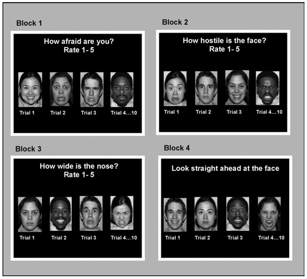
Procedures and stimuli have been described previously (7–9,29,32–34). Four epochs of 40 trials were presented (Figure 2): 32 trials showed different face emotions (eight fearful, eight angry, eight happy, eight neutral), and eight trials contained a fixation point. These 40 trials were divided into four blocks of 10 trials, in which eight faces and two fixation trials were presented in random order. In each block, participants completed one of four tasks that varied in attentional focus: rated subjective fear level to the face, rated the nose width on each face, rated the level of threat of each face, or passively viewed the face. Order of blocks was randomized across participants. Each block began with instructions (3000 msec) followed by 10 trials (4000 msec/trial). Intertrial intervals ranged from 750 to 1250 msec. Gray-scale face stimuli were from three sources (35–37). Stimuli were displayed with Avotec Silent Vision Glasses (Stuart, Florida). Ratings and reaction times (RT) were recorded with a five-key button box (MRI Devices Corporation, Waukesha, Wisconsin).
Figure 2.
Face-processing paradigm presented during functional magnetic resonance acquisition to all subjects. The paradigm consists of four tasks (afraid, nose width and hostility ratings, and passive viewing) across 160 trials. Reprinted with permission from (37).
Magnetic Resonance Imaging Data Acquisition and Processing
Whole-brain blood oxygen level–dependent (BOLD) functional magnetic resonance imaging (fMRI) data were acquired on a General Electric (Waukesha, Wisconsin) Signa 3-T scanner. Following sagittal localization and manual shimming, functional T2*-weighted images were acquired using an echo-planar single-shot gradient echo pulse sequence with matrix size of 64 × 64, repetition time (TR) of 2000 msec, echo time (TE) of 40 msec, field of view (FOV) of 240 mm, and voxels of 3.75 × 3.75 × 5.0 mm. Images were acquired in 23 contiguous axial slices per brain volume positioned parallel to the anterior commissure–posterior commissure line. Functional data were gathered in a single 14-min run. A high-resolution T1-weighted anatomic image was acquired to aid spatial normalization. A standardized magnetization-prepared gradient echo sequence (180 1-mm sagittal slices, FOV = 256, number of excitations = 1, TR = 11.4 msec, TE = 4.4 msec, matrix = 256 × 256, time to inversion = 300 msec, bandwidth = 130 Hz/pixel, 33 kHz/256 pixels) was used.
Reconstructed fMRI images were examined for excessive motion (> 3 mm in any plane) using MedX (Medical Numerics, Sterling, Virginia). Subsequent processing used SPM99 (University College, London, United Kingdom) and Matlab6 (Mathworks, Natick, Massachusetts). Functional data were corrected for slice timing and motion, coregistered to anatomic data, spatially normalized to a Montreal Neurologic Institute (MNI) T1-weighted template image, and resliced to 2-mm isotropic voxels. After inspecting images, event-related response amplitudes at the individual subject level for every face emotion were estimated in each attention task using the General Linear Model. Dividing each contrast image by subject-specific voxel time series means yielded percent fMRI signal change (38).
Statistical Analyses
Ratings and RT data during “how afraid” were examined using repeated-measures analyses of variance (ANOVAs) with two between-subjects factors (Diagnosis: patients, control subjects; Genotype: LALA homozygotes, S/LG carriers) and one within-subjects factor (Face Emotion: fearful, angry, happy, neutral). Greenhouse-Geisser (G-G) adjustment was applied in cases of unequal variances.
For group-level fMRI analyses, a random-effects model permitted population-level inferences (39). Analyses focused on the amygdala during “how afraid” using a region-of-interest approach (40). The boundaries of the amygdala were defined using standard anatomic criteria3 on a single MNI template and applied to all normalized brains at the group level. BOLD signal changes for each event type (fearful, angry, happy, neutral faces) during afraid ratings relative to fixations were averaged across all voxels in the left and right amygdala for each subject. Left and right amygdala values were analyzed separately with repeated-measures ANOVAs in SPSS-14, examining main effects and interactions of two between-subjects factors (Diagnosis: patients, controls; Genotype: LALA homozygotes, S/LG carriers) and one within-subjects factor (Face Emotion: fearful, angry, happy, neutral). The G-G correction was applied. Because amygdala values correlated significantly with age and ethnic ancestry scores, these were covariates in subsequent analyses. Voxelwise SPM analyses using small-volume Gaussian random field correction procedures for multiple comparisons confirmed significant Genotype-by-Diagnosis interactions in the amygdala during afraid ratings.
Results
Behavioral Data
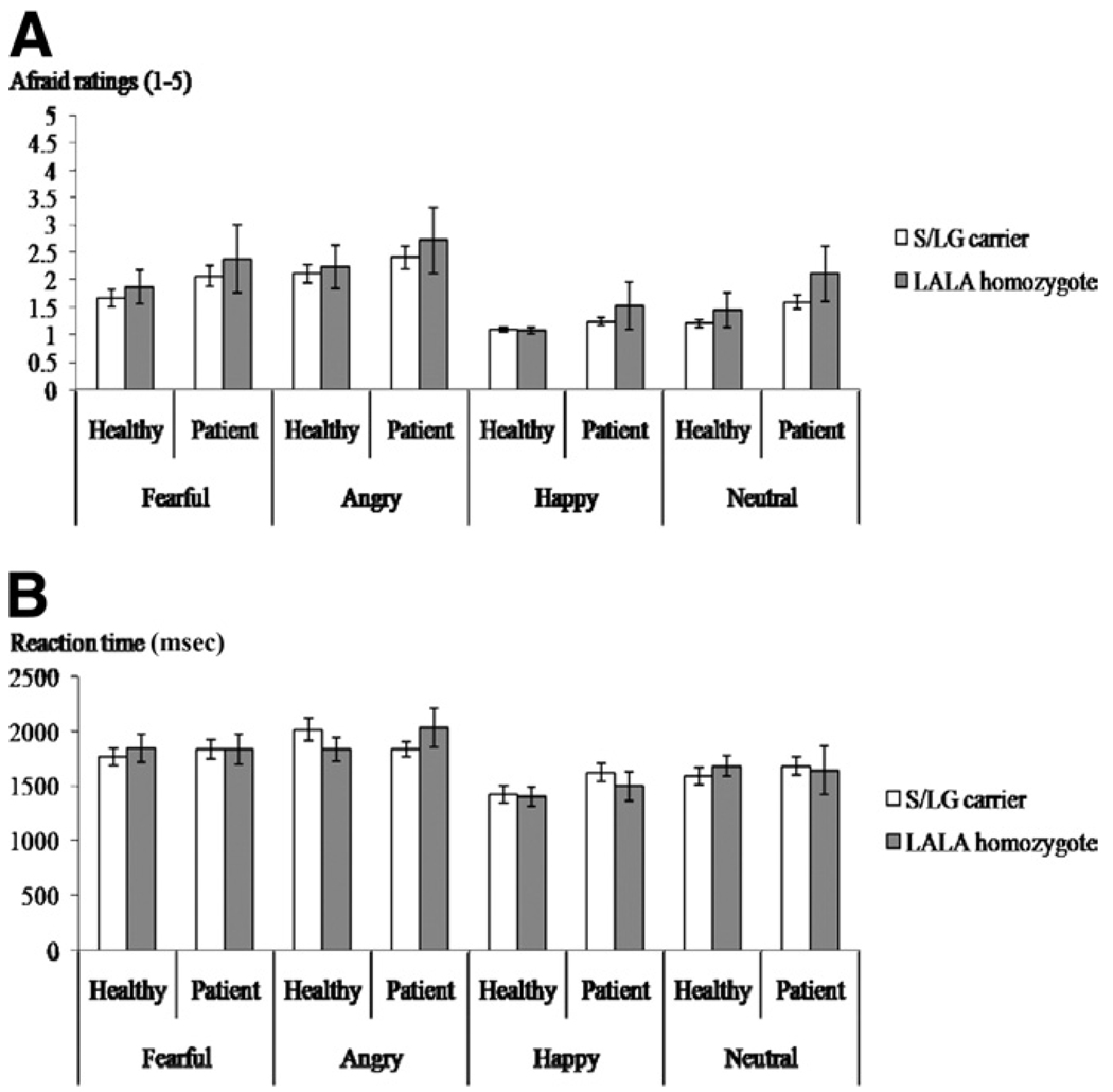
Ratings and RT data during “how afraid” are presented in Figure 3. Data for three healthy participants were not recorded. Significant Face Emotion [F(3,171) = 33.69, p < .001] and Diagnosis [F(1,57) = 4.99, p < .05] effects emerged on ratings. Angry faces received highest ratings (2.38), followed by fearful (2.00), neutral (1.56), and happy (1.24) faces. Patients gave higher ratings to face emotions (2.02) relative to control subjects (1.59). Similar Face-Emotion effects characterized RTs [F(3,171) = 23.29, p < .001]: rating angry faces was slowest (1932.92 msec) followed by fearful (1825.83 msec), neutral (1650.47 msec), and happy faces (1492.62 msec).
Figure 3.
(A) Afraid ratings of various face emotions (fearful, angry, happy, neutral) across healthy and anxious adolescents in each genotype group (S/LG carriers, LALA homozygotes). (B) Mean reaction times (msec) during afraid ratings of different face emotions (fearful, angry, happy, neutral) across patient and healthy adolescents belonging to each genotype group (S/LG carriers, LALA homozygotes). L, long; S, short allele.
fMRI Data
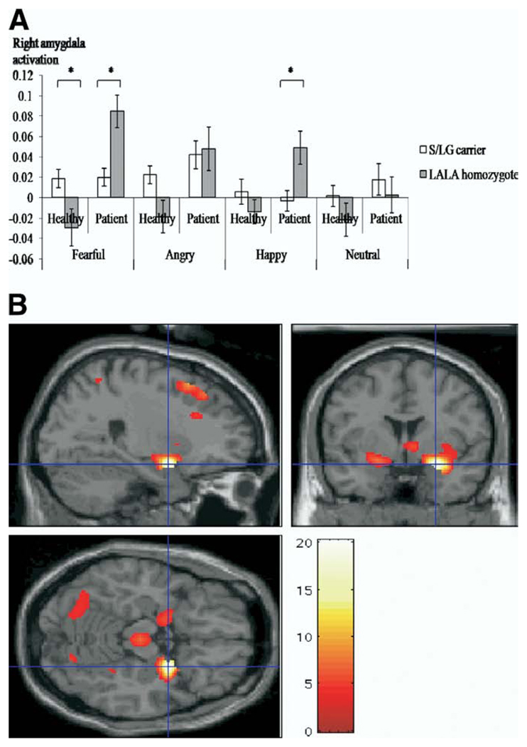
Significant effects of Diagnosis on both left and right amygdala responses indicated greater activity across Face Emotions (relative to fixations) among patients. Significant Genotype-by-Diagnosis and Genotype-by-Diagnosis-by-Fac1e-Emotion interactions characterized right [F(3,159) = 2.66, p < .05] but not left amygdala activity (Figure 4A). The three-way interaction was decomposed by examining Genotype and Diagnosis effects on right amygdala activity to each Face Emotion separately.
Figure 4.
(A) Bar graphs of activation in the right amygdala for the “how afraid” condition relative to the task null-event baseline in various face emotions for patient and healthy adolescents across combined genotype groups (S/LG carriers and LALA homozygotes). (B) The topography of peak activations in the right amygdala (Montreal Neurological Institute coordinates: 26, 2, −16) where the significant Genotype-by-Diagnosis interaction on afraid ratings of fearful faces emerged (p < .05). L, long; S, short allele.
Significant Genotype-by-Diagnosis interactions characterized fearful [F(1,59) = 18.65, p < .001] and happy [F(1,59) = 5.50, p < .05] faces. For fearful faces, this interaction was driven by differential amygdala responses across genotype in each diagnostic group. Among healthy subjects, S/LG carriers showed greater activity than LALA homozygotes [F(1,31) = 5.24, p < .05, Cohen’s d = .95]. In patients, greater activity occurred among LALA individuals than S/LG carriers [F(1,27) = 14.17, p < .01, Cohen’s d = 1.61]. For happy faces, the Genotype-by-Diagnosis interaction was explained by patient data only: LALA individuals manifested more amygdala activity than S/LG carriers [F(1,27) = 6.88, p < .05, Cohen’s d = 1.27].
Post hoc analyses contrasting amygdala responses to fearful faces across the three genotype groups (LA/LA, SLA/LALG, SS/SLG/LGLG) showed that SLA/LALG and SS/SLG/LGLG individuals were comparable in healthy subjects and patients, but they differed significantly from LA/LA individuals (Supplement 1). This justified pooling S- and LG-allele carriers. For happy faces, differences in amygdala responses across genotype groups were more apparent in patients, but these were inconsistent. Whereas SLA/LALG and SS/SLG/LGLG individuals showed similar responses, only SLA/LALG individuals differed significantly to LA/LA individuals (Supplement 2).
Voxelwise SPM analyses confirmed strong Genotype-by-Diagnosis interactions to afraid ratings of fearful faces in the right amygdala [F = 2.20, p < .001] (Figure 4B; Supplement 3). All regions where significant Genotype-by-Diagnosis interactions emerged to fearful faces are shown in Table 2. Weaker interactions characterized the right amygdala during afraid ratings of happy faces [F = 5.44, p < .05] (details on further request).
Table 2.
Voxls with Significant Genotype-by-Diagnosis Interactions During Afraid Ratings of Fearful Faces (p <.01)
| Brodmann Area | Region | Volume (mm) | x | y | z | F | p Value |
|---|---|---|---|---|---|---|---|
| Afraid fear relative to baseline (p < .01) | |||||||
| Right amygdala | 303 | 26 | 2 | −16 | 20.20 | <.001 | |
| 6 | Gyrus frontal superior | 135 | 22 | 16 | 56 | 10.60 | .002 |
| 8 | Gyrus frontal superior | 135 | 26 | 30 | 48 | 7.51 | .008 |
| Left amygdala | 57 | −16 | −4 | −10 | 9.78 | .003 | |
| Cerebellum | 48 | 36 | −68 | −22 | 8.91 | .004 | |
| 8 | Gyrus frontal superior | 11 | −6 | 42 | 42 | 8.26 | .006 |
| Brainstem | 3 | 4 | −20 | −14 | 7.16 | .01 | |
Parallel analyses employing a biallelic classification of 5-HTT genotypes (SS/SL vs. LL) on right amygdala activation yielded significant effects of Diagnosis, Face Emotion, and a two-way Diagnosis-by-Genotype interaction. Post hoc analyses showed significant Genotype-by-Diagnosis interactions for fearful [F(1,59) = 12.07, p < .01] and happy [F(1,59) = 6.97, p < .05] faces. For fearful faces, healthy SS/SL individuals showed greater amygdala activity than LL individuals [F(1,31) = 4.65, p < .05, Cohen’s d = .80]. Among patients, greater amygdala activity was found among LL than SL/SS-individuals [F(1,27) = 6.64, p < .05, Cohen’s d = .50]. For happy faces, LL individuals showed enhanced amygdala activity relative to SS/SL individuals [F(1,27) = 5.66, p < .05, Cohen’s d = .67]. Thus, results were broadly comparable to using a triallelic classification, but effect sizes for patient genotype differences were smaller. Amygdala responses to fearful and happy faces across the three genotype groups of the biallelic classification followed similar trends to the triallelic classification (Supplement 4 and Supplement 5).
To test specificity of results to the how afraid condition, analyses were repeated for data from other attention tasks, but no main or interaction effects emerged for left or right amygdala responses for triallelic or biallelic classifications. Modest sample sizes and low statistical power precluded testing a four-way Genotype-by-Diagnosis-by-Attention-by-Face-Emotion interaction.
To aid interpretation of fMRI results, we examined genotype and diagnosis effects on self-reported anxiety and depressive symptoms (42,43) among current subjects, as well as from healthy and anxious/depressed adolescents recruited for other NIMH studies (n > 230). Neither revealed significant effects of 5-HTT genotype on symptoms.
Discussion
Effects of 5-HTT gene variants on amygdala responses to emotional faces were studied in healthy and anxious/depressed adolescents during internal fear evaluation. A significant Genotype-by-Diagnosis-by-Face-Emotion interaction emerged on right amygdala activity, reflecting three key findings. First, in healthy adolescents, stronger amygdala responses to fearful faces characterized S/LG carriers, relative to LALA individuals. Second, this was opposite in patients in whom LALA individuals exhibited greater amygdala responses to fearful faces. Third, effects in patients extended to happy faces.
These data are the first to document conservation of gene–brain associations across typical development, supporting conceptualizations that S/LG alleles increase risks for psychopathology in healthy individuals (4), possibly through stress reactivity (11,44,45). However, gene–brain associations in affected adolescents differed from those in affected adults (4), with opposite gene–amygdala response patterns to fearful and happy faces. That these effects characterized happy faces as well may be because of ambiguity from discrepancies between stimulus valence and a potential threat context (9,46–48).
Although no theoretical accounts speak directly to these contrary findings in adolescent patients, three issues are relevant. First, literature on associations between 5-HTT gene variants and brain function or symptoms is mixed. A recent meta-analysis on adult gene–brain associations noted potential publication biases when three unpublished data sets reporting no association or associations in opposite directions were excluded (4). Moreover, far fewer studies have been conducted in adult patients, calling for more independent replications generally but in especially clinical groups. Data for gene–symptom associations in adolescents are also inconsistent over whether the S or L allelic variant predicts risk for psychopathology (16–19).
Second, some anxious responses to threat show developmental differences. Relative to healthy subjects, anxious adults exhibit selective attention toward threat stimuli (49), whereas anxious adolescents shift attention away from these stimuli (50). Whether these reflect distinct compensatory responses used by affected adolescents to regulate emotional arousal is unknown, but regardless, they illustrate developmental changes in clinical behaviors. Variable expression of S/LG alleles on brain function from adolescence to adulthood among affected individuals is thus feasible.
Finally, incomplete penetrance from reduced exposure to environmental factors in patient S/LG carriers could also explain lowered amygdala activity in this group.
In summary, we present new but preliminary data on the genetics of neural function in adolescents. Although current sample sizes constrain power to interpret gene–brain associations in relation to differences across risk alleles (biallelic vs. triallelic classification; “dose-response” vs. “threshold” effect), diagnosis (anxiety vs. depression), and attentional conditions (nose ratings, hostility ratings, passive viewing), notably our effect sizes of genotype differences are comparable, if not larger, than previous studies (4) using similar-sized samples (24–28). Because imaging genetics research is still in its infancy, any data clarifying these associations is informative. Furthermore, our data lay the groundwork for considering interactions among genes, brain function, and emotional processes across development.
Supplementary Material
Acknowledgments
We thank Jessica Jenness, Nina Shiffrin, Elizabeth Shroth, Veronica Temple, and Amber Williams for data processing assistance; Longina Akhtar and Gary Jenkins for assistance with genotyping; Dave Luckenbaugh for advice on statistical procedures; Harvey Iwamoto for programming and computer support; and Ellen Leibenluft, Ken Towbin, and Alan Zametkin for medical oversight. This study was supported by the Intramural Research Program of the National Institute of Mental Health, National Institutes of Health.
Footnotes
Definitions of trauma followed DSM-IV criteria for posttraumatic stress disorder, as having experienced, witnessed, or been confronted by an event or events that involved actual or threatened death or serious injury or a threat to the physical integrity of self or others.
Medication and/or recreational drugs.
Consistent with a prior publication (41), the amygdala was measured from the slice at the level of the mammillary bodies to its anterior boundary, including the uncus.
The authors report no biomedical financial interests or potential conflicts of interest.
Supplementary material cited in this article is available online.
References
- 1.Beesdo K, Bittner A, Pine DS, Stein MB, Hofler M, Lieb RH, et al. Incidence of social anxiety disorder and the consistent risk for secondary depression in the first three decades of life. Arch Gen Psychiatry. 2007;64:903–912. doi: 10.1001/archpsyc.64.8.903. [DOI] [PubMed] [Google Scholar]
- 2.Pine DS, Cohen P, Gurley D, Brook J, Ma Y. The risk for early-adulthood anxiety and depressive disorders in adolescents with anxiety and depressive disorders. Arch Gen Psychiatry. 1998;55:56–64. doi: 10.1001/archpsyc.55.1.56. [DOI] [PubMed] [Google Scholar]
- 3.Gross C, Hen R. The developmental origins of anxiety. Nat Rev Neuroscience. 2004;5:545–552. doi: 10.1038/nrn1429. [DOI] [PubMed] [Google Scholar]
- 4.Munafo MR, Brown SM, Hariri AR. Serotonin transporter (5-HTTLPR) genotype and amygdala activation: A meta-analysis. Biol Psychiatry. 2008;63:852–857. doi: 10.1016/j.biopsych.2007.08.016. [DOI] [PMC free article] [PubMed] [Google Scholar]
- 5.Thomas KM, Drevets WC, Dahl RE, Ryan ND, Birmaher B, Eccard CH, et al. Amygdala response to fearful faces in anxious and depressed children. Arch Gen Psychiatry. 2001;58:1057–1063. doi: 10.1001/archpsyc.58.11.1057. [DOI] [PubMed] [Google Scholar]
- 6.Killgore WD, Yurgelun-Todd DA. Social anxiety predicts amygdale activate on in adolescents viewing fearful faces. Neuroreport. 2005;16:1671–1675. doi: 10.1097/01.wnr.0000180143.99267.bd. [DOI] [PubMed] [Google Scholar]
- 7.McClure EB, Monk CS, Nelson EE, Parrish JM, Adler A, Blair RJR, et al. Abnormal attention modulation of fear circuit function in pediatric generalized anxiety disorder. Arch Gen Psychiatry. 2007;64:97–106. doi: 10.1001/archpsyc.64.1.97. [DOI] [PubMed] [Google Scholar]
- 8.Roberson-Nay R, McClure EB, Monk CS, Nelson EE, Guyer AE, Fromm SJ, et al. Increased amygdala activity during successful memory encoding in adolescent major depressive disorder: An FMRI study. Biol Psychiatry. 2006;60:966–973. doi: 10.1016/j.biopsych.2006.02.018. [DOI] [PubMed] [Google Scholar]
- 9.Perez-Edgar K, Roberson-Nay R, Hardin MG, Poeth K, Guyer AE, Nelson EE, et al. Attention alters neural responses to evocative faces in behaviorally inhibited adolescents. NeuroImage. 2007;35:1538–1546. doi: 10.1016/j.neuroimage.2007.02.006. [DOI] [PMC free article] [PubMed] [Google Scholar]
- 10.Monk CS, Telzer EH, Mogg K, Bradley BP, Mai X, Louro HM, et al. Amygdala and ventrolateral prefrontal cortex activation to masked angry faces in children and adolescents with generalized anxiety disorder. Arch Gen Psychiatry. 2008;65:568–576. doi: 10.1001/archpsyc.65.5.568. [DOI] [PMC free article] [PubMed] [Google Scholar]
- 11.Lesch KP, Bengel D, Heils A, Sabol SZ, Greenberg BD, Petri S, et al. Association of anxiety-related traits with a polymorphism in the serotonin transporter gene regulatory region. Science. 1996;274:1527–1531. doi: 10.1126/science.274.5292.1527. [DOI] [PubMed] [Google Scholar]
- 12.Hu XZ, Lipsky RH, Zhu G, Akhtar LA, Taubman J, Greenberg BD, et al. Serotonin transporter promoter gain-of-function genotypes are linked to obsessive-compulsive disorder. American J Hum Genet. 2006;78:815–826. doi: 10.1086/503850. [DOI] [PMC free article] [PubMed] [Google Scholar]
- 13.Hariri AR, Holmes A. Genetics of emotional regulation: The role of the serotonin transporter in neural function. Trends Cogn Sci. 2006;10:182–191. doi: 10.1016/j.tics.2006.02.011. [DOI] [PubMed] [Google Scholar]
- 14.Munafo MR, Clark TG, Moore LR, Payne E, Walton R, Flint J. Genetic polymorphisms and personality in healthy adults: A systematic review and meta-analysis. Mol Psychiatry. 8:471–484. doi: 10.1038/sj.mp.4001326. [DOI] [PubMed] [Google Scholar]
- 15.Sen S, Burmeister M, Ghosh D. Meta-analysis of the association between a serotonin transporter promoter polymorphism (5-HTTLPR) and anxiety-related personality traits. Am J Med Genet B Neuropsychiatr Genet. 2004;127B:85–99. doi: 10.1002/ajmg.b.20158. [DOI] [PubMed] [Google Scholar]
- 16.Ebstein RP, Levine J, Geller V, Auerbach J, Gritsenko I, Belmaker RH. Dopamine D4 receptor and serotonin transporter promoter in the determination of neonatal temperament. Mol Psychiatry. 1998;3:238–246. doi: 10.1038/sj.mp.4000363. [DOI] [PubMed] [Google Scholar]
- 17.Battaglia M, Ogliari A, Zanoni A, Citterio A, Pozzoli U, Giorda R, et al. Influence of the serotonin transporter promoter gene and shyness on children’s cerebral responses to facial expressions. Arch Gen Psychiatry. 2005;62:85–94. doi: 10.1001/archpsyc.62.1.85. [DOI] [PubMed] [Google Scholar]
- 18.Arbelle S, Benjamin J, Golin M, Kremer I, Belmaker RH, Ebstein RP. Relation of shyness in grade school children to the genotype for the long form of the serotonin transporter promoter region polymorphism. Am J Psychiatry. 2003;160:671–676. doi: 10.1176/appi.ajp.160.4.671. [DOI] [PubMed] [Google Scholar]
- 19.Jorm AF, Prior M, Sanson A, Smart D, Zhang Y, Easteal S. Association of a functional polymorphism of the serotonin transporter gene with anxiety-related temperament and behavior problems in children: A longitudinal study from infancy to the mid-teens. Mol Psychiatry. 2000;5:542–547. doi: 10.1038/sj.mp.4000782. [DOI] [PubMed] [Google Scholar]
- 20.Cicchetti D, Rogosch FA, Sturge-Apple ML. Gene-environment interaction. Dev Psychopathol. 2007;19:957–959. [PubMed] [Google Scholar]
- 21.Eley TC, Sugden K, Corsico A, Gregory AM, Sham P, McGuffin P, et al. Gene-environment interaction analysis of serotonin system markers with adolescent depression. Mol Psychiatry. 2004;9:908–915. doi: 10.1038/sj.mp.4001546. [DOI] [PubMed] [Google Scholar]
- 22.Fox NA, Nichols KE, Henderson HA, Rubin K, Schmidt L, Hamer D, et al. Evidence for a gene-environment interaction in predicting behavioral inhibition in middle childhood. Psychol Sci. 2005;16:921–926. doi: 10.1111/j.1467-9280.2005.01637.x. [DOI] [PubMed] [Google Scholar]
- 23.Kaufman J, Yang BZ, Douglas-Palumberi H, Houshyar S, Lipschitz D, Krystal JH, et al. Social supports and serotonin transporter gene moderate depression in maltreated children. Proc Natl Acad Sci U S A. 2004;101:17316–17321. doi: 10.1073/pnas.0404376101. [DOI] [PMC free article] [PubMed] [Google Scholar]
- 24.Smolka MN, Buhler M, Schumann G, Klein S, Hu XZ, Moayer M, et al. Gene-gene effects on central processing of aversive stimuli. Mol Psychiatry. 2007;12:307–317. doi: 10.1038/sj.mp.4001946. [DOI] [PubMed] [Google Scholar]
- 25.Dannlowski U, Ohrmann P, Bauer J, Kugel H, Baune BT, Hohoff C, et al. Serotonergic genes modulate amygdala activity in major depression. Genes Brain Behav. 2007;6:672–676. doi: 10.1111/j.1601-183X.2006.00297.x. [DOI] [PubMed] [Google Scholar]
- 26.Dannlowski U, Ohrmann P, Bauer J, Deckert J, Hohoff C, Kugel H, et al. 5-HTTLPR biases amygdala activity in response to masked facial expressions in major depression. Neuropsychopharmacology. 2008;33:418–424. doi: 10.1038/sj.npp.1301411. [DOI] [PubMed] [Google Scholar]
- 27.Furmark T, Tillfors M, Garpenstrand H, Marteinsdottir I, Langstrom B, Oreland L, et al. Serotonin transporter polymorphism related to amygdala excitability and symptom severity in patients with social phobia. Neurosci Lett. 2004;362:189–192. doi: 10.1016/j.neulet.2004.02.070. [DOI] [PubMed] [Google Scholar]
- 28.Domschke K, Braun M, Ohrmann P, Suslow T, Kugel H, Bauer J, et al. Association of the functional -1019C/G 5-HT1A polymorphism with prefrontal cortex and amygdala activation measured with 3T fMRI in panic disorder. Int J Neuropsychopharmacol. 2006;9:349–355. doi: 10.1017/S1461145705005869. [DOI] [PubMed] [Google Scholar]
- 29.Guyer AE, Monk CS, McClure-Tone EB, Nelson EE, Roberson-Nay R, Adler AD, et al. Adevelopmental examination of amygdala response to facial expressions. J Cogn Neurosci. 2008;20:1565–1582. doi: 10.1162/jocn.2008.20114. [DOI] [PMC free article] [PubMed] [Google Scholar]
- 30.Hodgkinson C, Xu K, Qiaoping Y, Pei-Hong S, Heinz E, Lobos EA, et al. Addictions biology: Haplotype based analysis for 130 candidate genes on a single array. Alcohol Alcohol. 43:505–515. doi: 10.1093/alcalc/agn032. [DOI] [PMC free article] [PubMed] [Google Scholar]
- 31.Kaufman J, Birmaher B, Brent D, Rao U, Flynn C, Moreci P, et al. Schedule for Affective Disorders and Schizophrenia for School-Age Children—Present and Lifetime Version (K-SADS-PL): Initial reliability and alidity data. J Am Acad Child Adolesc Psychiatry. 1997;36:980–988. doi: 10.1097/00004583-199707000-00021. [DOI] [PubMed] [Google Scholar]
- 32.Monk CS, McClure EB, Nelson EE, Zarahn E, Bilder RM, Leibenluft E, et al. Adolescent immaturity in attention-related brain engagement to emotional facial expressions. NeuroImage. 2003;20:420–428. doi: 10.1016/s1053-8119(03)00355-0. [DOI] [PubMed] [Google Scholar]
- 33.Pine DS, Klein RG, Mannuzza S, Moulton JL, 3rd, Lissek S, Guardino M, et al. Face-emotion processing in offspring at risk for panic disorder. J Am Acad Child Adolesc Psychiatry. 2005;44:664–672. doi: 10.1097/01.chi.0000162580.92029.f4. [DOI] [PubMed] [Google Scholar]
- 34.Rich BA, Vinton DT, Roberson-Nay R, Hommer RE, Berghorst LH, McClure EB, et al. Limbic hyperactivation during processing of neutral facial expressions in children with bipolar disorder. Proc Natl Acad Sci U S A. 2006;103:8900–8905. doi: 10.1073/pnas.0603246103. [DOI] [PMC free article] [PubMed] [Google Scholar]
- 35.Ekman P, Friesen WV. Pictures of Facial Affect. Palo Alto, CA: Consulting Psychology Press; 1976. [Google Scholar]
- 36.Gur RC, Ragland JD, Moberg PJ, Turner TH, Bilker WB, Kohler C, et al. Computerized neurocognitive scanning, I: methodology and validation in healthy people. Neuropsychopharmacology. 2001;25:766–776. doi: 10.1016/S0893-133X(01)00278-0. [DOI] [PubMed] [Google Scholar]
- 37.Tottenham N, Tanaka J, Leon AC, McCarry T, Nurse M, Hare TA, et al. The NimStim set of facial expressions: judgements from untrained research participants. Psychiatry Res. doi: 10.1016/j.psychres.2008.05.006. (in press) [DOI] [PMC free article] [PubMed] [Google Scholar]
- 38.Zarahn E, Aguirre G, D’Esposito M. A trial-based experimental design for fMRI. Neuroimage. 1997;6:122–138. doi: 10.1006/nimg.1997.0279. [DOI] [PubMed] [Google Scholar]
- 39.Holmes AP, Friston KJ. Generalisability, random effects and population inference. Neuroimage. 1998;7:S754. [Google Scholar]
- 40.Hariri AR, Mattay VS, Tessitore A, Kolachana B, Fera F, Goldman D, et al. Serotonin transporter genetic variation and the response of the human amygdala. Science. 2002;297:400–403. doi: 10.1126/science.1071829. [DOI] [PubMed] [Google Scholar]
- 41.Szeszko PR, Robinson D, Alvir JM, Bilder RM, Lencz T, Ashtari M, et al. Orbital frontal and amygdala volume reductions in obsessive compulsive disorder. Arch Gen Psychiatry. 1999;56:913–919. doi: 10.1001/archpsyc.56.10.913. [DOI] [PubMed] [Google Scholar]
- 42.Birmaher B, Khetarpal S, Brent D, Cully M, Balach L, Kaufman J, Neer SM. The Screen for Child Anxiety Related Emotional Disorders (SCARED): Scale construction and psychometric characteristics. J Am Acad Child Adolesc Psychiatry. 1997;36:545–553. doi: 10.1097/00004583-199704000-00018. [DOI] [PubMed] [Google Scholar]
- 43.Kovacs M. The Children’s Depression Inventory (CDI) Psychopharmacol Bull. 1985;21:995–998. [PubMed] [Google Scholar]
- 44.Barr CS, Newman TK, Shannon C, Parker C, Dvoskin RL, Becker ML, et al. Rearing condition and rh5-HTTLPR interact to influence limbichypothalamic-pituitary-adrenal axis response to stress in infant macaques. Biol Psychiatry. 2004;55:733–738. doi: 10.1016/j.biopsych.2003.12.008. [DOI] [PubMed] [Google Scholar]
- 45.Caspi A, Sugden K, Moffitt TE, Taylor A, Craig IW, Harrington H, et al. Influence of life stress on depression: moderation by a polymorphism in the 5-HTT gene. Science. 2003;301:386–389. doi: 10.1126/science.1083968. [DOI] [PubMed] [Google Scholar]
- 46.Monk CS, Klein RG, Telzer EH, Schroth EA, Mannuzza S, Moulton JL, 3rd, et al. Amygdala and nucleus accumbens activation to emotional facial expressions in children and adolescents at risk for major depression. Am J Psychiatry. 2008;165:90–98. doi: 10.1176/appi.ajp.2007.06111917. [DOI] [PubMed] [Google Scholar]
- 47.Guyer AE, Lau JYF, McClure EB, Parrish J, Shiffrin ND, Blair RJR, et al. Amygdala and Ventrolateral Prefrontal Cortex Function during Anticipated Peer Evaluation in Pediatric Social Anxiety. Arch Gen Psychiatry. doi: 10.1001/archpsyc.65.11.1303. (in press) [DOI] [PMC free article] [PubMed] [Google Scholar]
- 48.Dugas MJ, Buhr K, Ladouceur R. The role of intolerance of uncertainty in etiology and maintenance. In: Heimberg RG, Turk CL, Mennin DS, editors. Generalized Anxiety Disorder: Advances in Research and Practice. New York: Guilford Press; 2004. pp. 143–163. [Google Scholar]
- 49.Bradley BP, Mogg K, White J, Groom C, de Bono J. Attentional bias for emotional faces in generalized anxiety disorder. Br J Clin Psychol. 1999;38:267–278. doi: 10.1348/014466599162845. [DOI] [PubMed] [Google Scholar]
- 50.Monk CS, Nelson EE, McClure EB, Mogg K, Bradley BP, Leibenluft E, et al. Ventrolateral prefrontal cortex activation and attentional bias in response to angry faces in adolescents with generalized anxiety disorder. Am J Psychiatry. 2006;163:1091–1097. doi: 10.1176/ajp.2006.163.6.1091. [DOI] [PubMed] [Google Scholar]
Associated Data
This section collects any data citations, data availability statements, or supplementary materials included in this article.


