Mantle cell lymphoma (MCL) and multiple myeloma (MM) are aggressive hemopathies characterized by the presence of the cyclin D1 protein in 100% and 50% of the cases, respectively. The presence of cyclin D1, which is not expressed along the B-cell lineage, is causal in MCL and MM pathogenesis. It is generally admitted that the misexpression of cyclin D1 leads to uncontrolled cell proliferation. In turn, the selective suppression of cyclin D1 in MCL and MM has been envisaged as a therapeutic strategy. In a recent paper, Klier et al. reported that lentiviral shRNA-mediated downregulation of cyclin D1 in MCL cells led only to a moderate decrease of cell proliferation and had no effects on cell survival.1 This was likely due to a compensatory effect of cyclin D2 which was, in turn, up-regulated. We extend these observations by showing that: (i) such as in MCL cells, the stable downregulation of cyclin D1 impacts slightly on proliferation and not survival of multiple MM cells; (ii) does not amplify the apoptotic response towards bortezomib of MCL and MM cells.
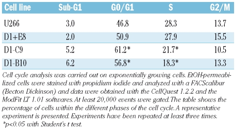
To understand the role of cyclin D1 in MCL and MM pathologies, we established stable JeKo-1 (MCL) and U266 (MM)-derived cell lines in which CCND1 was down-regulated by SureSilencing™ plasmids. Silencing of CCND1 was controlled by real time RT-PCR (Table 1). At the protein level, compared with the parental cell line (p), U266 D1-B10 and −C9 showed a downregulation of 15% and 50%, respectively; JeKo-1 D1–E4 and −G9 showed a downregulation of 48% and 20%, respectively (Figure 1A). No differences were seen in the two cell lines obtained after transfection of a scrambled shRNA (U266 D1+E8, JeKo-1 D1+G10). Proliferation curves of U266, JeKo-1 and derivatives were obtained after plating and counting cells during a one week period. The results obtained for JeKo-1 MCL line and derivatives were similar to those obtained by Klier et al. and are not described here.1 The downregulation of cyclin D1 in U266-derived cells had a modest but reproducible and statistically significant effect on cell proliferation (Figure 1B). From the proliferation curves we calculated a decrease of −25% and −35% for U266 D1–B10 and −C9, respectively, of total viable cells at the end of the culture. Importantly, the decrease in cell number correlated with cyclin D1 protein level reduction. Analysis of cell cycle distribution after PI-staining of exponentially growing cells indicated an increase in the percentage of cells within the G0/G1 fraction and a concomitant decrease within the S fraction in U266 (+14%/−7% for U266 D1-C9, +10%/−10% for −B10, Table 2) and JeKo-1-derivatives (data not shown) compared with parental cells. Those experiments also confirmed the absence of apoptotic cells in the cultures. Altogether, our data indicate that the knockdown of cyclin D1 slows down cell proliferation of MM and MCL cells by impairing G1 to S phase transition in agreement with the known function of cyclin D1. However, the effects of cyclin D1 knockdown are only modest. Since cyclin D2 is the only cyclin D-type expressed in mature B-cells2 and cyclin D2 can compensate cyclin D1 in vivo,3 we looked at the expression of cyclin D2 in U266 and derivatives. We observed an upregulation of cyclin D2 in U226-derivatives correlating with cyclin D1 downregulation (Figure 1C). We concluded that the slight effect of cyclin D1 extinction on cell proliferation and the absence of effect on cell survival is due to a compensatory effect of cyclin D2. Finally, since cyclin D1 downregulation could sensitize cells to apoptotic inducers, we analyzed the response of U266- and JeKo-1-derivatives treated with bortezomib, known to induce apoptosis in both MCL and MM cells.4,5 Bortezomib had a similar cytotoxic effect on cells independently of cyclin D1 level (Figure 1D).
Table 1.
Real-time quantitative RT-PCR analysis of cyclin D1 mRNA evel in U266 and derivatives.
Figure 1.
Cyclin D1 downregulation in MM and MCL cells has little effect on cell proliferation and no effects on bortezomib-mediated apoptosis. We established stable MCL- and MM-derived cell lines in which CCND1 was down-regulated by shRNA encoded by SureSilencing™ plasmids. Four shRNAs were tested; only three of them were efficient: D1–2, CTT CCT CTC CAG AGT GAT CAA (JeKo-1 D1–G9); D1–3, GCA TGT TCG TGG CCT CTA AGA (U266 D1-C9, JeKo-1 D1–E4); D1–4, GTG CCA CAG ATG TGA AGT TCA (U266 D1-B10). A “scrambled” shRNA with no homology with human sequences was also used: GGA ATC TCA TTC GAT GCA TAC (U266 D1+E8, JeKo-1 D1+G10). MM and MCL cell lines were transfected by electroporation, selected on neomycine resistance and cloned by limiting dilution to obtain stable shRNA-expressing cell clones. (A) Parental cells (p) and their derivatives (U266 on the left, JeKo-1 on the right) were analyzed by Western blot. Whole cell extracts were obtained from exponentially growing cells and separated by SDS-PAGE. Blots were incubated with rabbit anti-cyclin D1 (sc-718) and to control for gel loading, anti-β-tubulin (sc-9104) Abs from Santa Cruz Biotechnologies Inc. Blots were analyzed with the FluorSImager and the ratio cyclin D1/β-tubulin calculated with the QuantityOne software (Bio-Rad). Experiments were repeated at least three times; a representative one is shown. (B) U266 and their derivatives were plated (5x10³ cells/mL) in triplicate and viable cells counted every day for one week by trypan blue exclusion. Values represent the mean ± SD. *p<0.05 with the t test. (C) Blots were prepared as in (A), anti-cyclin D2 Ab was from Santa Cruz Biotechnologies Inc. (sc-593). CM and RAMOS cell lines were used as positive and negative controls, respectively. (D) Parental cells and their derivatives (104 cells) were plated in 96-well plates, treated with various concentrations of bortezomib (0–10 nM) for 24 h and cell viability was determined by the MTS assay (CellTiter 96 Aqueous One Solution®, Promega). Each determination was made in triplicate and the percentage of viability was deduced from the parental cells.
Table 2.
Cell cycle distribution of U266 and derivatives.
Together with data reported by Klier et al.,1 our findings question the relevance of specific cyclin D1 targeting in MM and MCL hemopathies. Since in these two hemopathies, the knockdown of cyclin D1 is compensated by the upregulation of cyclin D2, a more powerful strategy would be to inhibit both cyclin D1 and cyclin D2. This was achieved recently in MM cells by the plant cytokinin kinetin riboside.6 Indeed, kinetin riboside causes cell cycle arrest of primary myeloma cells and tumor lines and tumor cells selective apoptosis, in vitro and in vivo. However, in MCL cell lines, the double knockdown of cyclins D1 and D2 had no synergistic effect.1 A more powerful alternative strategy for MM and MCL pathologies would be to specifically inhibit cyclin-dependent kinase 4 or 6, the catalytic partners of both cyclins D1 and D2.
Acknowledgments
the authors would like to thank Anne Barbaras for technical help with cell culture and Marilyne Duval with cytometry.
Footnotes
Funding: this work was supported by grant from the Ligue Nationale Contre le Cancer – Comité du Calvados (to BS). GT was supported by the Ligue Nationale Contre le Cancer - Comité du Calvados, LR by the Conseil Régional de Basse-Normandie.
This paper is dedicated to the memory of LR.
References
- 1.Klier M, Anastasov N, Hermann A, Meindl T, Angermeier D, Raffeld M, et al. Specific lentiviral shRNA-mediated knockdown of cyclin D1 in mantle cell lymphoma has minimal effects on cell survival and reveals a regulatory circuit with cyclin D2. Leukemia. 2008;22:2097–105. doi: 10.1038/leu.2008.213. [DOI] [PubMed] [Google Scholar]
- 2.Chiles TC. Regulation and function of cyclin D2 in B lymphocytes subsets. J Immunol. 2004;173:2901–7. doi: 10.4049/jimmunol.173.5.2901. [DOI] [PubMed] [Google Scholar]
- 3.Carthon BC, Neumann CA, Das M, Pawlyk B, Li T, Geng Y, Sicinski P. Genetic replacement of cyclin D1 function in mouse development by cyclin D2. Mol Cell Biol. 2005;25:1081–8. doi: 10.1128/MCB.25.3.1081-1088.2005. [DOI] [PMC free article] [PubMed] [Google Scholar]
- 4.Bogner C, Peschel C, Decker T. Targeting the proteasome in mantle cell lymphoma: a promising therapeutic approach. Leuk Lymphoma. 2006;47:195–205. doi: 10.1080/10428190500144490. [DOI] [PubMed] [Google Scholar]
- 5.Podar K, Chauhan D, Anderson KC. Bone marrow microenvironment and the identification of new targets for myeloma therapy. Leukemia. 2009;23:10–24. doi: 10.1038/leu.2008.259. [DOI] [PMC free article] [PubMed] [Google Scholar]
- 6.Tiedemann RE, Mao X, Shi C-X, Zhu YX, Palmer SE, Sebag M, Marler R, Chesi M, Fonseca R, Bergsagel PL, Schimmer AD, Stewart AK. Identification of kinetin ribo-side as a repressor of CCND1 and CCND2 with preclinical antimyeloma activity. J Clin Invest. 2008;118:1750–64. doi: 10.1172/JCI34149. [DOI] [PMC free article] [PubMed] [Google Scholar]


