Cystic lymphangioma of the small bowel mesentery is a rare manifestation of an intra-abdominal tumour, particularly in adults.1–3 The clinical features of intra-abdominal lymphangioma are diverse, ranging from an asymptomatic abdominal tumour to symptoms of an acute abdomen. Therefore, a mass may be discovered incidentally during examination for an unrelated illness. We present the case of a adult with a cystic lymphangioma of the jejunal mesentery who had sudden onset of abdominal pain and tenderness with guarding.
Case report
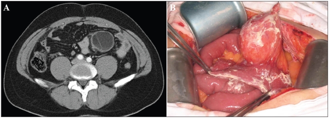
A 31-year-old man presenting with sudden onset of severe abdominal pain in the periumbilical area and elevated temperature (39.1ºC) was referred to our hospital. His medical and family history were unremarkable. He had no history of abdominal surgery. On physical examination, severe tenderness with guarding was evident in the whole abdomen. We palpated a 10-cm hard mass with marked tenderness in the left lower quadrant. Auscultation of the abdomen revealed no bowel sounds. Laboratory data showed leukocytosis of 14600/mm3 and an elevated C-reactive protein value of 82.86 nmol/L. A computed tomography (CT) scan of his abdomen revealed a low-density homogeneous tumour that was oval in shape and 8 × 6 × 6 cm in size in the left lower quadrant of the abdominal cavity; contrast medium revealed enhancement of the septum (Fig. 1). From these physical and radiographic findings, we diagnosed a cystic neoplasm in the mesentery, which was causing peritonitis.
Fig. 1.
(A) Computed tomography scan of the abdomen of a 31-year-old man showing a homogenous tumour, 8 × 6 × 6 cm in size. Contrast medium revealed enhancement of the septum. (B) At laparotomy, we found a yellowish cystic tumour located in the mesentery of the jejunum. We observed a small amount of milky fluid in the abdominal cavity.
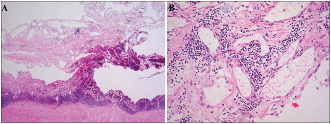
At laparotomy, we found a yellowish cystic tumour containing a milky fluid in the mesentery of the jejunum, about 90 cm distal to the Treiz ligament (Fig. 1). With careful dissection of the mesenteric arteries, we excised the tumour with a 20-cm length of the jejunum. Histological examination revealed that the tumour in the mesentery contained numerous dilated lymphatic spaces of varying sizes within collagenous stroma. These spaces were lined by flattened endothelial cells, and small collections of lymphocytes were scattered throughout the lesion (Fig. 2). The final diagnosis was cystic lymphangioma. The patient had an uneventful postoperative course, and there was no evidence of recurrence in the 9 months after surgery.
Fig. 2.
(A) Histological examination revealed that the tumour in the mesentery contained numerous dilated lymphatic spaces of varying sizes within collagenous stroma (hematoxylin and eosin stain, original magnification ×20). (B) Lymphatic spaces were lined by flattened endothelial cells, and small collections of lymphocytes were scattered throughout the lesion (hematoxylin and eosin stain, original magnification ×100).
Discussion
Lymphangiomas are uncommon benign tumours and occur mainly in children. As many as 90% of these tumours may manifest in children younger than 3 years, and the sex ratio is roughly equal in childhood.1,2 The most common sites are the head, neck and axillary lesion; intra-abdominal lymphangiomas are rare, accounting for only about 9% of all lymphangiomas.4
The etiology of lymphangiomas remains unclear. A well-established theory suggestes that lymphangiomas arise from sequestrations of lymphatic tissue during embryologic development.2 However, it is suggested that abdominal trauma, lymphatic obstruction, inflammatory process, surgery or radiation therapy may lead to the secondary formation of such a tumour.1
Histologically, lymphangiomas are classified into 3 types: simple capillary, cavernous and cystic lymphangiomas. The simple capillary lymphangioma is usually situated superficially in the skin and composed of small thin-walled lymphatics. The cavernous lymphangioma consists of larger lymphatics having a connection with normal adjacent lymphatics. The cystic lymphangioma consists of lymphatic spaces of various sizes that contain serous, chylous, bloody or purulent fluid, but has no connection with normal adjacent lymphatics.1,2
Patients with mesenteric lymphangiomas are usually asymptomatic until the tumours enlarge. Abdominal pain, palpable mass and distention seem to be the most common symptoms, but the clinical presentation varies. The mass is usually discovered only incidentally during examination or surgery for an unrelated illness; however, some patients may have acute clinical symptoms caused by compression of the adjacent structures or by complications such as infection, perforation, torsion and rupture.5
The appearance of a mesenteric lymphangioma on ultrasound is variable but it is most often described as a cystic mass with multiple thin septa. On CT scans, mesenteric lymphangiomas appear as uni- or multilocular masses containing septa of variable thickness; enhancement of the wall is revealed by contrast medium.1
The optimal treatment is radical excision, since incomplete resection may lead to recurrence.3–5 Although lymphangiomas are benign lesions, they often behave in an aggressively invasive manner and grow to an enormous size. Therefore, resection of adjacent organs may be required to accomplish complete excision. If radical surgery is not technically possible, injection of bleomycin or OK-432 into the tumour has been reported to be effective.4
Although mesenteric lymphangiomas are very rare, they can cause acute abdomen necessitating emergency surgery. Therefore, they should be considered in the differential diagnosis of acute abdomen in adults.
Footnotes
Competing interests: None declared.
References
- 1.Chen CW, Hsu SD, Lin CH, et al. Cystic lymphangioma of the jejunal mesentery in an adult: a case report. World J Gastroenterol. 2005;11:5084–6. doi: 10.3748/wjg.v11.i32.5084. [DOI] [PMC free article] [PubMed] [Google Scholar]
- 2.Rieker RJ, Quentmeier A, Weiss C, et al. Cystic lymphangioma of the small-bowel mesentery. Pathol Oncol Res. 2000;6:146–8. doi: 10.1007/BF03032366. [DOI] [PubMed] [Google Scholar]
- 3.Hanagiri T, Baba M, Shimabukuro T, et al. Lymphangioma in the small intestine: report of a case and review of the Japanese literature. Surg Today. 1992;22:363–7. doi: 10.1007/BF00308747. [DOI] [PubMed] [Google Scholar]
- 4.Tsukada H, Takaori K, Ishiguro S, et al. Giant cystic lymphangioma of the small bowel mesentery: report of a case. Surg Today. 2002;32:734–7. doi: 10.1007/s005950200138. [DOI] [PubMed] [Google Scholar]
- 5.Seki H, Ueda T, Kasuya T, et al. Lymphangioma of the jejunum and mesentery presenting with acute abdomen in an adult. J Gastroenterol. 1998;33:107–11. doi: 10.1007/s005350050053. [DOI] [PubMed] [Google Scholar]


