

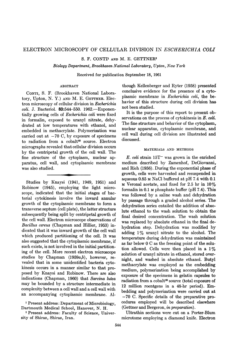
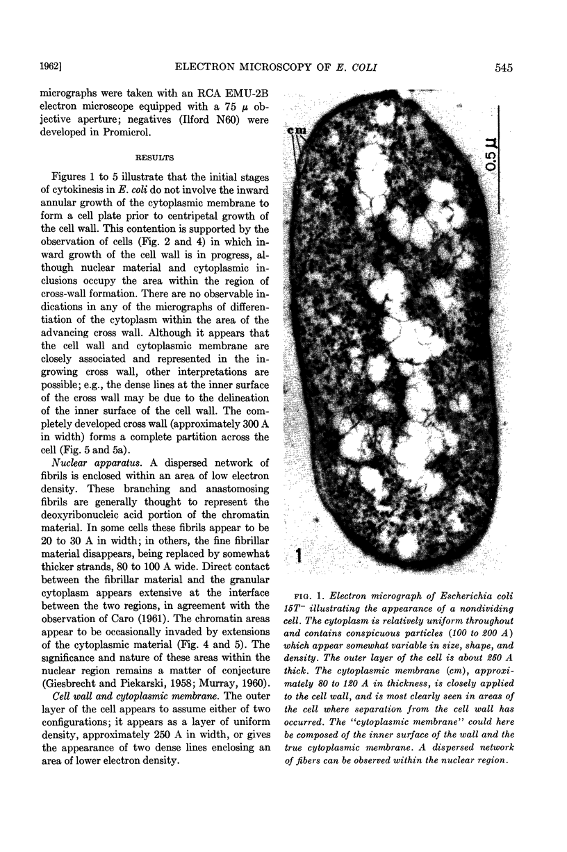
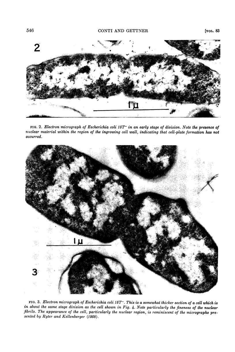
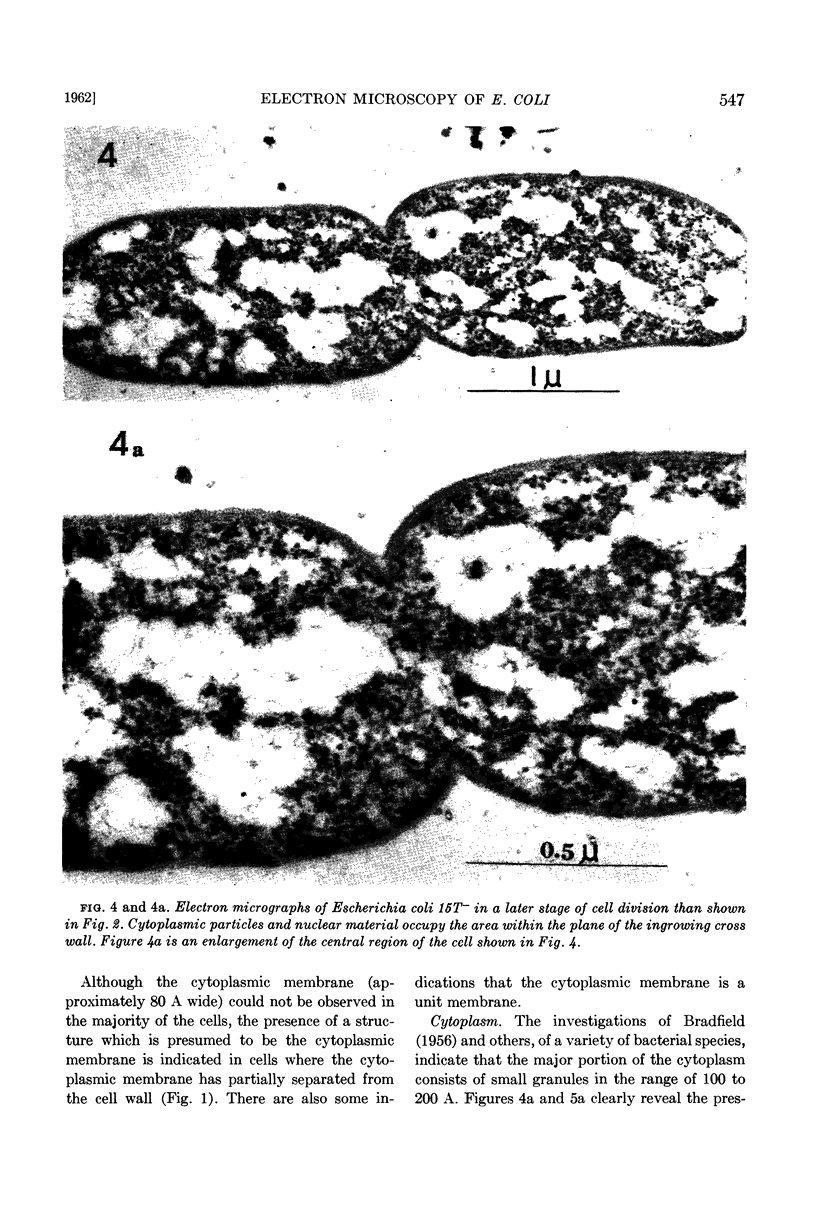
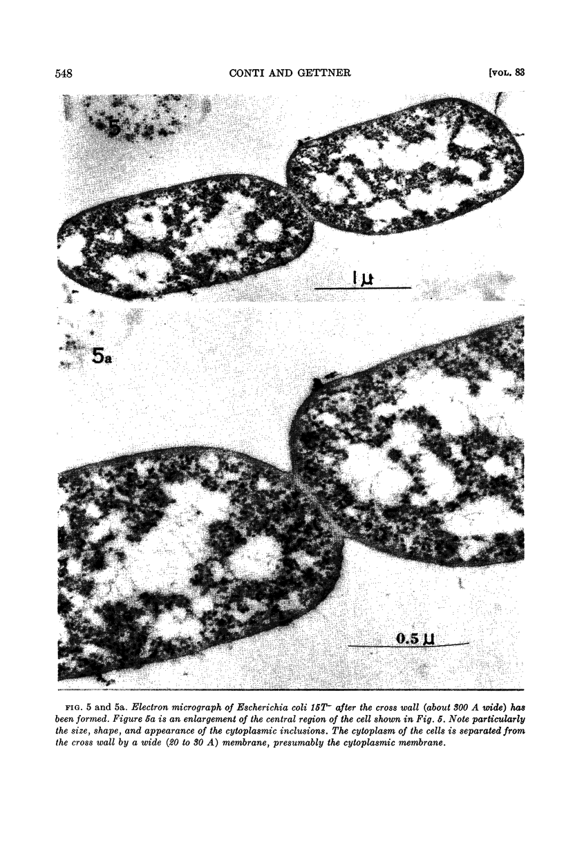
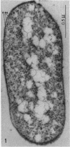
Abstract
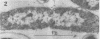
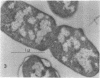
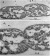
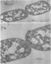
Conti, S. F. (Brookhaven National Laboratory, Upton, N. Y.) and M. E. Gettner. Electron microscopy of cellular division in Escherichia coli. J. Bacteriol. 83:544–550. 1962.—Exponentially growing cells of Escherichia coli were fixed in formalin, exposed to uranyl nitrate, dehydrated at low temperatures with ethanol, and embedded in methacrylate. Polymerization was carried out at −70 C, by exposure of specimens to radiation from a cobalt60 source. Electron micrographs revealed that cellular division occurs by the centripetal growth of the cell wall. The fine structure of the cytoplasm, nuclear apparatus, cell wall, and cytoplasmic membrane was also studied.
Full text
PDF
Images in this article
Selected References
These references are in PubMed. This may not be the complete list of references from this article.
- BEER M., HIGHTON P. J., McCARTHY B. J. Structure of ribosomes from Escherichia coli as revealed by their disintegration. J Mol Biol. 1960 Dec;2:447–449. doi: 10.1016/s0022-2836(60)80055-1. [DOI] [PubMed] [Google Scholar]
- CARO L. G. Localization of macromolecules in Escherichia coli. I. DNA and proteins. J Biophys Biochem Cytol. 1961 Mar;9:539–553. doi: 10.1083/jcb.9.3.539. [DOI] [PMC free article] [PubMed] [Google Scholar]
- CHAPMAN G. B. Electron microscope observations on the behavior of the bacterial cytoplasmic membrane during cellular division. J Biophys Biochem Cytol. 1959 Oct;6:221–224. doi: 10.1083/jcb.6.2.221. [DOI] [PMC free article] [PubMed] [Google Scholar]
- CHAPMAN G. B. Electron microscopy of cellular division in Sarcina lutea. J Bacteriol. 1960 Jan;79:132–136. doi: 10.1128/jb.79.1.132-136.1960. [DOI] [PMC free article] [PubMed] [Google Scholar]
- CHAPMAN G. B. Electron microscopy of ultrathin sections of bacteria. III. Cell wall, cytoplasmic membrane, and nuclear material. J Bacteriol. 1959 Jul;78(1):96–104. doi: 10.1128/jb.78.1.96-104.1959. [DOI] [PMC free article] [PubMed] [Google Scholar]
- CHAPMAN G. B., HILLIER J. Electron microscopy of ultra-thin sections of bacteria I. Cellular division in Bacillus cereus. J Bacteriol. 1953 Sep;66(3):362–373. doi: 10.1128/jb.66.3.362-373.1953. [DOI] [PMC free article] [PubMed] [Google Scholar]
- KELLENBERGER E., RYTER A. Cell wall and cytoplasmic membrane of Escherichia coli. J Biophys Biochem Cytol. 1958 May 25;4(3):323–326. doi: 10.1083/jcb.4.3.323. [DOI] [PMC free article] [PubMed] [Google Scholar]
- Knaysi G. Observations on the Cell Division of Some Yeasts and Bacteria. J Bacteriol. 1941 Feb;41(2):141–153. doi: 10.1128/jb.41.2.141-153.1941. [DOI] [PMC free article] [PubMed] [Google Scholar]
- RYTER A., KELLENBERGER E., BIRCHANDERSEN A., MAALOE O. Etude au microscope électronique de plasmas contenant de l'acide désoxyribonucliéique. I. Les nucléoides des bactéries en croissance active. Z Naturforsch B. 1958 Sep;13B(9):597–605. [PubMed] [Google Scholar]
- VAN ITERSON W., ROBINOW C. F. Observations with the electron microscope on the fine structure of the nuclei of two spherical bacteria. J Biophys Biochem Cytol. 1961 Jan;9:171–181. doi: 10.1083/jcb.9.1.171. [DOI] [PMC free article] [PubMed] [Google Scholar]
- ZAMENHOF S., DE GIOVANNI R., RICH K. Escherichia coli containing unnatural pyrimidines in its deoxyribonucleic acid. J Bacteriol. 1956 Jan;71(1):60–69. doi: 10.1128/jb.71.1.60-69.1956. [DOI] [PMC free article] [PubMed] [Google Scholar]