Abstract
Ca2+-independent pathways such as protein kinase C (PKC), extracellular-regulated kinases 1 and 2 (ERK1/2), and Rho kinase 1 and 2 (ROCK1/2) play important roles in modulating cerebral vascular tone. Because the roles of these kinases vary with maturational age, we tested the hypothesis that PKC differentially regulates the Ca2+-independent pathways and their effects on cerebral arterial contractility with development. We simultaneously examined the responses of arterial tension and intracellular Ca2+ concentration and used Western immunoblot analysis to measure ERK1/2, RhoA, 20 kDa regulatory myosin light chain (MLC20), PKC-potentiated inhibitory protein of 17 kDa (CPI-17), and caldesmon. Phorbol 12,13-dibutyrate (PDBu)-mediated PKC activation produced a robust contractile response, which was increased a further 20 to 30% by U-0126 (MEK inhibitor) in cerebral arteries of both age groups. Of interest, in the fetal cerebral arteries, PDBu leads to an increased phosphorylation of ERK2 compared with ERK1, whereas in adult arteries, we observed an increased phosphorylation of ERK1 compared with ERK2. Also, in the present study, RhoA/ROCK played a significant role in the PDBu-mediated contractility of fetal cerebral arteries, whereas in adult cerebral arteries, CPI-17 and caldesmon had a significantly greater role compared with the fetus. PDBu also led to an increased MLC20 phosphorylation, a response blunted by the inhibition of myosin light chain kinase only in the fetus. Overall, the present study demonstrates an important maturational shift from RhoA/ROCK-mediated to CPI-17/caldesmon-mediated PKC-induced contractile response in ovine cerebral arteries.
Keywords: protein kinase C-potentiated inhibitory protein of 17 kDa, development, extracellular signal-regulated kinase-1/2, RhoA, myosin light chain 20, caldesmon
in arterial smooth muscle, activated cell surface receptors are coupled to G proteins, which, in turn, may activate phospholipase C to promote the hydrolysis of phosphatidylinositol bisphosphate to diacylglycerol and inositol 1,4,5-trisphosphate. The latter can serve as an agonist for inositol 1,4,5-trisphosphate-dependent Ca2+ release from intracellular stores to augment intracellular Ca2+ concentration ([Ca2+]i; Ca2+-dependent pathway). Furthermore, diacylglycerol and Ca2+ activate protein kinase C (PKC), which can produce robust contractile responses not associated with an increase in [Ca2+]i (Ca2+-independent pathway). Both Ca2+-dependent and -independent pathways produce a contractile response by regulating thin-filament (actin) and thick-filament (myosin) interaction. Concerning myosin, the most studied is a 20 kDa regulatory myosin light chain (MLC20) filament pathway, the activity of which depends on the balance of activities of myosin light chain kinase (MLCK, which phosphorylates MLC20) and myosin light chain phosphatase (MLCP, which dephosphorylates MLC20) (18, 47). MLCP and MLCK activities are regulated by a complex signal transduction cascade modulated by PKC, mitogen-activated protein kinase (MAPK), Rho kinase (ROCK) pathways, and so forth.
PKC represents a family of a dozen serine/threonine kinases involved in numerous cellular signaling pathways, including cell growth and differentiation, gene expression, and apoptosis. In a previous study (36), we have reported that PKC activation induces robust contractile responses in ovine cerebral arteries, which are not associated with an increase in [Ca2+]i, and these responses differ significantly as a function of maturation. Other reports also have demonstrated PKC-mediated Ca2+-independent vascular contractions (17, 30, 55, 57), and PKC has been shown to modulate myogenic tone (13). Several studies from our and other laboratories have also examined various Ca2+-sensitization pathways downstream from PKC, such as mitogen-activated kinases and ROCK-dependent pathways (20, 21, 36, 40, 55, 58), which may modulate arterial contractility.
During the past decade, accumulating evidence from our and other laboratories has suggested that components of the MAPK cascade may be involved in vascular contraction via an altered sensitivity of the contractile machinery (7, 12, 15, 23, 56, 58). Key components of this MAPK cascade are the extracellular signal-regulated kinases (ERK1 and ERK2; ERK1/2) subfamily, the activation of which is dependent on dual phosphorylation of both a tyrosine (Try185) and a threonine (Thr187) residue (2, 3). Several studies have demonstrated that ERK1/2 phosphorylation and/or its upstream MAPK kinase (MEK) play a role in the contraction of numerous vessels, including rabbit femoral artery (43), sheep uterine artery (56), bovine carotid artery (8), pig carotid artery (1, 22), ferret aorta (7, 23), rat aorta, mesenteric, and tail arteries (51), as well as rat mesenteric artery smooth muscle cells (49). In adult ovine cerebral arteries, we have reported that PKC activation by phorbol 12,13-dibutyrate (PDBu) increased phosphorylated (p)-ERK1/2 levels, a response blocked by 1,4-diamino-2,3-dicyano-1,4-bis[2-aminophenylthio]butadiene (U-0126). In turn, U-0126 significantly augmented PDBu-induced contractile force (60). However, the role of MEK/ERK pathway and/or its interaction with PKC are not known with maturation and were examined in the present study.
When PKC is activated, along with the ERK1/2 pathway, RhoA, a monomeric G protein, binds to GTP and becomes activated. Active RhoA then modulates the Ca2+ sensitization through its effectors serine/threonine kinase, commonly referred to as ROCK. ROCK modulates the sensitivity of vascular smooth muscle contraction to [Ca2+]i by inhibiting MLCP activity, subsequently enhancing MLC20 phosphorylation (6, 28, 47, 48, 53).
Another important PKC-activated pathway is the PKC-potentiated inhibitory protein of 17 kDa (CPI-17), which upon phosphorylation of Thr38 is converted into a potent MLCP inhibitor (9, 10), leading to arterial smooth muscle contraction (27). CPI-17 expression is restricted in smooth muscle tissue and is present in several species (9, 53). Moreover, CPI-17 expression pattern in smooth muscle tissues correlates with the extent of PKC-induced contraction, implying that CPI-17 is critical to PKC-mediated Ca2+ sensitization (53). In a previous report, we observed that in adult ovine cerebral arteries, PKC activation leads to an increased CPI-17 phosphorylation, resulting in a decreased MLCP activity and an increased Ca2+ sensitivity (60). However, the role of CPI-17 phosphorylation with vascular smooth muscle cell maturation is not known. We also examined caldesmon, which is exclusively present in fully differentiated adult smooth muscle (16) and is known to interact with PKC (50).
Many aspects of these signaling pathways are unclear, and although there is evidence of cross talk among the pathways, much remains to be known regarding their interactions. Of importance, cerebral arteries also show significant differences in agonist-induced contraction with the development from fetal to adult life (32–36); however, little information is available regarding the effect of maturation on these pathways. Thus, in the present study, in fetal and adult sheep, we tested the hypothesis that PKC differentially regulates the Ca2+-independent pathways and their effects on cerebral arterial contractility with maturation.
METHODS
Experimental animals and tissues.
All experimental procedures were performed within the regulations of the Animal Welfare Act, the National Institutes of Health's Guide for the Care and Use of Laboratory Animals, and “The Guiding Principles in the Care and Use of Animals,” approved by the Council of the American Physiological Society, and were approved by the Animal Care and Use Committee of Loma Linda University. For these studies, we used cerebral arteries from near-term fetal (∼140 days) and nonpregnant adult sheep (≤2 yr) obtained from Nebeker Ranch (Lancaster, CA). The pregnant ewes were anesthetized with thiopental sodium (10 mg/kg iv), and anesthesia was maintained with an inhalation of 1% isoflurane in oxygen throughout surgery. After the fetus was delivered by hysterotomy, the fetuses and ewes were euthanized with an overdose of the proprietary euthanasia solution, Euthasol [pentobarbital sodium (100 mg/kg) and phenytoin sodium (10 mg/kg); Virbac, Fort Worth, TX]. Brains were removed from the nonpregnant adult and fetal sheep, following which we obtained the cerebral arteries for further analysis. Studies were performed in isolated vessels cleaned of adipose and connective tissue, and to avoid the complications of endothelial-mediated effects, we removed the endothelium by carefully inserting a small wire three times, as previously described (33, 35). The vessels were used immediately for the experiments.
Simultaneous measurement of [Ca2+]i and tension.
We have described this technique in several reports (31–33, 36). We cut the fetal or adult middle cerebral arteries into rings of 2 mm in length and incubated them at 25°C for 40 min with the acetoxymethyl ester of fura-2 (fura-2 AM; Molecular Probes, Eugene, OR), a fluorescent Ca2+ indicator (31–33). After loading the dye, we mounted the arterial segments on two tungsten wires (0.13 mm diameter; A-M Systems, Carlsborg, WA), attaching one wire to an isometric force transducer (Kent Scientific, Litchfield, CT) and the other to a post attached to a micrometer used to vary the resting tension in a 5-ml tissue bath mounted on a Jasco CAF-110 Intracellular Ca2+ analyzer (Jasco, Easton, MD). We then stabilized cerebral artery rings at 38°C for 40 min in an oxygenated standard Krebs solution containing (in mM) 122 NaCl, 25.6 NaHCO3, 5.56 dextrose, 5.17 KCl, 2.49 MgSO4, 1.60 CaCl2, 0.114 ascorbic acid, and 0.027 disodium EDTA. The bath chambers were continuously bubbled with 95% O2-5% CO2, and all the experiments were conducted at 38°C (core temperature of sheep). After stabilization, based on our previous studies, the optimum resting tension was 0.6 g for fetal and 0.7 g for adult cerebral artery, since at these tensions the contractility response to 125 mM KCl was maximum (35, 40). We then stimulated the isolated cerebral arterial rings from fetal and adult sheep with 125 mM KCl, and after the contractile force plateaued, 100 μM acetylcholine was applied. This procedure is commonly performed and allows for the evaluation of the relative quantity of contractile smooth muscle (37) and to determine the status of endothelium disruption (14). The contractile force due to 125 mM KCl was measured as gram tension (Fig. 1), before stimulation of the test compound for each arterial segment, and was used to normalize the arterial contraction with other agonist and antagonist for variation in the smooth muscle mass (33). The arteries that relaxed in the presence of acetylcholine were discarded from the study. Before treatment with PDBu (PKC agonist), with or without ERK antagonists (U-0126) or ROCK antagonist (Y-27632), we measured the KCl-induced contraction (Kmax) and increase in [Ca2+]i as a positive control. During all contractility experiments, we continuously digitized, normalized, and recorded contractile tensions and the fluorescence ratio of 340 to 380 nm using an online computer. In fetal and adult cerebral arteries, used to study PKC-ERK1/2-mediated contraction, we administered PDBu (3 × 10⁻⁶ M) to achieve near maximal response, a dose chosen based on our previous dose-response studies (36). A second set of arteries were incubated in U-0126, a selective inhibitor of mitogen-activated kinases (MEK1 and MEK2) (11) at a concentration of 2 × 10⁻⁵ M (60) (Promega, Madison, WI) for 30 min and then treated with PDBu (3 × 10⁻⁶ M). Similarly to ERK, to study the involvement of PKC-ROCK1/2-mediated contraction in vessels of both age groups, we measured the PDBu-induced contraction and intracellular Ca2+ change in the presence or absence of the Y-27632 (3 × 10⁻⁷ M) (19, 39).
Fig. 1.
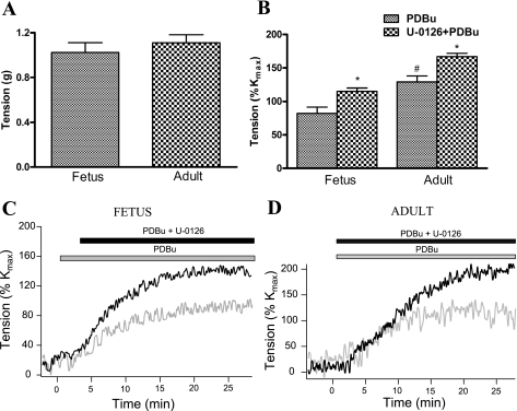
A: KCl-induced contractile responses in the ovine fetal and adult cerebral arteries. B: means ± SE of phorbol 12,13-dibutyrate (PDBu; 3 × 10⁻⁶ M)-induced contraction [as percent KCl-induced contraction (%Kmax)] in the presence and absence of U-0126 (10−5 M) in the fetal and adult cerebral arteries. PDBu-induced contractile response (gray bar) and effect of MEK inhibition by U-0126 (10−5 M) (black bar) in the fetal (C) and adult (D) cerebral arteries are shown. *P < 0.05, statistically significant differences between PDBu and PDBu + U-0126-mediated contractile response. #P < 0.05, statistically significant difference in PDBu-mediated contractile response between fetal and adult cerebral arteries.
Western immunoblot analysis.
Fetal and adult sheep cerebral arteries were isolated, cleaned in Na+-HEPES buffer (pH 7.5), and frozen rapidly in liquid nitrogen. Frozen samples were homogenized in the 1× cell lysing buffer (Cell Signaling Technology, Beverly, MA) containing 1× phosphatase and protease inhibitors cocktail (Sigma) and 1 mM phenylmethanesulfonyl fluoride. Nuclei and debris were pelleted by centrifugation at 1,000 g for 10 min. The supernatant was collected and stored at −80°C. SDS gel and Western blot analysis were performed by using p-ERK1/2 and total ERK1/2 antibody (Cell Signaling Technology) (58). We used α-actin as an internal control for uniform protein loading, as we have reported (59). For MEK1/2, CPI-17, and caldesmon, the methods for immunoblots were similar to those for ERK1/2, with appropriate antibodies. For MLC20 immunoblots, tissues were frozen in a freezing buffer [containing 5% trichloracetic acid, 10 mM dithiothreitol (DTT), 5 mM sodium fluoride (NaF), and 95% acetone] on dry ice. The tissues were then brought to room temperature in washing buffer (containing 10 mM DTT, 100% acetone, and 5 mM NaF) and washed three times. Proteins were extracted (0.04 g wet wt/ml) in extraction buffer containing 8.0 M urea, 20 mM Tris base, 23 mM glycine, 10 mM DTT, 10 mM EGTA, 10% glycerol, 0.05% bromphenol blue, and 5 mM NaF (pH 8.6) at room temperature for 2 h. Protein (6 μg) from each sample was loaded on a SDS gel and electrophoresed at 100 V for 3 h. The proteins were transferred to a nitrocellulose membrane and subjected to immunoblotting with phosphospecific MLC20 antibody (Ser19, 1:1,000; Cell Signaling Technology). The same blots were stripped and blotted for total MLC20 (1:300, Sigma). The bands were detected with enhanced chemiluminescence using a ChemiImager (Alpha-Innotech, San Leandro, CA). MLC20 phosphorylation was calculated by dividing the integrated density values of the phosphorylated band with the total MLC20 band and then normalized to control. The results are expressed as a fraction of control.
Rho-GTP activity assay.
We quantified RhoA activity by using Rhotekin-Rho binding domains beads pull-down assays kit (Cytoskeleton, Denver, CO) (5). Active Rho (Rho-GTP) binds with the Rho binding domain (RBD) fused to glutathione S-transferase bound to glutathione-coupled agarose beads, whereas inactive Rho (Rho-GDP) does not bind to RBD beads. We lysed treated cerebral arteries in lysis buffer containing 50 mM Tris·HCl (pH 7.5), 0.1 mM EDTA, 50 mM NaCl, 30 mM MgCl2, 1 mM phenylmethylsulfonyl fluoride, 1× protease inhibitor cocktail, and 1× phosphatase inhibitor cocktail (Sigma). The lysates were cleared of cellular debris by centrifugation at 3,000 g for 5 min at 4°C. The protein content in supernatants was measured using Bio-Rad Protein Assay Reagent (Bio-Rad, Hercules, CA). Cellular extract (30 μg) was used to assess total RhoA, and the remaining extract was incubated for 4 h at 4°C with Rhotekin-RBD beads. The beads were pelleted at 4°C, washed four times with cell lysis buffer, and resuspended in 20 μl of 2× Laemmli buffer (Boston Bioproducts, Worcester, MA). The samples were then subjected to 12% SDS-PAGE and analyzed using RhoA antibody by Western immunoblot assay.
Statistics.
We analyzed the data using unpaired, two-tailed Student's t-test and one-way ANOVA with Newman-Keuls post hoc test to determine significant differences between groups by the use of GraphPad Prism (GraphPad Software, San Diego, CA). The hypothesis was accepted at P < 0.05. For each study, n equaled 4 animals from which we obtained cerebral arteries.
RESULTS
PKC interacts differentially with MEK/ERK pathway in fetal and adult sheep cerebral arteries.
Figure 1A depicts 125 mM KCl-induced contractility response in the arteries isolated from fetus and adult sheep. We observed no significant difference in the contractile response to KCl. To understand better PKC-MEK/ERK interactions in agonist-induced contraction and the role of [Ca2+]i in fetal and adult cerebral arteries, we examined PDBu-induced (3 × 10⁻⁶ M) tension and [Ca2+]i in the absence or presence of the MEK inhibitor U-0126 (10−5 M). As seen in Fig. 1B, in the adult cerebral arteries, PDBu-induced tension was significantly higher than that achieved in fetal cerebral arteries, being ∼130% Kmax and ∼100% of Kmax, respectively. As seen in Fig. 1, C and D, in the presence of MEK inhibition, PDBu-induced tension was 20 to 30% higher in both adult and fetal cerebral arteries, being ∼166% Kmax and ∼120% of Kmax, respectively (P < 0.05 for each age group). Importantly, there was no change in [Ca2+]i in response to PDBu with or without U-0126 (data not shown).
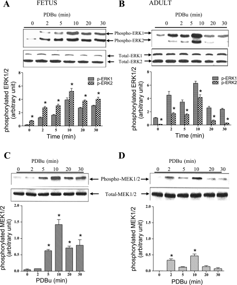
To explore further the role of PKC in modulating ERK-induced responses in cerebral arteries, we examined the time course of p-MEK and p-ERK1/2 levels following the addition of PDBu (3 × 10⁻⁶ M). As seen in Fig. 2A in fetal vessels, PDBu resulted in a significant increase in p-ERK1 (p44) and p-ERK2 (p42) by 2 and 5 min, with a further four- to fivefold increase above control, which peaked at ∼10 min (ERK phosphorylation correlated well with PDBu-induced contractile response). Total ERK1/2 remained constant. In adult cerebral arteries, Fig. 2B illustrates the increase in p-ERK1 and p-ERK2 levels at 2 min, which remained elevated at 10 min, and total ERK1/2 remained constant during this time. Of interest, in the fetus, ERK2 phosphorylation was significantly higher than ERK1, whereas in the adult vessels, the amount of p-ERK2 greatly exceeded the amount of p-ERK1 (P < 0.05). Figure 2C shows that in fetal cerebral arteries, PDBu induced an increase in p-MEK1/2 at 5 min, which increased further by 10 min and remained elevated. In adult cerebral arteries in contrast (Fig. 2D), PDBu stimulation was associated with small increases in p-MEK1/2 between 2 and 10 min, decreasing thereafter. Figure 2, C and D, shows the densitometric analysis as above (n = 4 for each age group).
Fig. 2.
Time course of PDBu (3 × 10⁻⁶ M)-induced activation of ERK1/2 and MEK1/2 in fetal and adult cerebral arteries. Western immunoblots of phosphorylated (p)-ERK1 (p44) and ERK2 (p42) in fetal (A, top) and adult (B, top) cerebral arteries at 2, 5, 10, 15, 20, and 30 min. Levels of total (t)-ERK1/2 also are shown. A and B, bottom: densitometry analysis of p-ERK1 and -2, expressed as means ± SE. Western immunoblot of PDBu-mediated increase in p-MEK1/2 in fetal (C, top) and adult (D, top) cerebral arteries, with t-MEK levels for each respective lane (fraction of control at time 0 = 1.0). C and D, bottom: densitometry analysis of p-MEK, expressed as means ± SE. *P < 0.05 compared with the control.
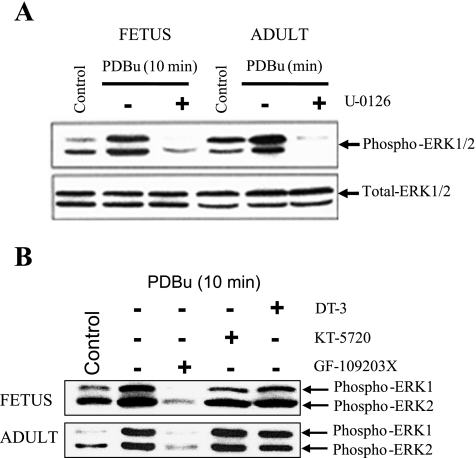
To investigate further the extent to which PDBu-mediated phosphorylation of ERK1/2 is through the activation of upstream MEK, we examined these responses in the absence or presence of the MEK inhibitor U-0126. Figure 3A shows Western immunoblots of p-ERK1/2 levels in fetal and adult ovine cerebral arteries in response to PDBu (3 × 10⁻⁶ M) in the absence or presence of U-0126 (10−5 M). As evident, in arteries of both age groups, U-0126 eliminated the PDBu-induced increases in both p-ERK1 and -2, whereas the total protein levels were unaffected. To rule out the possibility that the increase in the p-ERK1/2 levels occurred as a consequence of activation of other upstream kinases such as PKG and PKA, we conducted experiments in the presence and absence of the PKG inhibitor DT-3 (2.5 × 10⁻⁷ M), PKA inhibitor KT-5720 (10−6 M), and PKC inhibitor GF-109203X (5 × 10−6 M; n = 4). Figure 3B shows immunoblots of activated ERK1 (p44) and ERK2 (p42) after the addition of the PKG inhibitor DT-3 (2.5 × 10⁻⁷ M), the PKA inhibitor KT-5720 (10−6 M), and the PKC inhibitor GF-109203X (5 × 10−6 M) in the presence of PDBu. The results clearly demonstrate that PKC inhibition markedly attenuated p-ERK1 and -2 levels; however, PKA and PKG inhibition did not alter these (n = 4 each).
Fig. 3.
A: Western immunoblots of p-ERK1/2 levels at 10 min in fetal and adult cerebral arteries in response to PDBu (3 × 10⁻⁶ M) in the absence or presence of the MEK inhibitor U-0126 (10−5 M). B: Western immunoblot of PDBu-mediated increase (at 10 min) in p-ERK1 (p44) and p-ERK2 (p42) in fetal and adult cerebral arteries in the presence of the PKG inhibitor DT-3 (2.5 × 10⁻⁷ M), the PKA inhibitor KT-5720 (10−6 M), and the PKC inhibitor GF-109203X (5 × 10−6 M).
PKC interacts differentially with ROCK pathway in fetal and adult cerebral arteries.
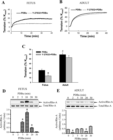
To examine the effect of PKC stimulation on the ROCK pathway, we measured PDBu-induced contractions in the presence and absence of the ROCK inhibitor (Y-27632, 3 × 10⁻⁷ M). As seen in Fig. 4A in fetal cerebral arteries in the presence of Y-27632, the PDBu (3 × 10⁻⁶ M)-induced maximum tension was reduced by ∼50%. However, in adult cerebral arteries, ROCK inhibition by Y-27632 was without an effect on PDBu-induced tension (Fig. 4B). In neither fetal nor adult cerebral arteries did [Ca2+]i change significantly in response to Y-27632 (3 × 10⁻⁷ M) or PDBu stimulation (data not shown). Figure 4C summarizes the responses for fetal and adult cerebral artery tensions with or without Y-27632 (n = 5 each). To confirm the observation that in fetal cerebral arteries RhoA/ROCKs are key effectors of PDBu-induced activation, we measured active RhoA (RhoA-GTP) in response to PDBu stimulation. Representative immunoblots of active RhoA with response to PDBu (3 × 10⁻⁶ M) are shown for both fetal (Fig. 4D) and adult (Fig. 4E) cerebral arteries. Strikingly, in the fetal arteries, PDBu increased phosphorylated RhoA significantly at 5 and 10 min; however, in adult vessels, active RhoA failed to increase. In both fetal and adult cerebral arteries, total RhoA levels appear to be of similar magnitude (n = 4 each).
Fig. 4.
PDBu-induced tensions and the role of Rho kinase (ROCK) in the fetal and adult cerebral arteries. Traces showing PDBu (3 × 10⁻⁶ M)-induced contraction in fetal (A) and adult (B) cerebral arteries in the absence (black line) and presence (gray line) of the ROCK inhibitor Y-27632 (3 × 10⁻⁷ M). C: means ± SE of PDBu (3 × 10⁻⁶ M)-induced contraction (as %Kmax) in the presence and absence of Y-27632 (10−5 M) in fetal and adult cerebral arteries. Western immunoblots of activated RhoA response to PDBu (3 × 10⁻⁶ M) in fetal (D, top) and adult (E, top) cerebral arteries are shown. D and E, bottom: densitometry analysis of active RhoA expressed as means ± SE. *P < 0.05, statistically significant differences between PDBu and PDBu + Y-27632-mediated contractile response. #P < 0.05, statistically significant difference in PDBu-mediated contractile response between fetal and adult cerebral arteries.
PKC interacts differentially with MLC20 in fetal and adult cerebral arteries.
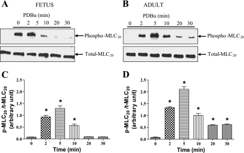
To explore further the mechanisms by which PKC induces Ca2+-independent cerebral arterial contraction via thick filament (myosin) regulation, we measured the extent to which PDBu (3 × 10⁻⁶ M) increased MLC20 phosphorylation. As seen in Fig. 5A, in the fetal cerebral arteries, Western immunoblot density of p-MLC20 levels increased severalfold at 2 to 5 min and thereafter declined to near-control values. Similarly, in the adult cerebral arteries (Fig. 5B), PDBu increased the phosphorylation of MLC20 between 2 and 5 min, declining thereafter. Figure 5, C and D, presents histograms of the time course in both fetal and adult cerebral arteries, respectively, of p-MLC20 normalized to total MLC20 for each respective lane (n = 4 each).
Fig. 5.
Time course of PDBu-induced activation of myosin light chain 20 (MLC20) in fetal and adult cerebral arteries. Western immunoblots of p-MLC20 at 2, 5, 10, 20, and 30 min after stimulation with PDBu in fetal (A) and adult (B) are shown. Densitometry analysis of p-MLC20 in fetus (C) and adult (D) is shown, expressed as means ± SE. *P < 0.05 compared with control.
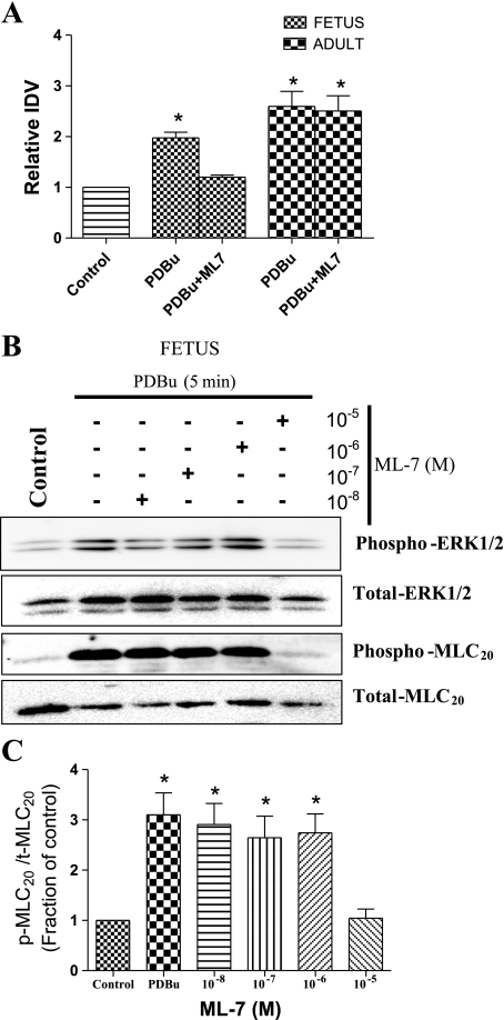
To examine the mechanisms of MLC20 activation as a consequence of PKC stimulation, we conducted the experiments in adult and fetal sheep cerebral arteries in the presence and absence of the MLCK inhibitor (ML-7). In fetal cerebral arteries, Fig. 6A depicts the increase in the p-MLC20 level in response to PDBu at 5 min, with a blunted response in the presence of ML-7 (10−5 M). In contrast, in adult cerebral arteries, ML-7 failed to inhibit MLC20 phosphorylation (Fig. 6A). Furthermore, in a ML-7 dose-dependent study in fetal arteries, we found no significant decrease in the active MLC20 at doses of 10−8 M to 10−6 M of ML-7, whereas a complete blockage of p-MLC20 occurred at 10−5 M (Fig. 6, B and C). We also used these membranes to estimate the p-ERK1/2 level in an effort to identify any specific/nonspecific upstream kinase inhibition by ML-7. Surprisingly, we observed a significant decrease in p-ERK level at 10−5 M ML-7 concentration (Fig. 6B) (n = 4 each).
Fig. 6.
Role of myosin light chain kinase (MLCK) in PDBu-mediated MLC20 activation in the fetal and adult cerebral arteries. A: densitometric analysis of MLC20 phosphorylation in response to 3 × 10⁻⁶M PDBu alone and 3 × 10⁻⁶M PDBu + 10−5 M ML-7 in the fetal and adult cerebral arteries as detected by Western immunoblot analysis. IDV, integrated density values. B and C: Western immunoblots (B) and densitometry analysis as means ± SE (C) of ML-7 concentration-response relations for p-MLC20 in presence of PDBu in fetal cerebral arteries incubated with combinations of various concentration (10−8 M to 10−5 M) of MLCK antagonist, i.e., ML-7 for 30 min before stimulation with PDBu (3 × 10⁻⁶ M). *P < 0.05.
PKC interacts differentially with CPI-17 and caldesmon in fetal and adult sheep cerebral arteries.
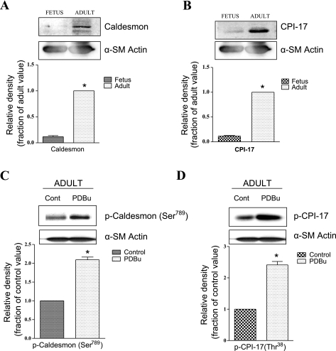
To explore further the downstream PKC activation pathway(s), we examined the effect of PDBu on two cytosolic proteins, CPI-17 and caldesmon, which are known to be involved in smooth muscle contraction (54, 60). As shown in Fig. 7, A and B, fetal cerebral arteries have very low levels of both caldesmon and total CPI-17 compared with adult arteries. Importantly, in adult cerebral arteries, PDBu increased the phosphorylation levels of both caldesmonser789 and CPIThr38 (Fig. 7, C and D) (n = 4 each). This increase did not occur in fetal vessels, however (data not shown).
Fig. 7.
Basal and PDBu-stimulated levels of t- and p-caldesmon and -PKC-potentiated inhibitory protein of 17 kDa (CPI-17) levels in adult and fetal cerebral arteries. A and B: Western immunoblots (top) and densitometry analysis (bottom) of caldesmon (A) and CPI-17 (B) in control fetal and adult cerebral arteries. α-SM, α-smooth muscle. C and D: Western immunoblot (top) and densitometric analysis (bottom) of p-caldesmon and p-CPI-17, respectively, in adult cerebral vessels. *P < 0.05.
DISCUSSION
In the present study of ovine cerebral arteries, we have demonstrated the following. First, in the adult, PKC activation produces significantly higher contractility compared with that of the fetus. Second, MEK inhibition significantly augmented the PKC-mediated contractile force in both fetal and adult vessels, suggesting a negative feedback by ERK1/2 on PKC activation. Third, in both fetus and adult, PKC stimulation resulted in an increased phosphorylation of both MEK and ERK1/2. In the fetal arteries, ERK2 was more phosphorylated than ERK1, whereas the reverse was the case in the adult. Fourth, in both age groups, MEK inhibition blocked the PKC-mediated phosphorylation of ERK1/2. Fifth, in the fetus but not in the adult, the RhoA/ROCK pathway plays a key role in PKC-mediated contractility. Sixth, also, in arteries of the fetus but not in the adult, MLCK inhibition by ML-7 inhibited MLC20 phosphorylation. Finally, in the adult but not in the fetus, CPI-17 and caldesmon phosphorylation were demonstrated to be important regulatory pathways.
In the developing fetus as in the adult, hypoxia is associated with a significant increase in cerebral blood flow, and this response is acquired early in life (90–103-day-old fetal lamb) (24). However, in previous in vivo studies, we have demonstrated that despite the acclimatization to high altitude-induced long-term hypoxia during fetal life, flow compensations in response to acute hypoxia are not adequate to sustain oxygen delivery for the cerebral metabolic rate (4, 26, 41). Consequently, the fetal brain is more vulnerable (as compared with adults) to cerebral hypoxia/ischemia. The present study demonstrated no difference in KCl responses, suggesting that the depolarization-mediated contractile apparatus is functionally similar to the adult. Thus it would appear that the differences in the Ca2+ sensitization arm (PKC, ERKs, and ROCK) may be responsible for the increased vulnerability of fetal cerebral circulation.
During the past two decades, we and others have demonstrated that the kinases, such as PKC (21, 36, 42, 60), ERKs (55, 58, 60), and ROCK (20), each plays important roles in modulating Ca2+-independent contraction. Nonetheless, the role of MEK/ERK1/2 and RhoA/ROCK pathways in modulating PKC-induced contraction in cerebral arteries with maturation remains poorly understood. In the present study, we have demonstrated several important aspects of these interactions. For instance, PDBu-mediated PKC activation produces significantly higher contraction in the adult cerebral arteries compared with fetal arteries (Fig. 1). This may be due to more proliferative than contractile smooth muscle phenotypes in fetal cerebral smooth muscle cells compared with those in the adult. Our data also indicate that in both age groups, the PDBu-mediated response commenced at 2 to 5 min and peaked at ∼10 min. This agrees with the similar time course of MLC20 phosphorylation as a result of PDBu-mediated stimulation (Fig. 5). Thus it is evident that the thick filament (myosin) plays an important role in PKC-mediated Ca2+-independent contractility.
To study the mechanism(s) of PKC-induced MLC20 phosphorylation, in a previous study in adult cerebral arteries we observed a significant increase in PDBu-mediated contraction in the presence of ERK inhibition (60). Similar results were observed in the sheep uterine artery (56). In the present study, in both fetal and adult cerebral arteries, ERK inhibition increased the PKC-mediated contractile force starting at 2 to 5 min, supporting the idea of the negative feedback of PKC by ERK, and this is consistent with our previous studies (56, 60). Also in a previous study, we demonstrated that ERK1/2 activity inhibition abolished any increase in [Ca2+]i, whereas only slightly decreasing the phenylephrine-induced tension (58). This response pattern (change in tension with no change in [Ca2+]i) was similar to that which we have demonstrated following the PKC stimulation by PDBu (36) and emphasizes the importance of the PKC Ca2+-independent pathway. We also demonstrated that PKC inhibition by staurosporine relieved the U-0126-mediated suppression of [Ca2+]i increase in response to norepinephrine (58). Together, these findings suggest that ERK1/2 negatively regulate PKC-mediated contractility and that PKC activation leads to the phosphorylation of ERK1/2, which limits the phosphorylation of MLC20 by the sensitization of MLCP or MLCK. In the present study, in adult vessels, however, MLCK inhibition failed to inhibit PDBu-induced MLC20 phosphorylation. This suggests that ERK1/2 activation decreases the [Ca2+]i sensitization through MLCP or through some other PKC-related mechanism, but not by MLCK. We speculate that, following ERK activation, the inhibition of PKC-induced tonic and sustained contraction occurs as a protective mechanism against an intense cerebral artery vasospasm. Consistent with this speculation, ERK has been shown to play an important role in pathological conditions such as cerebral vasospasm and brain ischemic injury (29).
In the present study, we also observed that in fetal cerebral artery, the response to PDBu-mediated PKC stimulation was associated with significantly higher activation of ERK2 compared with ERK1. In adult cerebral arteries, the reverse was the case. At present, with limited understanding of the role of specific ERK isoforms, a straightforward rationale for such a major change in the signaling pathway with maturation is unclear. Further study with the specific inhibition of ERK1 or ERK2 by small interfering RNA or specific inhibitory peptides is required.
In addition to the MEK/ERK1/2 pathway, the inhibitory signal for Ca2+ sensitization is communicated by RhoA to a Rho kinase that phosphorylates the myosin 110–130 regulatory subunit and inhibits the catalytic activity of MLCP, resulting in an increased MLC20 phosphorylation, contraction, and cell motility (48). In the present study, our data suggest that whereas in the fetus RhoA/ROCK activation plays an important role in cerebral arterial contractility, this is not the case for the adult. Also, in response to PDBu, in fetal cerebral arteries, active RhoA increased significantly at 5 and 10 min (Fig. 4D), but this was not manifest in the adult (Fig. 4E). Furthermore, our results demonstrate that ROCK inhibition with its specific inhibitor Y-27632 caused a decrease in PDBu-induced contractility in fetal, but not in adult, cerebral arteries (Fig. 4). In fetal arteries, the increased active RhoA levels commenced at 5 min (Fig. 4) at about the same time as PDBu-mediated contractility (Fig. 1B). We speculate that during fetal life, PKC activation, in turn, activates the RhoA/ROCK pathway to regulate further the cerebral circulation. Of note, in a manner similar to ERK, ROCK has been shown to play an important role in pathological conditions such as cerebral vasospasm and brain ischemic injury (45, 46). ROCK inhibition also has been suggested to play a neuroprotective effect on ischemic brain damage (44). The present findings reinforce the idea that whereas RhoA/ROCK plays a key role in the regulation of cerebral cerebrovascular tone in the fetus, such is not the case for the adult (Fig. 4). We speculate that PDBu-stimulated RhoA/ROCK in fetal, compared with adult, cerebral arteries could be an important therapeutic target in ischemic injury in preterm birth.
In both fetal and adult cerebral arteries, PDBu increased MLC20 phosphorylation (Figs. 5 and 6) without changes in [Ca2+]i. In adult cerebral arteries, PDBu-induced MLC20 phosphorylation was not blocked by the MLCK inhibitor ML-7 (10−5 M; Fig. 6A). This suggests that PKC phosphorylation modulates MLC20 activation through MLCP rather than MLCK. Surprisingly, in fetal cerebral arteries, ML-7 at a very high dose (10−5 M) inhibited PKC-induced MLC20 phosphorylation; however, we also noticed at this very high dose that ML-7 inhibited ERK1/2 phosphorylation (Fig. 6). It is possible that at this high dose, ML-7 also inhibits other pathways and kinases, which may be responsible for reduced MLC20 phosphorylation, rather than a direct inhibitory effect on MLCK. In the present study, PDBu demonstrated essentially no effect on CPI-17 phosphorylation in fetal cerebral arteries, whereas it significantly increased CPI-17 phosphorylation in the adult (Fig. 7B), even in the presence of U-0126 compound (data not shown). In adult arteries, CPI-17 appears to increase Ca2+ sensitivity by inhibiting MLCP, thus increasing MLC20 phosphorylation (47). In addition, in fetal but not in adult vessels, PDBu-induced contraction was decreased significantly by the ROCK inhibitor Y-27632 (Fig. 4). This suggests that in the adult cerebral arteries, PKC may act directly to phosphorylate CPI-17 in the absence of the activation of RhoA/ROCK.
As noted above, in addition to its influence on ERK activation (15), PKC has been demonstrated to phosphorylate the thin filament-associated protein caldesmon (25, 38, 52, 55). PKC-mediated phosphorylation of caldesmon also significantly decreases its ability to inhibit actin-myosin ATPase (50). Similar to CPI-17, in contrast to the adult in which PDBu increased caldesmonSer789 phosphorylation, in fetal cerebral arteries PDBu stimulation failed to demonstrate any such increase in caldesmonSer789 phosphorylation (Fig. 7C). Our results are consistent with a previous report that caldesmon is exclusively active in adults (16). Although the major site of ERK-dependent phosphorylation in caldesmon is at Ser789, PKC may phosphorylate other sites. In the present study, we have demonstrated that PKC activation by PDBu phosphorylates both ERK and caldesmonSer789. In a previous report we have shown that U-0126 inhibition of ERK phosphorylation blocked caldesmonSer789 phosphorylation and yet increased PDBu-induced contraction (60). This suggests that caldesmonSer789 phosphorylation was not directly involved in releasing the inhibitory effect on actin-myosin ATPase. In fact, the ERK-specific phosphorylation of caldesmonSer789 may inhibit other PKC-mediated phosphorylation sites on caldesmon, a finding agreeing with that in uterine arteries of pregnant sheep (56).
Perspective.
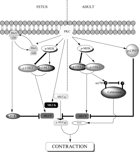
Figure 8 illustrates some key components of the PKC-mediated pathways in the two age groups. Overall, we accept the hypothesis that the signal transduction pathways differ significantly in fetal and adult cerebral arteries. A challenge for the future will be to clarify the role and importance of specific PKC, ERK1/2, and ROCK1/2 isoforms and their interactions. From a clinical perspective, the vulnerability of the immature cerebral vessels in the fetus/newborn with attendant dysregulation of cerebral blood flow may, in part, be a function of poor development of the Ca2+-dependent and Ca2+-sensitive arms of the agonist-induced signal transduction cascade. An important observation of the present study in fetal vessels was the greater activation of ERK2 and the involvement of RhoA/ROCK pathway, with the lack of participation by CPI-17 or caldesmon. From a therapeutic point of view, these specific pathways may be appropriate to correct the dysregulation of cerebral blood flow in the fetus and the premature infant and hopefully their profound neurological sequelae.
Fig. 8.
Proposed signal transduction pathways in fetal and adult cerebral arteries for PKC-induced contraction. In fetal cerebral arteries, PKC stimulation produces activation of MEK/ERK as well as RhoA/ROCK pathway and can also phosphorylate MLCK directly. In contrast, PKC stimulation in adult cerebral arteries activates MEK/ERK, CPI-17, and caldesmon, which further modulate contractile response. MLCP, myosin light chain phosphotase.
GRANTS
This work was supported by National Institutes of Health Grants HD/HL-03807 and HD-31226 (to L. D. Longo).
DISCLOSURES
No conflicts of interest are declared by the author (s).
ACKNOWLEDGMENTS
We thank Brenda Kreutzer for preparing the manuscript.
REFERENCES
- 1.Adam LP, Franklin MT, Raff GJ, Hathaway DR. Activation of mitogen-activated protein kinase in porcine carotid arteries. Circ Res 76: 183–190, 1995 [DOI] [PubMed] [Google Scholar]
- 2.Alessandrini A, Crews CM, Erikson RL. Phorbol ester stimulates a protein-tyrosine/threonine kinase that phosphorylates and activates the ERK-1 gene product. Proc Natl Acad Sci USA 89: 8200–8204, 1992 [DOI] [PMC free article] [PubMed] [Google Scholar]
- 3.Anderson NG, Maller JL, Tonks NK, Sturgill TW. Requirement for integration of signals from two distinct phosphorylation pathways for activation of MAP kinase. Nature 343: 651–653, 1990 [DOI] [PubMed] [Google Scholar]
- 4.Bishai JM, Blood AB, Hunter CJ, Longo LD, Power GG. Fetal lamb cerebral blood flow (CBF) and oxygen tensions during hypoxia: a comparison of laser Doppler and microsphere measurements of CBF. J Physiol 546: 869–878, 2003 [DOI] [PMC free article] [PubMed] [Google Scholar]
- 5.Crow T, Xue-Bian JJ, Dash PK, Tian LM. Rho/ROCK and Cdk5 effects on phosphorylation of a beta-thymosin repeat protein in hermissenda. Biochem Biophys Res Commun 323: 395–401, 2004 [DOI] [PubMed] [Google Scholar]
- 6.Deng JT, Sutherland C, Brautigan DL, Eto M, Walsh MP. Phosphorylation of the myosin phosphatase inhibitors, CPI-17 and PHI-1, by integrin-linked kinase. Biochem J 367: 517–524, 2002 [DOI] [PMC free article] [PubMed] [Google Scholar]
- 7.Dessy C, Kim I, Sougnez CL, Laporte R, Morgan KG. A role for MAP kinase in differentiated smooth muscle contraction evoked by α-adrenoceptor stimulation. Am J Physiol Cell Physiol 275: C1081–C1086, 1998 [DOI] [PubMed] [Google Scholar]
- 8.Epstein AM, Throckmorton D, Brophy CM. Mitogen-activated protein kinase activation: an alternate signaling pathway for sustained vascular smooth muscle contraction. J Vasc Surg 26: 327–332, 1997 [DOI] [PubMed] [Google Scholar]
- 9.Eto M, Ohmori T, Suzuki M, Furuya K, Morita F. A novel protein phosphatase-1 inhibitory protein potentiated by protein kinase C. Isolation from porcine aorta media and characterization. J Biochem 118: 1104–1107, 1995 [DOI] [PubMed] [Google Scholar]
- 10.Eto M, Senba S, Morita F, Yazawa M. Molecular cloning of a novel phosphorylation-dependent inhibitory protein of protein phosphatase-1 (CPI17) in smooth muscle: its specific localization in smooth muscle. FEBS Lett 410: 356–360, 1997 [DOI] [PubMed] [Google Scholar]
- 11.Favata MF, Horiuchi KY, Manos EJ, Daulerio AJ, Stradley DA, Feeser WS, Van Dyk DE, Pitts WJ, Earl RA, Hobbs F, Copeland RA, Magolda RL, Scherle PA, Trzaskos JM. Identification of a novel inhibitor of mitogen-activated protein kinase kinase. J Biol Chem 273: 18623–18632, 1998 [DOI] [PubMed] [Google Scholar]
- 12.Gerthoffer WT, Yamboliev IA, Shearer M, Pohl J, Haynes R, Dang S, Sato K, Sellers JR. Activation of MAP kinases and phosphorylation of caldesmon in canine colonic smooth muscle. J Physiol 495: 597–609, 1996 [DOI] [PMC free article] [PubMed] [Google Scholar]
- 13.Gokina NI, Osol G. Temperature and protein kinase C modulate myofilament Ca2+ sensitivity in pressurized rat cerebral arteries. Am J Physiol Heart Circ Physiol 274: H1920–H1927, 1998 [DOI] [PubMed] [Google Scholar]
- 14.Greenberg B, Kishiyama S. Endothelium-dependent and -independent responses to severe hypoxia in rat pulmonary artery. Am J Physiol Heart Circ Physiol 265: H1712–H1720, 1993 [DOI] [PubMed] [Google Scholar]
- 15.Horowitz A, Menice CB, Laporte R, Morgan KG. Mechanisms of smooth muscle contraction. Physiol Rev 76: 967–1003, 1996 [DOI] [PubMed] [Google Scholar]
- 16.Huber PA. Caldesmon. Int J Biochem Cell Biol 29: 1047–1051, 1997 [DOI] [PubMed] [Google Scholar]
- 17.Hunter T. Signaling—2000 and beyond. Cell 100: 113–127, 2000 [DOI] [PubMed] [Google Scholar]
- 18.Ihara E, MacDonald JA. The regulation of smooth muscle contractility by zipper-interacting protein kinase. Can J Physiol Pharmacol 85: 79–87, 2007 [DOI] [PubMed] [Google Scholar]
- 19.Ishizaki T, Uehata M, Tamechika I, Keel J, Nonomura K, Maekawa M, Narumiya S. Pharmacological properties of Y-27632, a specific inhibitor of rho-associated kinases. Mol Pharmacol 57: 976–983, 2000 [PubMed] [Google Scholar]
- 20.Janssen LJ, Tazzeo T, Zuo J, Pertens E, Keshavjee S. KCl evokes contraction of airway smooth muscle via activation of RhoA and Rho-kinase. Am J Physiol Lung Cell Mol Physiol 287: L852–L858, 2004 [DOI] [PubMed] [Google Scholar]
- 21.Jiang MJ, Morgan KG. Intracellular calcium levels in phorbol ester-induced contractions of vascular muscle. Am J Physiol Heart Circ Physiol 253: H1365–H1371, 1987 [DOI] [PubMed] [Google Scholar]
- 22.Katoch SS, Moreland RS. Agonist and membrane depolarization induced activation of MAP kinase in the swine carotid artery. Am J Physiol Heart Circ Physiol 269: H222–H229, 1995 [DOI] [PubMed] [Google Scholar]
- 23.Khalil RA, Morgan KG. PKC-mediated redistribution of mitogen-activated protein kinase during smooth muscle cell activation. Am J Physiol Cell Physiol 265: C406–C411, 1993 [DOI] [PubMed] [Google Scholar]
- 24.Kurth CD, Wagerle LC. Cerebrovascular reactivity to adenosine analogues in 0.6–0.7 gestation and near-term fetal sheep. Am J Physiol Heart Circ Physiol 262: H1338–H1342, 1992 [DOI] [PubMed] [Google Scholar]
- 25.Kyriakis JM, Avruch J. Mammalian mitogen-activated protein kinase signal transduction pathways activated by stress and inflammation. Physiol Rev 81: 807–869, 2001 [DOI] [PubMed] [Google Scholar]
- 26.Lee SJ, Hatran DP, Tomimatsu T, Pena JP, McAuley G, Longo LD. Fetal cerebral blood flow, electrocorticographic activity, and oxygenation: responses to acute hypoxia. J Physiol 587: 2033–2047, 2009 [DOI] [PMC free article] [PubMed] [Google Scholar]
- 27.Li L, Eto M, Lee MR, Morita F, Yazawa M, Kitazawa T. Possible involvement of the novel CPI-17 protein in protein kinase C signal transduction of rabbit arterial smooth muscle. J Physiol 508: 871–881, 1998 [DOI] [PMC free article] [PubMed] [Google Scholar]
- 28.Liao JK, Seto M, Noma K. Rho kinase (ROCK) inhibitors. J Cardiovasc Pharmacol 50: 17–24, 2007 [DOI] [PMC free article] [PubMed] [Google Scholar]
- 29.Lin CL, Dumont AS, Tsai YJ, Huang JH, Chang KP, Kwan AL, Hong YR, Howng SL. 17β-Estradiol activates adenosine A2a receptor after subarachnoid hemorrhage. J Surg Res . In press. [DOI] [PubMed] [Google Scholar]
- 30.Lohn M, Kampf D, Gui-Xuan C, Haller H, Luft FC, Gollasch M. Regulation of arterial tone by smooth muscle myosin type II. Am J Physiol Cell Physiol 283: C1383–C1389, 2002 [DOI] [PubMed] [Google Scholar]
- 31.Long W, Zhang L, Longo LD. Cerebral artery KATP- and KCa-channel activity and contractility: changes with development. Am J Physiol Regul Integr Comp Physiol 279: R2004–R2014, 2000 [DOI] [PubMed] [Google Scholar]
- 32.Long W, Zhang L, Longo LD. Cerebral artery sarcoplasmic reticulum Ca2+ stores and contractility: changes with development. Am J Physiol Regul Integr Comp Physiol 279: R860–R873, 2000 [DOI] [PubMed] [Google Scholar]
- 33.Long W, Zhao Y, Zhang L, Longo LD. Role of Ca2+ channels in NE-induced increase in [Ca2+]i and tension in fetal and adult cerebral arteries. Am J Physiol Regul Integr Comp Physiol 277: R286–R294, 1999 [DOI] [PubMed] [Google Scholar]
- 34.Longo LD, Ueno N, Zhao Y, Pearce WJ, Zhang L. Developmental changes in α1-adrenergic receptors, IP3 responses, and NE-induced contraction in cerebral arteries. Am J Physiol Heart Circ Physiol 271: H2313–H2319, 1996 [DOI] [PubMed] [Google Scholar]
- 35.Longo LD, Ueno N, Zhao Y, Zhang L, Pearce WJ. Ne-induced contraction, α1-adrenergic receptors, and Ins(1,4,5)P3 responses in cerebral arteries. Am J Physiol Heart Circ Physiol 270: H915–H923, 1996 [DOI] [PubMed] [Google Scholar]
- 36.Longo LD, Zhao Y, Long W, Miguel C, Windemuth RS, Cantwell AM, Nanyonga AT, Saito T, Zhang L. Dual role of PKC in modulating pharmacomechanical coupling in fetal and adult cerebral arteries. Am J Physiol Regul Integr Comp Physiol 279: R1419–R1429, 2000 [DOI] [PubMed] [Google Scholar]
- 37.McDonald TF, Pelzer S, Trautwein W, Pelzer DJ. Regulation and modulation of calcium channels in cardiac, skeletal, and smooth muscle cells. Physiol Rev 74: 365–507, 1994 [DOI] [PubMed] [Google Scholar]
- 38.Morgan KG, Gangopadhyay SS. Invited review: Cross-bridge regulation by thin filament-associated proteins. J Appl Physiol 91: 953–962, 2001 [DOI] [PubMed] [Google Scholar]
- 39.Narumiya S, Ishizaki T, Uehata M. Use and properties of ROCK-specific inhibitor Y-27632. Methods Enzymol 325: 273–284, 2000 [DOI] [PubMed] [Google Scholar]
- 40.Pearce WJ, Hull AD, Long DM, Longo LD. Developmental changes in ovine cerebral artery composition and reactivity. Am J Physiol Regul Integr Comp Physiol 261: R458–R465, 1991 [DOI] [PubMed] [Google Scholar]
- 41.Pena JP, Tomimatsu T, Hatran DP, McGill LL, Longo LD. Cerebral blood flow and oxygenation in ovine fetus: responses to superimposed hypoxia at both low and high altitude. J Physiol 578: 359–370, 2007 [DOI] [PMC free article] [PubMed] [Google Scholar]
- 42.Rasmussen H, Forder J, Kojima I, Scriabine A. TPA-induced contraction of isolated rabbit vascular smooth muscle. Biochem Biophys Res Commun 122: 776–784, 1984 [DOI] [PubMed] [Google Scholar]
- 43.Ratz PH. Regulation of ERK phosphorylation in differentiated arterial muscle of rabbits. Am J Physiol Heart Circ Physiol 281: H114–H123, 2001 [DOI] [PubMed] [Google Scholar]
- 44.Rikitake Y, Kim HH, Huang Z, Seto M, Yano K, Asano T, Moskowitz MA, Liao JK. Inhibition of Rho kinase (ROCK) leads to increased cerebral blood flow and stroke protection. Stroke 36: 2251–2257, 2005 [DOI] [PMC free article] [PubMed] [Google Scholar]
- 45.Sato M, Tani E, Fujikawa H, Kaibuchi K. Involvement of Rho-kinase-mediated phosphorylation of myosin light chain in enhancement of cerebral vasospasm. Circ Res 87: 195–200, 2000 [DOI] [PubMed] [Google Scholar]
- 46.Satoh S, Utsunomiya T, Tsurui K, Kobayashi T, Ikegaki I, Sasaki Y, Asano T. Pharmacological profile of hydroxy fasudil as a selective rho kinase inhibitor on ischemic brain damage. Life Sci 69: 1441–1453, 2001 [DOI] [PubMed] [Google Scholar]
- 47.Somlyo AP, Somlyo AV. Ca2+ sensitivity of smooth muscle and nonmuscle myosin II: modulated by G proteins, kinases, and myosin phosphatase. Physiol Rev 83: 1325–1358, 2003 [DOI] [PubMed] [Google Scholar]
- 48.Somlyo AP, Somlyo AV. Signal transduction by G-proteins, rho-kinase and protein phosphatase to smooth muscle and non-muscle myosin II. J Physiol 522: 177–185, 2000 [DOI] [PMC free article] [PubMed] [Google Scholar]
- 49.Touyz RM, El Mabrouk M, He G, Wu XH, Schiffrin EL. Mitogen-activated protein/extracellular signal-regulated kinase inhibition attenuates angiotensin II-mediated signaling and contraction in spontaneously hypertensive rat vascular smooth muscle cells. Circ Res 84: 505–515, 1999 [DOI] [PubMed] [Google Scholar]
- 50.Umekawa H, Hidaka H. Phosphorylation of caldesmon by protein kinase C. Biochem Biophys Res Commun 132: 56–62, 1985 [DOI] [PubMed] [Google Scholar]
- 51.Watts S. Serotonin activates the mitogen-activated protein kinase pathway in vascular smooth muscle: use of the mitogen-activated protein kinase kinase inhibitor PD098059. J Pharmacol Exp Ther 279: 1541–1550, 1996 [PubMed] [Google Scholar]
- 52.Winder SJ, Allen BG, Fraser ED, Kang HM, Kargacin GJ, Walsh MP. Calponin phosphorylation in vitro and in intact muscle. Biochem J 296: 827–836, 1993 [DOI] [PMC free article] [PubMed] [Google Scholar]
- 53.Woodsome TP, Eto M, Everett A, Brautigan DL, Kitazawa T. Expression of CPI-17 and myosin phosphatase correlates with Ca2+ sensitivity of protein kinase C-induced contraction in rabbit smooth muscle. J Physiol 535: 553–564, 2001 [DOI] [PMC free article] [PubMed] [Google Scholar]
- 54.Xiao D, Longo LD, Zhang L. α1-Adrenoceptor-mediated phosphorylation of MYPT-1 and CPI-17 in the uterine artery: role of ERK/PKC. Am J Physiol Heart Circ Physiol 288: H2828–H2835, 2005 [DOI] [PubMed] [Google Scholar]
- 55.Xiao D, Pearce WJ, Longo LD, Zhang L. ERK-mediated uterine artery contraction: role of thick and thin filament regulatory pathways. Am J Physiol Heart Circ Physiol 286: H1615–H1622, 2004 [DOI] [PubMed] [Google Scholar]
- 56.Xiao D, Zhang L. ERK MAP kinases regulate smooth muscle contraction in ovine uterine artery: effect of pregnancy. Am J Physiol Heart Circ Physiol 282: H292–H300, 2002 [DOI] [PubMed] [Google Scholar]
- 57.Yeon DS, Kim JS, Ahn DS, Kwon SC, Kang BS, Morgan KG, Lee YH. Role of protein kinase C- or RhoA-induced Ca2+ sensitization in stretch-induced myogenic tone. Cardiovasc Res 53: 431–438, 2002 [DOI] [PubMed] [Google Scholar]
- 58.Zhao Y, Long W, Zhang L, Longo LD. Extracellular signal-regulated kinases and contractile responses in ovine adult and fetal cerebral arteries. J Physiol 551: 691–703, 2003 [DOI] [PMC free article] [PubMed] [Google Scholar]
- 59.Zhao Y, Xiao H, Long W, Pearce WJ, Longo LD. Expression of several cytoskeletal proteins in ovine cerebral arteries: developmental and functional considerations. J Physiol 558: 623–632, 2004 [DOI] [PMC free article] [PubMed] [Google Scholar]
- 60.Zhao Y, Zhang L, Longo LD. PKC-induced ERK1/2 interactions and downstream effectors in ovine cerebral arteries. Am J Physiol Regul Integr Comp Physiol 289: R164–R171, 2005 [DOI] [PubMed] [Google Scholar]


