

Abstract
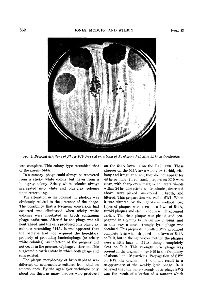
Jones, Lois M. (University of Wisconsin, Madison), C. R. McDuff, and J. B. Wilson. Phenotypic alterations in the colonial morphology of Brucella abortus due to a bacteriophage carrier state. J. Bacteriol. 83:860–866. 1962.—In the course of examining a number of Brucella cultures with a brucellaphage, it was observed that B. abortus cultures of intermediate colonial morphology, which had a blue-gray colonial appearance, were not lysed within 24 hr; in 48 hr they had developed sticky white growth in the area of the phage drop. When this growth was streaked on agar plates, both white and blue-gray colonies developed. White colonies which were sticky always carried phage and upon restreaking always gave rise to both white and blue-gray colonies. White colonies which were not sticky were rough and phage resistant. Blue-gray colonies produced only blue-gray colonies, did not carry phage, and were similar to the parent in their response to phage. When sticky white colonies were incubated for 6 hr or more in phage antiserum, all phage was eliminated and only blue-gray colonies developed. It was believed that the sticky white colonies were carrier clones in which lysis was delayed until after cell division, thus resulting in the establishment of a colony containing some phage-free progeny. With the accumulation of phage, the colony became sticky. This effect may be caused by the action of bacteriophage enzymes on the cell walls.
Brucellaphage had an extremely slow rate of adsorption on a culture of intermediate colonial morphology. A phage mutant which was more strongly lytic for cultures of intermediate colonial morphology was selected from the original phage. The adsorption rate of this phage was more rapid and the latent period shorter. A serological difference between phages could not be demonstrated.
Full text
PDF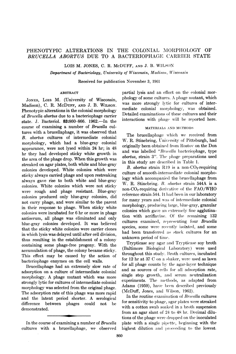


Images in this article

Selected References
These references are in PubMed. This may not be the complete list of references from this article.
- DROZHEVKINA M. S., KHARITONOVA T. I. Lizogeniia u brutsell. Vopr Virusol. 1958 Mar-Apr;3(2):93–97. [PubMed] [Google Scholar]
- DUBROVSKAIA I. I., OSTROVSKAIA N. N. [Changes in the process of preservation of the chemical composition of a variant of Brucella obtained under the influence of bacteriophage]. Biokhimiia. 1960 May-Jun;25:511–516. [PubMed] [Google Scholar]
- FRASER D. K. Host range mutants and semitemperate mutants of bacteriophage T3. Virology. 1957 Jun;3(3):527–553. doi: 10.1016/0042-6822(57)90008-9. [DOI] [PubMed] [Google Scholar]
- LI K., BARKSDALE L., GARMISE L. Phenotypic alterations associated with the bacteriophage carrier state of Shigella dysenteriae. J Gen Microbiol. 1961 Mar;24:355–367. doi: 10.1099/00221287-24-3-355. [DOI] [PubMed] [Google Scholar]
- McDuff C. R., Jones L. M., Wilson J. B. CHARACTERISTICS OF BRUCELLAPHAGE. J Bacteriol. 1962 Feb;83(2):324–329. doi: 10.1128/jb.83.2.324-329.1962. [DOI] [PMC free article] [PubMed] [Google Scholar]
- NELSON E. L., PICKETT M. J. The recovery of L forms of Brucella and their relation to Brucella phage. J Infect Dis. 1951 Nov-Dec;89(3):226–232. doi: 10.1093/infdis/89.3.226. [DOI] [PubMed] [Google Scholar]
- PARNAS J., FELTYNOWSKI A., BULIKOWSKI W. Anti-brucella phage. Nature. 1958 Dec 6;182(4649):1610–1611. doi: 10.1038/1821610a0. [DOI] [PubMed] [Google Scholar]
- WAHL R., BLUM-EMERIQUE L. Les bacteries semi-résistantes au bactériophage I. Techniques; souches et phage utilisés. lyse sur milieu gélosé. Ann Inst Pasteur (Paris) 1952 Jan;82(1):29–43. [PubMed] [Google Scholar]