Abstract
A microtiter alamarBlue assay was adapted and optimized for Mycobacterium avium subsp. paratuberculosis. Using cell concentrations ranging from 104 to 108 CFU/ml, a minimum incubation time to indicate viability was obtained after 24 h. Rifampin (rifampicin) was used to demonstrate that this method has applications for high-throughput screening against M. avium subsp. paratuberculosis.
Mycobacterium avium subsp. paratuberculosis is a chronic enteric pathogen which is widely distributed throughout the food chain (4, 16). Its association with Johne's disease in cattle is economically significant, with the United States alone suffering losses of $1.5 billion a year (6). Furthermore, its potential to cause human disease is disconcerting and controversial (7, 10, 11, 13). Therefore, the identification and development of novel anti-M. avium subsp. paratuberculosis agents are urgently required. In order to facilitate this, it is important to have available rapid anti-M. avium subsp. paratuberculosis assays permitting high-throughput analysis. A microtiter alamarBlue assay (currently untested for M. avium subsp. paratuberculosis) is a reliable means of determining cellular viability in bacteria (9). This rapid and inexpensive assay lends itself to a high-throughput screening format and has been shown to be applicable to some species of mycobacteria (3, 15). Furthermore, its correlation with other more expensive methods for determining mycobacterium viability is high, between 93 and 100% (1, 5, 15). These include the Mycobacteria Growth Indicator Tube, the Bactec radiometric method, and luciferase reporter systems. This study set out to establish and optimize a microtiter alamarBlue assay for a broad range of M. avium subsp. paratuberculosis titers and to evaluate applications for this assay, including high-throughput screening of novel anti-M. avium subsp. paratuberculosis compounds, and antibiotic resistance profiling of M. avium subsp. paratuberculosis.
M. avium subsp. paratuberculosis (CIT03) was isolated from the feces of an infected cow, as described by Ristow et al. (12), and cultivated on Herrold's egg yolk medium agar supplemented with mycobactin J (2 μl/ml), amphotericin B (50 μg/ml), vancomycin (50 μg/ml), and nalidixic acid (50 μg/ml) for 16 weeks at 37°C. The identity of M. avium subsp. paratuberculosis was confirmed using acid-fast staining, mycobactin dependency, and PCR analysis as previously described (2, 8, 14). To generate sufficient biomass, M. avium subsp. paratuberculosis was subsequently grown in Middlebrook 7H9 broth (MB broth), supplemented with oleic acid, albumin, dextrose, catalase (10%; Becton Dickinson), glycerol (0.2%), and mycobactin J (0.2%). This generally took 16 weeks.
Prior to the assay, 10 ml of the 16-week culture was centrifuged at 15,000 rpm for 20 min. The pellet was washed in fresh MB broth and resuspended in 10 ml of fresh supplemented MB broth containing 0.2% mycobactin J. The turbidity was adjusted to match McFarland standard no. 1 (3 × 10⁸ CFU/ml). From this suspension, a series of 1:5 dilutions ranging from 3 × 10⁸ to 9.6 × 10⁴ CFU/ml was set up in MB broth (5-ml volumes), using sterile Falcon tubes. The microtiter plate was organized into rows B, C, D, E, F, and G. Two-hundred-microliter aliquots of 3 × 10⁸ CFU/ml M. avium subsp. paratuberculosis were added to 10 wells in row B. Two-hundred-microliter aliquots of 6 × 10⁷ CFU/ml were added to row C, 1.2 × 10⁷ CFU/ml to row D, 2.4 × 10⁶ CFU/ml to row E, 4.8 × 10⁵ CFU/ml to row F, and 9.6 × 10⁴ CFU/ml to row G. This assay was allowed to progress over a period of 11 days. Twenty microliters (10% of the volume in the well) of a fresh alamarBlue reagent (AbD Serotec) was added, with mixing, to each column on each sampling day. Plates were covered and resealed with Parafilm and incubated at 37°C after the addition of the dye. Absorbance readings at 570 and 600 nm were then taken at 6 h, 24 h, and 48 h for each column. The assay was performed in triplicate, and percent reduction values of alamarBlue were determined using the appropriate formula (www.biokom.com.pl/files/alamarblue.pdf).
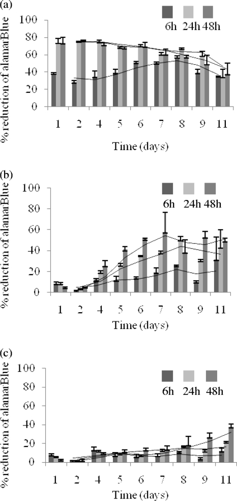
In terms of optimization, this assay examined the influence of cell numbers, the cellular incubation time, and the optimal incubation time with alamarBlue. All three had a significant impact on color development and percent reduction of the dye. For the highest concentration of cells (3 × 10⁸ CFU/ml) (Fig. 1a), a strong reduction of the dye was observed after 1 day of cellular incubation. Further incubation of the cells or incubation with the dye did not result in an appreciable increase in dye reduction values. Indeed, a decrease in the percent reduction was noted, most likely due to buffering agents reaching their maximum buffering efficiencies in the reagent mix (www.abdserotec.com/about/alamarblue). At the mid-range cellular levels (2.4 × 10⁶ CFU/ml) (Fig. 1b), detectable dye reduction occurred after 2 days. The reduction was substantial after day 4. At the lowest concentration of cells (9.6 × 10⁴ CFU/ml) (Fig. 1c), a noticeable change in dye reduction was observed after 9 days. While longer incubation with alamarBlue led to greater dye reduction with a given cell titer, as shown with each suspension at 6, 24, and 48 h, the 24-h reading was considered sufficient to give a clear indication of viability.
FIG. 1.
Optimization of alamarBlue conditions using 3 × 10⁸ CFU/ml (a), 2.4 × 10⁶ CFU/ml (b), and 9.6 × 10⁴ CFU/ml (c) over 11 days. Following the addition of alamarBlue, readings were taken at 6, 24, and 48 h.
Percent reduction of the dye was standardized to 10, 20, 40, and 60% for each concentration of cells (Table 1). These values serve as definitive indicators of metabolic activity and may be used for multiple applications, such as comparing the relative viabilities of strains, or the influence of media composition or environmental stress on M. avium subsp. paratuberculosis. In particular, we feel that this assay is suited to comparing the relative efficacies of multiple anti-M. avium subsp. paratuberculosis compounds and/or antibiotic resistance profiling in a high-throughput screening format. The success of this assay requires strict adherence to specific cell numbers, growth phases, and their equivalent incubation times.
TABLE 1.
Required time taken for M. avium subsp. paratuberculosis to reduce alamarBlue
| M. avium subsp. paratuberculosis cell concn (CFU/ml) | Time (h) required to reach indicated % reductiona |
|||||||
|---|---|---|---|---|---|---|---|---|
| 10 |
20 |
40 |
60 |
|||||
| Cellular | alamarBlue | Cellular | alamarBlue | Cellular | alamarBlue | Cellular | alamarBlue | |
| 3 × 10⁸ | 24 | 6 | 24 | 6 | 24 | 24 | 24 | 24 |
| 6 × 10⁷ | 24 | 6 | 24 | 24 | 24 | 48 | 96 | 24 |
| 1.2 × 10⁷ | 24 | 24 | 96 | 24 | 96 | 24 | 144 | 48 |
| 2.4 × 10⁶ | 96 | 6 | 96 | 48 | 120 | 48 | N/A | N/A |
| 4.8 × 10⁵ | 96 | 6 | 144 | 48 | 216 | 48 | 264 | 48 |
| 9.6 × 10⁴ | 96 | 6 | 216 | 48 | >264 | N/A | N/A | N/A |
N/A, not applicable.
Traditionally, detection of viability with the alamarBlue assay has been achieved on the basis of the pink-blue color change (1, 5, 9, 15). However, this method for M. avium subsp. paratuberculosis is also quantitative, as it determines the threshold percent reduction values required for visual color change of the medium (<12% blue, 12 to 18% purple, and >20% pink). Visually, all wells with calculated values below 12% after the addition of the dye were consistently blue, most probably due to insufficient metabolism needed to indicate viability. All wells which were purple were recorded as having percent reduction values of 12 to 18%. Wells with values over 20% were consistently pink, which is indicative of cellular metabolism. The time taken to indicate definitive viability varied significantly for each dilution of cells, ranging from 1 day plus 6 h with the dye (3 × 10⁸ CFU/ml) to 9 days plus 48 h with the dye (9.6 × 10⁴ CFU/ml), as seen in Table 1.
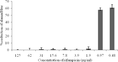
To demonstrate the application of the assay, it was used to assess the susceptibility of M. avium subsp. paratuberculosis to rifampin (rifampicin). Rifampin was prepared in dimethyl sulfoxide at double the maximum concentration required. The assay was carried out using 6 × 10⁷ CFU/ml over a period of 4 days, plus a 24-h incubation with alamarBlue in supplemented MB broth containing 0.2% mycobactin J. The final antibiotic concentration ranged from 125 to 0.48 μg/ml. The MIC90 of rifampin for M. avium subsp. paratuberculosis was determined as 1.92 μg/ml, which correlates well with other studies (17) (Fig. 2). This result highlights the assay's potential as a high-throughput screening platform for antibiotic resistance profiling and the identification of novel anti-M. avium subsp. paratuberculosis agents.
FIG. 2.
Determination of the MIC90 of rifampin against M. avium subsp. paratuberculosis
Given the association of M. avium subsp. paratuberculosis with both animal and human diseases, such rapid, quantitative, and inexpensive (approximately 10 euro cents per sample) viability assays will be very useful for rapid screening of anti-M. avium subsp. paratuberculosis compounds and antibiotic profiling.
Acknowledgments
This research was funded by a Strand I research grant from the Technological Strand Research Program awarded through the Institute of Technology Ireland initiative.
Footnotes
Published ahead of print on 16 October 2009.
REFERENCES
- 1.Bastian, I., L. Rigouts, J. C. Palomino, and F. Portaels. 2001. Kanamycin susceptibility testing of Mycobacterium tuberculosis using Mycobacterium Growth Indicator Tube and a colorimetric method. Antimicrob. Agents Chemother. 45:1934-1936. [DOI] [PMC free article] [PubMed] [Google Scholar]
- 2.Bull, T. J., E. J. McMinn, K. S. Boumedine, A. Skull, D. Durkin, P. Neild, G. Rhodes, R. Pickup, and J. H. Taylor. 2003. Detection and verification of Mycobacterium avium subsp. paratuberculosis in fresh ileocolonic mucosal biopsy specimens from individuals with and without Crohn's disease. J. Clin. Microbiol. 41:2915-2923. [DOI] [PMC free article] [PubMed] [Google Scholar]
- 3.Collins, L. A., and S. G. Franzblau. 1997. Microplate Alamar Blue assay versus BACTEC 460 for high-throughput screening of compounds against Mycobacterium tuberculosis and Mycobacterium avium. Antimicrob. Agents Chemother. 41:1004-1009. [DOI] [PMC free article] [PubMed] [Google Scholar]
- 4.Eltholth, M. M., V. R. Marsh, S. Van Winden, and F. J. Guitian. 2009. Contamination of food products with Mycobacterium avium paratuberculosis: a systematic review. J. Appl. Microbiol. 107:1061-1071. [DOI] [PubMed] [Google Scholar]
- 5.Franzblau, S. G., R. S. Witzig, J. C. McLaughlin, P. Torres, G. Madico, A. Hernandez, M. T. Degnan, M. B. Cook, V. K Quenzer, R. M. Ferguson, and R. H. Gilman. 1998. Rapid, low-technology MIC determination with clinical Mycobacterium tuberculosis isolates by using the microplate AlamarBlue assay. J. Clin. Microbiol. 36:362-366. [DOI] [PMC free article] [PubMed] [Google Scholar]
- 6.Harris, N. B., and R. G. Barletta. 2001. Mycobacterium avium subsp. paratuberculosis in veterinary medicine. Clin. Microbiol. Rev. 14:489-512. [DOI] [PMC free article] [PubMed] [Google Scholar]
- 7.Hermon-Taylor, J., T. J. Bull, J. M. Sheridan, J. Cheng, M. L. Stellakis, and N. Sumar. 2000. Causation of Crohn's disease by Mycobacterium avium subspecies paratuberculosis. Can. J. Gastroenterol. 14:521-539. [DOI] [PubMed] [Google Scholar]
- 8.Kirschher, P., B. Springer, U. Vogle, A. Meier, A. Wrede, M. Kiekenbeck, F. C. Bange, and E. C. Bottger. 1993. Genotypic identification of mycobacteria of nucleic acid sequence determination: report of a 2-year experience in a clinical laboratory. J. Clin. Microbiol. 31:2882-2889. [DOI] [PMC free article] [PubMed] [Google Scholar]
- 9.Leonard, B., J. Coronel, M. Siedner, L. Grandjean, L. Caviedes, P. Navarro, R. J. Gilman, and D. J. A. Moore. 2008. Inter- and intra-assay reproducibility of microplate alamarBlue assay results for isoniazid, rifampicin, ethambutol, streptomycin, ciprofloxacin, and capreomycin drug susceptibility testing of Mycobacterium tuberculosis. J. Clin. Microbiol. 46:3526-3529. [DOI] [PMC free article] [PubMed] [Google Scholar]
- 10.Mendoza, J. L., R. Lana, and M. Díaz-Rubio. 2009. Mycobacterium avium subspecies paratuberculosis and its relationship with Crohn's disease. World J. Gastroenterol. 15:417-422. [DOI] [PMC free article] [PubMed] [Google Scholar]
- 11.Nacy, C., and M. Buckley. 2008. Mycobacterium avium paratuberculosis: infrequent human pathogen or public health theat? Report from a colloquium, 15 to 17 June 2007, Salem, MA. American Academy of Microbiology, Washington, DC. [PubMed]
- 12.Ristow, P., M. G. Siva, L. Fonseca, and W. Lilenbaum. 2006. Evaluation of MAP faecal culture protocol and media. Pesqui. Vet. Bras. 26:1-4. [Google Scholar]
- 13.Scanu, A. M., T. J. Bull, S. Cannas, J. D. Sanderson, L. A. Sechi, G. Dettori, S. Zanetti, and J. Hermon-Taylor. 2007. Mycobacterium avium subspecies paratuberculosis infection in cases of irritable bowel syndrome and comparison with Crohn's disease and Johne's disease: common neural and immune pathogenicities. J. Clin. Microbiol. 45:3883-3890. [DOI] [PMC free article] [PubMed] [Google Scholar]
- 14.Semret, M., C. Turenne, P. de Haas, D. M. Collins, and M. A. Behr. 2006. Differentiating host-associated variants of Mycobacterium avium by PCR for detection of large sequence polymorphisms. J. Clin. Microbiol. 44:881-887. [DOI] [PMC free article] [PubMed] [Google Scholar]
- 15.Shawar, M. R., D. J. Humble, J. M. Van Dalfsen, C. K. Stover, M. J. Hickey, S. Steele, L. A. Mitscher, and W. Baker. 1997. Rapid screening of natural products for antimycobacterial activity by using luciferase-expressing strains of Mycobacterium bovis BCG and Mycobacterium intracellulare. Antimicrob. Agents Chemother. 41:570-574. [DOI] [PMC free article] [PubMed] [Google Scholar]
- 16.Wells, J. E., J. M. Bosilevac, N. Kalchayanand, T. M. Arthur, S. D. Shackelford, T. L. Wheeler, and M. Koohmaraie. 2009. Prevalence of Mycobacterium avium subsp. paratuberculosis in ileocecal lymph nodes and on hides and carcasses from cull cows and fed cattle at commercial beef processing plants in the United States. J. Food Prot. 72:1457-1462. [DOI] [PubMed] [Google Scholar]
- 17.Zanetti, S., P. Molicotti, S. Cannas, S. Ortul, N. Ahmed, and L. A. Sachi. 2006. “In vitro” activities of antimicrobial agents Mycobacterium avium subsp. paratuberculosis linked to Crohn's disease and paratuberculosis. Ann. Clin. Microbiol. Antimicrob. 5:27. [DOI] [PMC free article] [PubMed] [Google Scholar]


