

Abstract
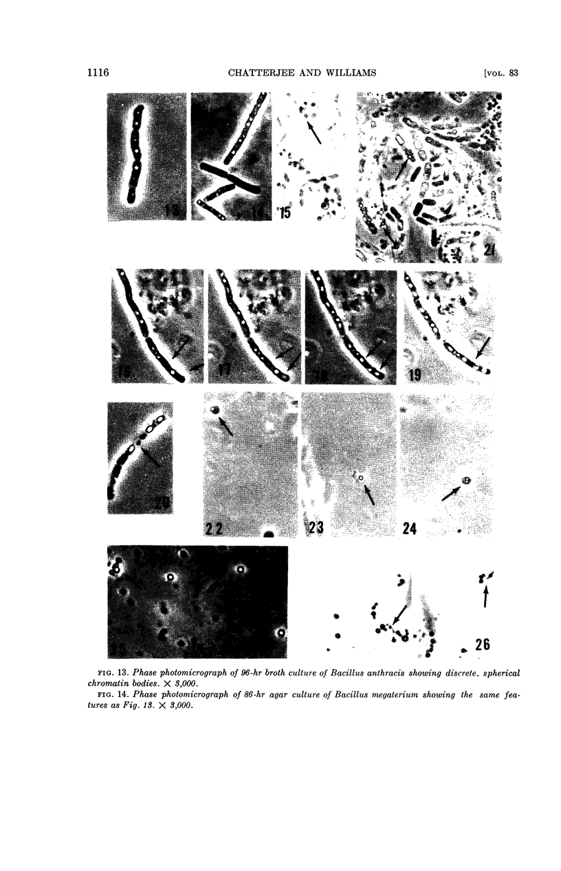
Chatterjee, B. R. (Baylor University College of Medicine, Houston, Tex.) and Robert P. Williams. Cytological observations on the chromatin bodies of two Bacillus species. J. Bacteriol. 83:1112–1118. 1962.—The nature of the chromatin bodies in Bacillus anthracis and B. megaterium has been studied intracellularly as well as after isolation from the bacilli. Preparations were examined by phase-contrast microscopy in the living state and by acid-Giemsa and Feulgen staining. In cells from young cultures, chromatin bodies occupy a continuous, long, central area in the bacteria. As the cells mature, the bodies lose their continuity and divide into two halves. In cells grown for 24 hr or longer, the chromatin bodies become more complex, and appear as multiple, spherical, interconnected constellations. Nuclear staining reveals that only the peripheral area, whether in cells from young or older cultures, takes up the stain, leaving a clear, central core. Discrete chromatin bodies are liberated from cells grown for longer periods after disintegration of the bacterial cell wall. These discrete bodies have been isolated from whole bacteria by treating washed, buffered saline suspensions of old cells with high concentrations of lysozyme followed by digestion with purified pancreatic lipase. The isolated chromatin bodies retain the same structure and appearance as inside the cells and show the same staining characteristics. Observations suggest that the chromatin bodies are discrete and circumscribed in nondividing, resting-phase bacteria.
Full text
PDF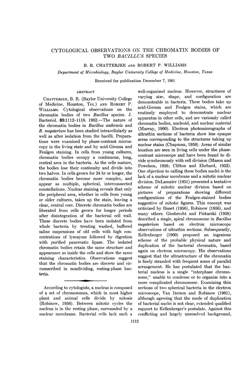
Images in this article
Selected References
These references are in PubMed. This may not be the complete list of references from this article.
- CHAPMAN G. B. Electron microscopy of ultrathin sections of bacteria. III. Cell wall, cytoplasmic membrane, and nuclear material. J Bacteriol. 1959 Jul;78(1):96–104. doi: 10.1128/jb.78.1.96-104.1959. [DOI] [PMC free article] [PubMed] [Google Scholar]
- CLIFTON C. E., EHRHARD H. B. Nuclear changes in living cells of a variant of Bacillus anthracis. J Bacteriol. 1952 Apr;63(4):537–543. doi: 10.1128/jb.63.4.537-543.1952. [DOI] [PMC free article] [PubMed] [Google Scholar]
- DeLAMATER E. D. A new cytological basis for bacterial genetics. Cold Spring Harb Symp Quant Biol. 1951;16:381–412. doi: 10.1101/sqb.1951.016.01.029. [DOI] [PubMed] [Google Scholar]
- DeLAMATER E. D. A staining and dehydrating procedure for the handling of microorganisms. Stain Technol. 1951 Jul;26(3):199–204. doi: 10.3109/10520295109113208. [DOI] [PubMed] [Google Scholar]
- FITZ-JAMES P. C. The duplication of bacterial chromatin; interpretations of some cytological and chemical studies of the germinating spores of Bacillus cereus and Bacillus megaterium. J Bacteriol. 1954 Oct;68(4):464–473. doi: 10.1128/jb.68.4.464-473.1954. [DOI] [PMC free article] [PubMed] [Google Scholar]
- MASON D. J., POWELSON D. M. Nuclear division as observed in live bacteria by a new technique. J Bacteriol. 1956 Apr;71(4):474–479. doi: 10.1128/jb.71.4.474-479.1956. [DOI] [PMC free article] [PubMed] [Google Scholar]
- SARDA L., MARCHIS-MOUREN G., CONSTANTIN M. J., DESNUELLE P. Sur quelques essais de purification de la lipase pancréatique. Biochim Biophys Acta. 1957 Feb;23(2):264–274. doi: 10.1016/0006-3002(57)90328-1. [DOI] [PubMed] [Google Scholar]
- SPIEGELMAN S., ARONSON A. I., FITZJAMES P. C. Isolation and characterization of nuclear bodies from protoplasts of Bacillus megaterium. J Bacteriol. 1958 Jan;75(1):102–117. doi: 10.1128/jb.75.1.102-117.1958. [DOI] [PMC free article] [PubMed] [Google Scholar]
- VAN ITERSON W., ROBINOW C. F. Observations with the electron microscope on the fine structure of the nuclei of two spherical bacteria. J Biophys Biochem Cytol. 1961 Jan;9:171–181. doi: 10.1083/jcb.9.1.171. [DOI] [PMC free article] [PubMed] [Google Scholar]