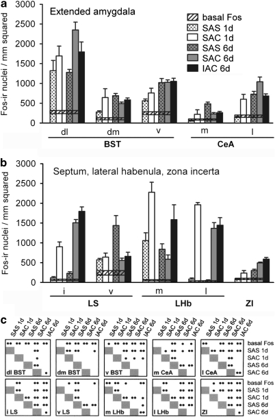
Figure 7.
Graphs illustrating the density of Fos-immunoreactive nuclei in the extended amygdala (a), including the bed nucleus of stria terminalis (BST) and central nucleus of the amygdala (CeA), and in (b) the lateral septum (LS), lateral habenula (LHb), and zona incerta (ZI), for the experimental groups as described in the text. Conventions in (a–c) are as in Figure 6. dl,dorsolateral division; dm,dorsomedial division; i,intermediate division; IAC,investigator-administered cocaine; l,lateral division; m,medial division; SAC,self-administered cocaine; SAS,self-administered saline; v,ventral division.

