

Abstract
Voltage-gated sodium channels are key to the initiation and propagation of action potentials in electrically excitable cells. Molecular characterization has shown there to be nine functional members of the family, with a high degree of sequence homology between the channels. This homology translates into similar biophysical and pharmacological properties. Confidence in some of the channels as drug targets has been boosted by the discovery of human mutations in the genes encoding a number of them, which give rise to clinical conditions commensurate with the changes predicted from the altered channel biophysics. As a result, they have received much attention for their therapeutic potential. Sodium channels represent well-precedented drug targets as antidysrhythmics, anticonvulsants and local anaesthetics provide good clinical efficacy, driven through pharmacology at these channels. However, electrophysiological characterization of clinically useful compounds in recombinant expression systems shows them to be weak, with poor selectivity between channel types. This has led to the search for subtype-selective modulators, which offer the promise of treatments with improved clinical efficacy and better toleration. Despite developments in high-throughput electrophysiology platforms, this has proven very challenging. Structural biology is beginning to offer us a greater understanding of the three-dimensional structure of voltage-gated ion channels, bringing with it the opportunity to do real structure-based drug design in the future. This discipline is still in its infancy, but developments with the expression and purification of prokaryotic sodium channels offer the promise of structure-based drug design in the not too distant future.
Keywords: voltage-gated sodium channel, subtype-selectivity, local anaesthetic, toxin, structural biology
Introduction
The α-subunit of the mammalian voltage-gated sodium channel is a 260 kDa protein comprising four homologous transmembrane domains (DI-DIV), each of which is made up of six segments (S1-S6) traversing the membrane in the form of α-helices. The channel pore is formed by the S5 and S6 helices, and the re-entrant P-loop which links these segments (Catterall et al., 2005). In recombinant systems, the α-subunit alone is capable of forming a fully functional channel. To date, nine functional members of the family have been described (Nav1.1–Nav1.9), each sharing the same predicted common secondary structure. Table 1 summarizes some key features of the channel family (this review uses nomenclature conforming to the BJP's Guide to Receptors and Channels; Alexander et al., 2008). The majority of the channels are sensitive to block by low concentrations of tetrodotoxin (<30 nM), while Nav1.5, 1.8 and 1.9 are only blocked by high concentrations (>1 µM) of the toxin (Hille, 1992). Genetic knockout of any of the genes SCN1A, SCN2A and SCN8A[(which code for the α-subunits of Nav1.1, 1.2 and Nav1.6 and are thought to be the major channels expressed in the adult central nervous system (CNS)] are lethal, indicating the essential and non-redundant function of the individual channels (Catterall et al., 2008). Although all the properties for gating and ion permeation are encapsulated in the α-subunit, the pore-forming domain is also associated with a number of accessory proteins. Four β-subunits have been identified (β1–4) which are thought to be composed of a single transmembrane-spanning single α-helix (Tseng et al., 2007). The C-terminus on the intracellular side is short, while the extracellular N-terminus is large, and believed to form an immunoglobin domain, similar in sequence to a family of cell adhesion molecules (Isom and Catterall, 1996). β1 and β3 subunits bind covalently to the α-subunit via a disulphide bridge, whereas β2 and β4 bind much more weakly. For some channels, the β-subunits play an important role in changing the biophysical properties of the channel and also the insertion of the α-subunit in the plasma membrane (Tseng et al., 2007). In addition to the β-subunits, a plethora of cytosolic proteins combine to anchor the channel to the cell cytoskeleton. In this way, there is tight control of sodium channel expression on both the intra- and extracellular sides of the channel (for reviews see Lai and Jan, 2006; Cusdin et al., 2008) and this is likely to be important in creating sites of special hyperexcitability in cells, for example in the axon initial segment (Van Wart et al., 2007). Each of the sodium channel subtypes exhibits its own distinct biophysical properties. A combination of expression pattern and activation and inactivation characteristics bestow on the host cell, a fine control of excitability.
Table 1.
Voltage-gated sodium channel nomenclature, chromosomal location and tissue distribution
| Channel nomenclature | Gene | Chromosomal location (human) | Tetrodotoxin sensitivity | Major tissue expression | Effect of mutation |
|---|---|---|---|---|---|
| Nav1.1 | SCN1A | 2q24 | ✓ | CNS, PNS | Epilepsy |
| Nav1.2 | SCN2A | 2q23–24 | ✓ | CNS, PNS | Epilepsy |
| Nav1.3 | SCN3A | 2q24 | ✓ | CNS, PNS | None reported |
| Nav1.4 | SCN4A | 17q23–25 | ✓ | Skeletal muscle | Myotonia, periodic paralysis |
| Nav1.5 | SCN5A | 3p21 | ✗ | Heart | Long QT, Brugada syndrome, progressive familial heart block |
| Nav1.6 | SCN8A | 12q13 | ✓ | CNS, PNS | Cerebellar atrophy |
| Nav1.7 | SCN9A | 2q24 | ✓ | PNS (SNS and PAs) | Increased and decreased pain sensitivity |
| Nav1.8 | SCN10A | 3p21–24 | ✗ | PNS (PAs) | None reported |
| Nav1.9 | SCN11A | 3p21–24 | ✗ | PNS (PAs) | None reported |
| Nax | SCN6/7A | 2q21–23 | Non-functional | Glia | – |
CNS, central nervous system; PAs, primary afferent neurones; PNS, peripheral nervous system; SNS, sensory nervous system.
Genetic mutations in sodium channel genes give rise to channelopathies and clinical disease
A number of clinical conditions have been traced back to mutations in sodium channel genes, which in turn give rise to alterations in channel function precipitating a clinical phenotype. These channelopathies provide tremendous insights into the normal physiological function of individual channels. For the drug hunter, these mutations are invaluable in boosting confidence in rationale for a given target and can provide an indication of what the clinical pharmacology may look like for a selective modulator. Conversely, the channelopathies may also highlight the penalty, that is, the potential side effects, if non-selective agents exhibit pharmacology at ‘off-target’ sodium channels. Mutations in sodium channel genes giving rise to channelopathies have been identified in heart, brain, skeletal muscle and also peripheral nerves. Table 1 indicates the major conditions arising from particular sodium channel gene mutations, and these are discussed in more detail below.
Cardiac arrhythmias
Nav1.5 is responsible for the rising phase of the cardiac action potential, so it is not surprising that mutations in the SCN5A gene resulting in altered function of the channel have profound effects on cardiac function. To date, more than 160 mutations in SCN5A have been described, leading to range of cardiac abnormalities and these mutations are spread widely across the channel with particular clusters around the intracellular loops, C-terminus and S4 transmembrane segment in DIV (Zimmer and Surber, 2008). Long QT is a condition resulting from prolonged ventricular repolarization, predisposing patients to toursades de pointes which in extreme cases causes sudden death. Most inherited long QT is caused by mutations in the potassium channel genes responsible for cardiac repolarization. However, up to 10% of long QT patients have mutations in SCN5A arising from more than 80 distinct mutations in the gene (Napolitano et al., 2005). The majority of these mutations result in impaired ability of the channels to close, leading to persistent sodium currents and a prolongation of the ventricular action potential (Zimmer and Surber, 2008). Brugada syndrome can also result in sudden death caused by ventricular fibrillation. Approximately 20% of Brugada patients carry mutations in SCN5A (Schulze-Bahr et al., 2003). The majority of the mutations are missense which produce a truncated channel protein. These loss of function mutations alter the action potential waveform, causing an imbalance of excitability in regions of the heart, leading to venticular arrhythmias (Tan et al., 2003). In patients with compromised cardiac conduction due to ischaemia, drugs such as encainide or flecainide which target Nav1.5 are contraindicated, as they have been shown to significantly increase mortality due to promotion of lethal arrhythmias (Echt et al., 1991). In addition, a number of studies have reported that intentional drug overdose with amitriptyline (which is a relatively potent modulator of sodium channels, Dick et al., 2007) induces changes in ECG waveform and a Brugada-like syndrome (Bolognesi et al., 1997; Rouleau et al., 2001; Goldgran-Toledano et al., 2002; Akhtar and Goldschlager, 2006). Clinical genetics and the pharmacological effects of sodium channel modulators highlight the cardiac risk associated with drugs which exhibit off-target effects on Nav1.5. Given the potentially catastrophic consequences of inadvertent pharmacology at Nav1.5, selectivity at this channel must be paramount in the profile of novel sodium channel modulators.
Primary periodic paralysis
Periodic paralyses are a collection of heterogeneous disorders resulting in sudden onset of flaccid paralysis which is episodic, and from which the patients make a full recovery. Primary periodic paralysis is an autosomal dominant condition occurring from a genetic defect in either sodium, calcium or potassium ion channels. The disease is classified as either hyper- or hypokalaemic dependent on circulating potassium concentrations and whether the condition improves or triggers the muscle weakness. Approximately 20% of hypo- and 50% of hyperkalaemic periodic paralyses are caused by mutations in the SCN4A gene which encodes for the Nav1.4 channel (Venance et al., 2006). The clinical symptomatology results from abnormal biophysical properties of the channel. The majority of the SCN4A missense mutations occur in the regions of the channel associated with fast inactivation, which results in an impairment of the channels ability to move into a non-conducting state. This leads to the Nav1.4 channels passing more current, resulting in either myotonia due to the initiation of pathologic bursts of action potentials or a flaccid paralysis resulting from depolarizing block, rendering the fibres inexcitable (Cannon, 2000).
Epilepsy
SCN1A is the gene which encodes for the Nav1.1 channel. To date, more than 200 mutations have been identified in the Nav1.1 channel leading to a number of inherited epilepsies (Ragsdale, 2008). The mutations result in either gain or loss of function and a spectrum of disease severities (Mulley et al., 2005). An additional population of patients have epilepsy driven by a mutation in the SCN1B gene which is responsible for the β1 subunit. This results in a modification to the extracellular domain of the β-subunit, impairing the interaction with the α-subunit and reducing the level of channel expression in the membrane (Wallace et al., 1998). A much smaller number of mutations have been found in SCN2A (coding for Nav1.2). The clinical symptoms are less marked and the convulsions usually do not continue into adulthood. The condition is associated with a decrease in the magnitude of whole-cell currents resulting from a decrease in channel density in the membrane (Misra et al., 2008). Therefore, both gain and loss of function mutations in SCN1A and SCN2A (Lossin et al., 2003; Ragsdale, 2008) can give rise to epilepsy, and in fact severe myoclonic epilepsy in infancy (SMEI) which is a very severe form of the disease, is caused by loss of function of the Nav1.1 channel (Fujiwara et al., 2003). This apparent paradox hints at the level of complexity governing brain excitability. Mice with homozygous null or heterozygous mutations in SCN1A exhibited spontaneous epileptic seizures and upon examination were found to have reduced (∼75%) sodium current density in hippocampal GABAergic interneurons (Yu et al., 2006). The inference being that loss of inhibitory tone could provoke SMEI in carriers of mutant SCN1A. The situation is yet more complex because pyramidal neurones also express Nav1.1 yet their sodium currents appear to be unaffected by Nav1.1 ablation (Yu et al., 2006). A possible explanation could lie in the observation that Nav1.1 is found at the axon initial segment of parvalbumin-positive (presumed GABAergic) neurones of the developing neocortex, while the channel is more localized to somata and axons in hippocampal neurones and at much lower levels in the pyramidal cells (Ogiwara et al., 2007). Epilepsy can be viewed as a disorder of network excitability (Ragsdale, 2008). At the single cell level, aberrations in sodium channel density in localized regions of the neurone can produce abnormal (either increased or decreased) patterns of firing, resulting from the complex interaction of sodium channels at the single neurone level. Many of these changes are likely to occur during embryological development, with some neurones being able to compensate for sodium channel loss with the increased expression of another family member (Yu et al., 2006), while other cells may not have the capacity to bring about this change. The net effect is dependent on the cellular location of expression, whether the neurone is inhibitory or excitatory and how this activity is shunted onto neurones within the network.
Thankfully, despite epilepsy being a common condition, most sufferers are well controlled with anticonvulsant drugs. However, up to 30% of patients do not respond to the drugs currently available (Hemming et al., 2008). Thus, a greater understanding of the cellular pathology underpinning epilepsy, together with more effective pharmacological agents to treat the condition, is needed. It seems feasible that subtype selective modulators of sodium channels could provide useful therapeutic entities in this regard, but we always need to be mindful of the complex interplay of brain sodium channels. Are the current crop of anticonvulsants which target sodium channels reasonably well tolerated because of a lack of selectivity, and will targeting a single subtype unmask unwanted side-effects?
Familial hemiplegic migraine
Recently, mutations in SCN1A have also been linked to familial hemiplegic migraine (Castro et al., 2009). Individuals carrying an L263V mutation in the Nav1.1 channel suffered from migraine and also epileptic attacks, at times independent of each other. The mutation occurs in the S5 domain of DI, an area important for ion selectivity and gating of the channel (Zhao et al., 2004). No functional data were presented although other missense mutations in the S5 segment result in a range of epileptic phenotypes (Meisler and Kearney, 2005). These findings support the notion that familial hemiplegic migraine and epilepsy share at least in part, a common pathological link.
Ataxia
The Nav1.6 channel has been linked to cerebellar atrophy, behavioural deficits and ataxia. In a single case, mutations in the SCN8A gene resulted in complete loss of Nav1.6 function (Trudeau et al., 2006), implicating the involvement of the channel in cognitive and behavioural processing. The mutation inserts a translation termination codon into the pore loop of domain IV, deleting the C-terminal cytoplasmic domain with loss of channel function. The ataxia3 mouse exhibits severe movement disorders, ataxia and tremor which in part, resembles the human phenotype. The mice carry a single amino acid substitution (S21P) located on the N-terminal cytoplasmic domain of Nav1.6 which results in reduced intracellular trafficking of the channel and a decrease in channel density at Nodes of Ranvier (Sharkey et al., 2009).
Pain
A number of preclinical studies have implicated Nav1.3, 1.7, 1.8 and 1.9 which are expressed in DRG neurones, in nociceptive processing (for review see Dib-Hajj et al., 2009). In rodents, Nav1.3 is expressed in fetal development, but expression is lost within a few days after birth (Waxman et al., 1994). Following peripheral nerve damage expression of the channel re-emerges in DRGs and given the biophysical properties of the channel, it has been suggested that this channel may be a significant contributor to ectopic activity (Dib-Hajj et al., 1999). However, mice which are null mutants for Nav1.3 exhibit normal responsiveness to neuropathic challenge (Nassar et al., 2006). Either antisense or genetic ablation of 1.8 or 1.9 in rodents produces deficits in nociceptive processing in response to either inflammatory or neuropathic challenge (Akopian et al., 1999; Lai et al., 2000; Priest et al., 2005). However, the picture is confusing with interventions being effective in some models but not others. Plasticity in channel expression is likely to contribute to this as a number of studies using traditional global knockout approaches have demonstrated clear up-regulation of Nav channels, presumably to compensate for the loss of another (Akopian et al., 1999; Yu et al., 2006; Vega et al., 2008). The value of global knockout approaches to investigate sodium channel function and translation to man is also questionable in light of the findings with Nav1.7. SCN9A encodes Nav1.7 and global gene deletion in mice results in pups which die shortly after birth (Nassar et al., 2004). However, nociceptor-specific Nav1.7 knockout mice are viable and show deficits in response to inflammatory (Nassar et al., 2004) but not neuropathic insult (Nassar et al., 2005). This contrasts markedly with recent findings from clinical genetic analyses of patients who exhibit rare and unusual pain phenotypes. These individuals have provided an invaluable insight into the role of Nav1.7 in sensory processing. Congenital indifference to pain (CIP) is a rare autosomal recessive disorder characterized by the inability to recognize a painful stimulus (Nagasako et al., 2003). In the past, individuals who suffered from indifference to pain would often earn a living performing feats of amazing pain tolerance to paying audiences (e.g. Dearborn, 1932). CIP individuals have normal autonomic and central function but are unable to perceive or interpret the quality or intensity of a painful challenge. Some patients also report anosmia (loss of the sense of smell, Losa et al., 1989; Hirsch et al., 1995), although the reasons for this are unclear. Tactile sensitivity in these individuals is retained to the extent that patients with CIP can distinguish between the sharp and blunt ends of a pin (Ford and Wilkins, 1938). Some cases of CIP are caused by a hereditary sensory autonomic neuropathy, of which a number have been identified, leading to defects in C- and A-δ primary afferent fibres (Nagasako et al., 2003). Retrospective genotyping of some families with CIP has led to the identification of mutations in the SCN9A gene (Cox et al., 2006; Goldberg et al., 2007). These mutations lead to non-sense or frameshift mutations of the Nav1.7 channel and a complete loss of function. Observations such as these indicate that the Nav1.7 channel has a pivotal role in pain processing in man, in the transduction and/or the conduction events in the nociceptors.
Gain of function mutations associated with the SCN9A gene have also been identified in patients suffering from a heightened sense of pain. Erythermalgia is a rare but extremely painful disorder, characterized by a very severe burning sensation usually in the legs, and coupled with profound erythema in the affected area (Babb et al., 1964; Drenth and Michiels, 1992; Drenth et al., 2008). Electrophysiological investigation of erythermalgia mutants in recombinant expression systems indicate that, in comparison to wild-type whole-cell currents, the activation threshold for the mutant channels is shifted in the hyperpolarizing direction. Some of the mutants also give rise to an increase in the magnitude of sodium currents (Cummins et al., 2004; Cheng et al., 2008).
Paroxysmal extreme pain disorder (PEPD) is another condition linked to mutations in SCN9A. The disease is a rare autosomal-dominant disorder with a diffuse, but extreme pain reported from the rectal, ocular and mandibular areas (Fertleman et al., 2007). In addition to the pain, patients present with syncopes, bradycardia and asystole leading some to suggest that the condition can be regarded as a type of ‘reflex epilepsy’ (Schubert and Cracco, 1992). PEPD mutant Nav1.7 channels have characteristic electrophysiological signatures when expressed recombinantly. Following a depolarizing step, wild-type Nav1.7 channels activate and inactivate in less than 5 ms. In comparison, PEPD mutant channels either inactivate over hundreds of ms, or either fail to inactivate at all. They may also have more negative activation thresholds (Fertleman et al., 2006; Jarecki et al., 2008).
Collectively, the mutations give rise to dramatic changes in the biophysical properties of the channel. At one extreme, the mutations can render the channels completely non-functional and unable to pass current. While at the other end of the spectrum, point mutations produce significant changes in the activation threshold and rate of inactivation of the channels. The cellular changes are consistent with either the inability to sense pain at all, or an exaggerated and extremely unpleasant sensation of pain. It is very unusual to have extremes of clinical pain phenotype which are so well explained at the molecular level. It is therefore understandable why the Nav1.7 channel has generated so much interest as an analgesic target. However, there are a number of unanswered questions. Why the clinical symptoms of erythermalgia and PEPD are different, given that in both conditions gain of function mutations in Nav1.7 underpin the hyperexcitability. Many CIP patients report anosmia (Losa et al., 1989; Hirsch et al., 1995), but the cellular and anatomical basis of this has yet to be determined. Most cases of erythermalgia are reported in early life, but some mutations seem to result in symptoms emerging in later life (Han et al., 2009). It is not clear why this should be or what the trigger factors are for emergence of the disease in affected individuals. However, we are beginning to gain a better understanding of how the biophysical changes that occur in the mutations can affect neuronal excitability and how this relates to symptoms (Dib-Hajj et al., 2009; Han et al., 2009).
Despite the excellent confidence in rationale for Nav1.7 and a number of other sodium channel subtypes, it has proven to be extremely challenging to identify compounds which target some channels in the family while sparing others. The architecture of the channel, the high degree of conservation of sequence in key parts of the α-subunit and homology between voltage-gated channels in general have all contributed to very few isoform-selective compounds moving into clinical development. The reasons are discussed below.
Sodium channel pharmacology
Voltage-gated sodium channels represent a well-precedented drug target class as local anaesthetics (LAs; e.g. lidocaine), the classical anticonvulsants (lamotrigine, carbamazepine, phenytoin) and antidysrhythmics (mexiletine, flecainide) exhibit their pharmacology through modulation of these channels. Compounds such as these are very effective in treating clinical hyperexcitability states driven through the aberrant activity of sodium channels, in conditions such as epilepsy and neuropathic pain, or to produce complete nerve block in the case of the LAs. These drugs were discovered using empirical means, often in isolated tissue preparations which were designed to model some component of the clinical condition being targeted. Therefore in the past, drugs were discovered due to pharmacological activity in, for example, isolated heart or brain slice preparations, and this was very successful in identifying drugs to treat arrhythmias or epilepsy (Borchard et al., 1982; Piredda et al., 1986).
The definitive characterization of the molecular target as the voltage-gated sodium channel often occurred much later, with drugs like procaine and cocaine being used for many years (e.g. Harris and Goldberg, 1931) before the underlying pharmacology became apparent. The discovery of these drugs also pre-dates by a very considerable time, the cloning and expression of the individual channel subtypes, which came many years later (Noda et al., 1986). When the clinically useful sodium channel modulators were eventually profiled in recombinant systems using electrophysiological methods, they were, in general, shown to be weak and poorly selective between channel subtypes. Although empiricism led to the drugs of yesterday, today the bar is now considerably higher as we seek to identify subtype-selective drugs with the improved efficacy and reduced side-effect profile that the patient demands.
In toxins, nature has provided some potent modulators of sodium channels, which has given rise to their classification as either resistant (Nav1.5, 1.8 and 1.9) or sensitive (at nanomolar potency; Nav1.1, 1.2, 1.3, 1.4, 1.7) to block by the pufferfish toxin, tetrodotoxin (TTX). The mechanism of block exhibited by TTX and that of drug molecules is completely different. Drug molecules usually have molecular weights of less than 500, often significantly so. This is because the chemical space in which drug-like molecules can be found is limited due to properties such as solubility, permeability and pharmacokinetics becoming problematic when molecular weight exceeds 500 (Lipinski et al., 1997). Drugs like lamotrigine and lidocaine (which have molecular weights of ∼256 and 350 respectively) are able to penetrate deep into the channel either via passage through the hydrophilic pore or through the hydrophobic cell membrane, and bind to a common binding site on the inner face (Hille, 1977). This site is believed to be shared and overlapping for the sodium channel modulating anticonvulsants, the antidysrhythmics (of Class 1b; lidocaine and 1c; flecainide, encainide) and LAs (Fozzard et al., 2005). This common binding domain for small molecules has become known as the ‘local anaesthetic’ binding site. Historically, displacement of radiolabelled batrachotoxin (BTX) binding was used as the mainstay for the identification of novel sodium channel modulators. The BTX binding site overlaps that for the LAs and other small molecule modulators (Willow and Catterall, 1982; Postma and Catterall, 1984; Nau and Wang, 2004). Site-directed mutagenesis has further refined this binding site and demonstrated the key residues which form part of the ligand binding domain (Ragsdale et al., 1994; 1996;). Figure 1 provides the sequence alignments in the region around the LA binding site for rat Nav1.2. Isoleucine, phenylalanine and tyrosine residues at positions 1760, 1764 and 1771 respectively are conserved across all channels. These residues have been identified as key determinates of state-dependent binding of LAs (Ragsdale et al., 1994; 1996;).
Figure 1.
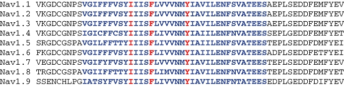
Multiple sequence alignment of human voltage-gated sodium channel α-subunits at the region of the local anaesthetic binding site. Blue text denotes the putative transmembrane α-helix; red identifies the key residues for local anaesthetic binding (Ragsdale et al., 1994; 1996;). Note the identical sequence in this region and the high degree of homology extending beyond the transmembrane domain into the flanking loop sequences.
A number of excellent reviews are available which discuss the various molecular models for LA binding to the channel and how these compounds alter channel gating (Fozzard et al., 2005; Lipkind and Fozzard, 2005). Beyond this region, sequence identity is very similar even into the flanking sites beyond the trans-membrane domain and including some of the loop region. The architecture of the channel dictates its pharmacology and although compounds which can access the LA binding site may well exhibit pharmacology, the sequence identity in the region means that pharmacological selectivity between channel subtypes is likely to be very challenging if compounds bind to this region. Nonetheless, it is hoped that molecular modelling of the LA site will offer an insight into channel pharmacology at the molecular level, eventually offering the possibility of designing drugs which target some isoforms and channel states while sparing others. Given that the BTX and LA sites are overlapping, BTX binding is probably not a good method for identifying compounds which will target some sodium channels and spare others. A recent study has produced some interesting data that may conflict with this view. Using a mutagenesis approach, the Nav1.8-selective modulator A-803467 was shown to bind to a different site than that occupied by the LA tetracaine (Browne et al., 2009). There was some overlap between the sites recognized by the two compounds, but this could suggest that isoform selectivity can be achieved at sites close to that for LA binding. A possible explanation for this could be the shortcomings in the two-dimensional view of the channel which cannot take into account the potential influence of amino acids in adjacent domains, which could affect the structure of an otherwise highly conserved region. The LA site seems crucial for small molecule pharmacology, but time will tell whether this is a fruitful site to target to enable isoform selectivity.
TTX is sufficiently large to produce true channel block by occlusion of the pore. TTX has a guanidinium group which fits well with a relatively superficial binding site at the extracellular end of the pore (for review, see Narahashi, 2008). The remainder of the molecule in its entirety is too large to penetrate deeper into the channel, but is large enough to reach down to the selectivity filter and prevent sodium flux. Early molecular models provided a useful explanation for TTX and saxitoxin binding to the sodium channel and also an understanding at the molecular level, for toxin selectivity. TTX forms a hydrophobic interaction with aromatic rings of phenylalanine or tyrosine in TTX-sensitive channels such as Nav1.2 or 1.4, but is unable to do so with the cysteine residue in Nav1.5 (Lipkind and Fozzard, 1994). This provides an explanation for the TTX-resistance of the Nav1.5 channel. TTX bound to a channel does not inhibit the ability of the channels to gate, which under block, operates normally in response to depolarization (Keynes and Rojas, 1974). However, there is some evidence from binding studies that when the TTX site is occupied by the toxin, there is decreased affinity of BTX for its binding site deeper in the channel (Brown, 1986). The reason for this phenomenon is unclear.
A combination of site-directed mutagenesis, binding and electrophysiological approaches has identified at least six distinct binding domains, in addition to the TTX and LA binding sites, in or around the pore region of the α-subunit. The majority of these sites have been identified using peptide and non-peptide toxin molecules and alkaloids as pharmacological probes, coupled with electrophysiology to characterize their pharmacology (Nicholson, 2007). Table 2 lists some of the toxins, their putative binding domains and also pharmacological action. Figure 2 identifies these binding domains in a 3D model of a sodium channel.
Table 2.
Toxin and alkaloid binding sites on the α-subunit, the pharmacological effect and the putative binding site
| Site | Pharmacological probe | Effect/binding domains |
|---|---|---|
| 1 | tetrodotoxin, saxitoxin | Occlusion of the pore IS5-S6, IIS5-S6, IIIS5-S6, IVS5-S6 |
| 2 | batrachotoxin, veratridine, aconitine | Promotes persistent activation IS6, IVS6 |
| 3 | scorpion α-toxin, sea anemone toxins | Destabilization of inactivated state, increased Popen, IS5-IS6, IVS3-S4, IVS5-S6 |
| 4 | scorpion β-toxin | Shifts activation in hyperpolarizing direction IIS1-S2, IIS3–4 |
| 5 | brevetoxins, ciguatoxin | Enhanced activation and block of inactivation IS6, IVS5 |
| 6 | δ-conotoxin | Slows inactivation (sub-site of Site 3) IVIS3–4 |
| 7 | pyrethroids | Inhibits inactivation, shifts voltage-dependence of activation IIIS6 |
‘I’ refers to the domain, and ‘S’ the transmembrane segment.
Figure 2.

Homology model for a sodium channel with toxin binding domains shown. Top view (left) and side view (right). Site 1: (tetrodotoxin), blue; Site 2: (batrachotoxin), red; Site 3 (scorpion α-toxin), orange; Site 4 (scorpion β-toxin), yellow; Site 5 (brevetoxins), white; Site 6 (δ-conotoxin), orange; and Site 7 (pyrethroids), white. See main text for details on model.
Pharmacological selectivity by TTX has defined the TTX-sensitive and resistant members of the family. Despite a concerted effort to screen toxins and venoms from a number of animal species, few fractions have been identified which show significant selectivity between isoforms. However, a number of toxins exhibit preference for some channels over others (Table 3). It seems likely that for many of these molecules, their size may well preclude access to sites crucial for selectivity. In some instances, they do provide useful tools for probing channel structure and function. Given the limitations of toxins as drug molecules, small molecules probably offer the best chance of achieving subtype selectivity.
Table 3.
Some toxins reported to demonstrate sodium channel subtype-selectivity
| Toxin (species) | Major pharmacology, subtype selectivity | Reference |
|---|---|---|
| ι-conotoxin RXIA (Cone snail) | Shifts voltage-dependence of activation in hyperpolarizing direction rNav1.6 > rNav1.2 > rNav1.7. Ineffective v rNav1.1, rNav1.3, rNav1.4, rNav1.5, rTTX-R | Fiedler et al., 2008 |
| μ-conotoxin TIIIA (Cone snail) | Current block rNav1.2 ∼ = rNav1.4 Ineffective v rNav1.3, hNav1.5, hNav1.7, hNav1.8 | Lewis et al., 2007 |
| μ-conotoxin KIIIA (Cone snail) | Current block rNav1.2 > rNav1.4 > rNav1.7 >/= rNav1.1 > rNav1.3 > rNav1.5 | Khoo et al., 2009 |
| μ-conotoxin GIIIA/B (Cone snail) | Current block rNav1.4 >> hNav1.4 > hNav1.5 | Chahine et al., 1998; Cummins et al., 2002 |
| μO-conotoxin MrVIB (Cone snail) | Current block hNav1.8 > rNav1.2, rNav1.3, hNav1.5 | Ekberg et al., 2006 |
| ProTxII (Tarantula) | Current block hNav1.7 > hNav1.6 > hNav1.5 > hNav1.8 | Schmalhofer et al., 2008 |
| Ceratotoxin1 Ceratotoxin2 Phrixotoxin (Tarantula) | Current block, depolarizing shift in V-dependence of activation rNav1.2 > hNav1.5 > rNav1.1 > rNav1.4 rNav1.2 > rNav1.3 > rNav1.1 = rNav1.4 > hNav1.5 rNav1.2 > rNav1.3 > hNav1.5 > rNav1.4 > rNav1.1 | Bosmans et al., 2006 |
| Jingzhaotoxin-XI (Tarantula) | Channel block, small depolarising shift in threshold of activation in Nav1.5 TTX-sensitive INa (rat DRG) > rNav1.5 (cardiac myocyte) | Liao et al., 2006 |
| Huwentoxin-IV (Tarantula) | Current block, no effect on V-dependence of activation hNav1.7 > rNav1.2 > rNav1.3 >> hNav1.5 > rNav1.4 | Xiao et al., 2008 |
Table shows the major pharmacological actions and selectivity of a range of toxins reported to show isoform-specificity at voltage-gated sodium channels. Symbol h, human; r, rat. TTX-R and TTX-S refer to the tetrodotoxin-resistant and tetrodotoxin-sensitive whole-cell sodium currents recorded from rat dorsal root ganglion neurones, respectively.
Biophysical modulation, state/use dependency, etc
The current selection of clinically useful sodium channel modulators exhibits poor potency and selectivity between channel subtypes. The channel family also has a critical role in many physiological processes, so why are these drugs reasonably well tolerated? Put another way, why do anticonvulsants not have serious side effects on the heart, or antidysrhythmics on CNS function? Over 50 years ago, it became clear that rapidly firing nerve fibres were more sensitive to block by the LA procaine than those firing at a slower rate (Matthews and Rushworth, 1957), and this eventually became known as ‘use-dependent’ (or phasic) block (Courtney et al., 1978). Small molecule sodium channel modulators frequently possess state- and use-dependency. A compound showing state-dependent pharmacology exhibits a higher binding affinity for one channel state over another. For the LAs, affinities are highest for the open and inactivated states of the channel and lowest for the resting state (Courtney et al., 1978; Chernoff, 1990). This seems to be true for most of the drug-like molecules described to date. Use-dependency occurs when neurones fire frequently and the probability of a channel being in an inactivated state (i.e. one for which the drug has a higher affinity and that can be pharmacologically modified) is highest. Block can therefore accumulate as LAs have a slower dissociation rate from inactivated channels, compared with those in the resting state (Chernoff, 1990). This causes the accumulation of ‘functionally’ blocked channels and this is a dynamic process in that ‘block changes inactivation and inactivation changes block’ (Hille, 1977). The channels are not truly blocked in the sense that the pore is occluded (as occurs with TTX), but the binding of the drug to the channel renders it less able to move from a non-conducting (inactivated) to a conducting (open) state.
The ‘on’ and ‘off’ rates of drug molecules are clearly very important in determining the potency of compounds in the context of use-dependency, and this in turn is governed by the physicochemical properties of the drug (Chernoff and Strichartz, 1990). These observations form the central tenets of the ‘modulated receptor hypothesis’ (Hille, 1977). However, when the LAs are used to produce a local nerve block, such as may be used prior to third molar tooth extraction, very high concentrations of the drug are infiltrated close to the nerve. For lidocaine, typically 2% solutions are used (Porto et al., 2007) which is a concentration in the tens of millimoles, applied to the nerve. Under these circumstances, it is difficult to gauge the importance or benefit of use-dependent activity. Clearly, use-dependence is crucial in those drugs which are systemically available. Lamotrigine has a greater than 200-fold affinity for inactivated channels compared with those at rest. The compound binds slowly to fast-inactivated channels and the kinetics of lamotrigine binding to inactivated channels is faster than the development of the slow inactivated state (Kuo and Lu, 1997). These features mean that lamotrigine needs a sustained depolarization in a neurone to produce significant block. Long depolarizations are unusual in normal physiological systems but are characteristic of epileptic discharge (De Curtis and Avanzini, 2001) which probably underpins why the drug is very effective at controlling seizures, while being generally well tolerated.
From our current perspective, it seems unlikely that a strongly use-dependent but non-selective channel modulator will be able to deliver the next generation of therapies for conditions such as epilepsy or pain. Isoform-specific modulators offer the promise of therapies with a significant advantage over current standard of care, which will be essential if the drugs are to gain approval. Given the tremendous therapeutic potential of sodium channel modulators, it is no surprise that this has prompted a high degree of interest from the pharmaceutical industry. The patent literature contains over 150 sodium channel modulator applications published in the last 2 years, coming from over 50 different organizations (Pfizer internal data), which reflects the level of activity in this area. Table 4 summarizes some of the pharmacologies reported for small molecule isoform-specific sodium channel modulators. Because of state-dependency of compounds, IC50 values are very dependent on the protocol used to generate the data which makes comparisons between studies difficult. Nonetheless, some of the compounds show good isoform selectivity. Most of the activity has been directed towards pain as a therapeutic indication and a number of these compounds have been shown to exhibit antinociceptive activity in preclinical models. A-803467, which is selective for Nav1.8, reverses nociceptive responses following neuropathic and inflammatory challenge (Jarvis et al., 2007). The imidazopyridines are Nav1.7 preferring, and also demonstrate efficacy in neuropathic models (London et al., 2008). This in vivo efficacy is encouraging and suggests that targeting either Nav1.7 or 1.8 in isolation, can produce a robust antinociceptive effect in preclinical models.
Table 4.
Drug-like molecules reported to exhibit isoform selectivity
| Compound/class | Pharmacology, subtype selectivity | Reference |
|---|---|---|
| A-803467 [5-(4-Chlorophenyl-N-(3,5-dimethoxyphenyl) furan-2-carboxamide) | hNav1.8 primary pharmacology ∼8 nM IC50. 100× selectivity over hNav1.2, hNav1.3, hNav1.5, hNav1.7 | Jarvis et al., 2007 |
| 1-benzazepin-2-ones | Most potent compound in series ∼30 nM IC50 v hNav1.7. Some compounds selective (<10×) hNa1.5 < hNav1.8 | Williams et al., 2007 |
| Imidazopyridines | Most potent compound ∼60 nM IC50 v hNav1.7. Selectivity <10× over hNav1.8 | London et al., 2008 |
| Furan piperazines | hNav1.8 >mNav1.8, most potent compound ∼100 nM. Selectivity <10× over hNav1.2 and hNav1.5 | Drizin et al., 2008 |
| 5-Aryl-2-furfuramides | hNav1.8 IC50 < 10 nM for most potent compound >100× selectivity over hNav1.2, hNav1.3, hNav1.5, hNav1.7 | Kort et al., 2008 |
| 4,9-anhydro-TTX | IC50 ∼78 nM at mNav1.6. >15× selectivity over rNav1.2, rNav1.3, rNav1.4, hNav1.5, hNav1.7, rNav1.8 | Rosker et al., 2007 |
Table shows primary pharmacology and selectivity profiles for a number of drug-like molecules. Symbols m, mouse; r, rat; h, human.
The opportunity offered by structural biology
At the heart of the search for novel chemical entities is the screen. At one end of the spectrum, a full high throughput screen could involve the basic pharmacological profiling of a million compounds. This is useful in identifying a ‘hit’ which undergoes a more detailed interrogation of pharmacology using more complex methods. This is performed on a much smaller number of compounds from which the drug is ultimately found. The pharmaceutical industry will continue to search for compounds in this way, believing that larger and larger compound libraries offer an improved chance of finding the elusive ‘hit’. However, in the 10 year period between 1991 and 2000, the average success rate (from first in man to registration) for new chemical entities in all therapeutic areas combined, was 11% (Kola and Landis, 2004). Overall, the Pharma industry needs to reduce attrition and improve its success rate. Can a more detailed understanding of the three-dimensional architecture of the channel offer a smarter way to discover subtype selective compounds?
As a science, structural biology is in its relative infancy and obtaining information on the three-dimensional structure of membrane proteins has proven to be extremely challenging. However, significant advances have been made in recent years. Pioneering work from the Mackinnon laboratory in elucidating the crystal structure of a mammalian potassium channel (Long et al., 2005) has opened up the exciting possibility of obtaining the crystal structure of a mammalian sodium channel in the future. This is still some way off, but the identification and characterization of the bacterial channel NaChBac has provided useful impetus. One of the stumbling blocks to obtaining structures of mammalian sodium channels is their stability under solubilization with detergents and difficulty in synthesizing high levels of functionally active channel. It is now possible to produce large amounts of purified, functional NaChBac (Nurani et al., 2008).
NaChBac has been isolated from Bacillus halodurans, and shares a number of structural features with eukaryotic channels, underpinning its importance to the field. The channel is presumed to be made from a tetramer comprising four major domains, each of which is made up of six-transmembrane spanning segments. However, unlike the mammalian equivalent, NaChBac has a tetramer made from four identical repeating domains (Nurani et al., 2008), and more closely resembles the structural motif of potassium channels in this respect. The pore region shows greatest similarity to that of Cav channels, despite exhibiting a high degree of selectivity for sodium ions. In keeping with this observation, the NaChBac channel is much more sensitive to block with the L-type calcium channel modulator nifedipine (IC50∼2.2 µM), than to tetrodotoxin (no block at 30 µM; Ren et al., 2001). The gating kinetics of the NaChBac currents are also very different than that for mammalian Nav channels, with the rate of activation, inactivation and recovery from inactivation being approximately one-tenth that of Navs (Ren et al., 2001).
Despite these significant functional differences, structural modelling of the NaChBac channel has provided insights into the binding of lamotrigine and batrachotoxin (Cronin et al., 2003). More recently, NaChBac has provided models which attempt to explain the gating mechanisms and conformational changes which occur in the channel (Shafrir et al., 2008a,b;). At present, the NaChBac channel probably offers the best chance of producing a crystal structure for a mammalian sodium channel. When this happens, it will transform our understanding of the three-dimensional structure of the channel and greatly enhance our ability to make subtype-selective compounds.
The textbook depiction of the sodium channel is one where the channel, by necessity, is cut and flattened showing the classical ‘four times six’ transmembrane-spanning domains. This 2-dimensional view is useful up to a point, but the 3-dimensional view is much more information-rich, as even at low resolution, it allows us to visualize parts of the structure which come into close proximity with each other. In this way, it forces us to think in three dimensions as we consider the gating mechanisms and pharmacology of the channel. For example, the 3-dimensional structure of the electric eel sodium channel has been determined at 19 Å resolution using cryo-electron microscopy and single-particle image analysis (Sato et al., 2001). The channel appears to be bell-shaped with a large number of cavities and holes in the structure. This provides a low-resolution 3-dimensional map of the channel but clearly falls short of the detail needed to understand the pharmacology of the channel. Mutational analyses have identified the key residues for binding of lamotrigine and enabled a model to be generated based on the binding of the compound to the eel channel (Cronin et al., 2003). In a similar way, site-directed mutagenesis of the cockroach sodium channel has revealed the residues critical for the binding of pyrethroid insecticides and enabled the modelling of a pyrethroid-bound channel (Du et al., 2009). These types of study are producing a gradual improvement in the resolution of the modelling, for a number of compounds bound to the channel.
These analyses are beginning to provide images of what the channel may look like and are the first steps on a journey which will hopefully lead to the co-crystallization of the channel with pharmacologically relevant drugs. This would lead to true ‘structure-based’ drug design. At present, this may seem far-fetched for the sodium channel, but exciting progress has been made in understanding the architecture of another difficult target; G-protein coupled receptors (Cherezov et al., 2007; Rasmussen et al., 2007; Warne et al., 2008), and this may give a hint to what is possible in the future.
We are currently modelling the Nav channel based on the Kv1.2 work of the MacKinnon lab (Long et al., 2005) to try and gain insight into the key binding domains for compounds with known pharmacology. Unfortunately, the resolution to allow true docking of molecules into the channel structure is still not currently available. However, site-directed mutagenesis has identified the critical binding domains for a number of toxins and small molecules, and has formed the basis of what we currently understand as the well-recognized binding sites on the channel (Catterall et al., 2005). Using the crystal structure of Kv1.2 (Long et al., 2007), a homology model of a sodium channel was created using Modeller. The intracellular loops between domains 1 and 2 and domains 2 and 3, as well as the C- and N-terminal parts were omitted during the model construction. All extracellular loops were included, but relative orientations of the large loop has not been optimized yet and these loops are only shown for reference. The structure of lidocaine (Figure 3) was manually docked into the vestibule/LA site.
Figure 3.

Binding of lidocaine to a 3D model of a sodium channel. Top view (left) and side view (right). Lidocaine (white circle) is bound in the vestibule/local anaesthetic binding site, below the selectivity filter domain (yellow circle).
Solution structure determination of toxin molecules could offer an opportunity to gain a better insight into the interaction of a toxin molecule and its binding site on a channel. This approach could provide a better understanding of the structure–function relationship for a toxin at the molecular level. In turn, this could also provide valuable information concerning the channel structure in the region of the binding domain. A good example of this is work involving μ-conotoxin GIIIA, which has been used to build a model to enable a better understanding of the outer vestibule of the Nav1.4 channel (Choudhary et al., 2007). Mutational analyses of residues on both the channel and the toxin assist in defining the key binding domains, resulting in the generation of a preliminary docking arrangement. A greater understanding of why certain toxins exhibit some selectivity while others do not may lead to the identification of unique binding domains. If this can be achieved, it opens up the possibility of designing small molecules which mimic the key pharmacologically important structural features of the toxin. However, many of the toxin molecules are large and peptidic in nature, and it may prove impossible for small molecular weight drug molecules to span the distances required to bring about modulation of the channel. Nonetheless, small molecules offer the benefit of being able to permeate further into the channel, maybe offering the opportunity to interfere with the gating or inactivation machinery in sites which may not be available to large toxin molecules.
A greater understanding of the architecture of the voltage-gated sodium channel can potentially assist in the identification of compounds with particular pharmacological properties. This would lead to true ‘structure-based’ drug design. This is still a long way off but progress on elucidating the 3D structure of other membrane proteins like the β-adrenoceptors offers hope that this is not an impossible task. Binding sites within the protein may be identified, opening up the future potential of guiding medicinal chemistry design towards (or away from, depending on the specific ion channel) better interactions between small molecules and the ion channel of interest.
Glossary
Abbreviations:
- BTX
batrachotoxin
- CIP
congenital indifference to pain
- LA
local anaesthetic
- PEPD
paroxysmal extreme pain disorder
- SMEI
severe myoclonic epilepsy in infancy
- TTX
tetrodotoxin
Statement of conflict of interest
Both authors are full-time employees of Pfizer Global Research & Development.
References
- Akhtar M, Goldschlager NF. Brugada electrocardiographic pattern due to tricyclic antidepressant overdose. J Electrocardiol. 2006;39:336–339. doi: 10.1016/j.jelectrocard.2006.02.005. [DOI] [PubMed] [Google Scholar]
- Akopian AN, Souslova V, England S, Okuse K, Ogata N, Ure J, et al. The tetrodotoxin-resistant sodium channel SNS has a specialized function in pain pathways. Nat Neurosci. 1999;2:541–548. doi: 10.1038/9195. [DOI] [PubMed] [Google Scholar]
- Alexander SPH, Mathie A, Peters JA. Guide to receptors and channels (GRAC), 3rd edition (2008 revision) Br J Pharmacol. 2008;153(Suppl. 2):S1–S209. doi: 10.1038/sj.bjp.0707746. [DOI] [PMC free article] [PubMed] [Google Scholar]
- Babb RR, Alarcon-Segovia D, Fairbairn JF., 2nd Erythermalgia. Review of 51 cases. Circulation. 1964;29:136–141. doi: 10.1161/01.cir.29.1.136. [DOI] [PubMed] [Google Scholar]
- Bolognesi R, Tsialtas D, Vasini P, Conti M, Manca C. Abnormal ventricular repolarization mimicking myocardial infarction after heterocyclic antidepressant overdose. Am J Cardiol. 1997;79:242–245. doi: 10.1016/s0002-9149(96)00727-8. [DOI] [PubMed] [Google Scholar]
- Borchard U, Bösken R, Greeff K. Characterization of antiarrhythmic drugs by alternating current induced arrhythmias in isolated heart tissues. Arch Int Pharmacodyn Ther. 1982;256:253–268. [PubMed] [Google Scholar]
- Bosmans F, Rash L, Zhu S, Diochot S, Lazdunski M, Escoubas P, et al. Four novel tarantula toxins as selective modulators of voltage-gated sodium channel subtypes. Mol Pharmacol. 2006;69:419–429. doi: 10.1124/mol.105.015941. [DOI] [PubMed] [Google Scholar]
- Brown GB. 3H-batrachotoxinin-A benzoate binding to voltage-sensitive sodium channels: inhibition by the channel blockers tetrodotoxin and saxitoxin. J Neurosci. 1986;6:2064–2070. doi: 10.1523/JNEUROSCI.06-07-02064.1986. [DOI] [PMC free article] [PubMed] [Google Scholar]
- Browne LE, Blaney FE, Yusaf SP, Clare JJ, Wray D. Structural determinants of drugs acting on the nav1.8 channel. J Biol Chem. 2009;284:10523–10536. doi: 10.1074/jbc.M807569200. [DOI] [PMC free article] [PubMed] [Google Scholar]
- Cannon SC. Spectrum of sodium channel disturbances in the nondystrophic myotonias and periodic paralyses. Kidney Int. 2000;57:772–779. doi: 10.1046/j.1523-1755.2000.00914.x. [DOI] [PubMed] [Google Scholar]
- Castro MJ, Stam AH, Lemos C, de Vries B, Vanmolkot KR, Barros J, et al. First mutation in the voltage-gated Nav1.1 subunit gene SCN1A with co-occurring familial hemiplegic migraine and epilepsy. Cephalalgia. 2009;29:308–313. doi: 10.1111/j.1468-2982.2008.01721.x. [DOI] [PubMed] [Google Scholar]
- Catterall WA, Goldin AL, Waxman SG. International Union of Pharmacology. XLVII. Nomenclature and structure-function relationships of voltage-gated sodium channels. Pharmacol Rev. 2005;57:397–409. doi: 10.1124/pr.57.4.4. [DOI] [PubMed] [Google Scholar]
- Catterall WA, Dib-Hajj S, Meisler MH, Pietrobon D. Inherited neuronal ion channelopathies: new windows on complex neurological diseases. J Neurosci. 2008;28:11768–11777. doi: 10.1523/JNEUROSCI.3901-08.2008. [DOI] [PMC free article] [PubMed] [Google Scholar]
- Chahine M, Sirois J, Marcotte P, Chen L, Kallen RG. Extrapore residues of the S5-S6 loop of domain 2 of the voltage-gated skeletal muscle sodium channel (rSkM1) contribute to the mu-conotoxin GIIIA binding site. Biophys J. 1998;75:236–246. doi: 10.1016/s0006-3495(98)77510-1. [DOI] [PMC free article] [PubMed] [Google Scholar]
- Cheng X, Dib-Hajj SD, Tyrrell L, Waxman SG. Mutation I136V alters electrophysiological properties of the Na(v)1.7 channel in a family with onset of erythromelalgia in the second decade. Mol Pain. 2008;4:1. doi: 10.1186/1744-8069-4-1. [DOI] [PMC free article] [PubMed] [Google Scholar]
- Cherezov V, Rosenbaum DM, Hanson MA, Rasmussen SG, Thian FS, Kobilka TS, et al. High-resolution crystal structure of an engineered human beta2-adrenergic G protein-coupled receptor. Science. 2007;318:1258–1265. doi: 10.1126/science.1150577. [DOI] [PMC free article] [PubMed] [Google Scholar]
- Chernoff DM. Kinetic analysis of phasic inhibition of neuronal sodium currents by lidocaine and bupivacaine. Biophys J. 1990;58:53–68. doi: 10.1016/S0006-3495(90)82353-5. [DOI] [PMC free article] [PubMed] [Google Scholar]
- Chernoff DM, Strichartz GR. Kinetics of local anesthetic inhibition of neuronal sodium currents. pH and hydrophobicity dependence. Biophys J. 1990;58:69–81. doi: 10.1016/S0006-3495(90)82354-7. [DOI] [PMC free article] [PubMed] [Google Scholar]
- Choudhary G, Aliste MP, Tieleman DP, French RJ, Dudley SC., Jr Docking of mu-conotoxin GIIIA in the sodium channel outer vestibule. Channels (Austin) 2007;1:344–352. doi: 10.4161/chan.5112. [DOI] [PMC free article] [PubMed] [Google Scholar]
- Courtney KR, Kendig JJ, Cohen EN. The rates of interaction of local anesthetics with sodium channels in nerve. J Pharmacol Exp Ther. 1978;207:594–604. [PubMed] [Google Scholar]
- Cox JJ, Reimann F, Nicholas AK, Thornton G, Roberts E, Springell K, et al. An SCN9A channelopathy causes congenital inability to experience pain. Nature. 2006;444:894–898. doi: 10.1038/nature05413. [DOI] [PMC free article] [PubMed] [Google Scholar]
- Cronin NB, O'Reilly A, Duclohier H, Wallace BA. Binding of the anticonvulsant drug lamotrigine and the neurotoxin batrachotoxin to voltage-gated sodium channels induces conformational changes associated with block and steady-state activation. J Biol Chem. 2003;278:10675–10682. doi: 10.1074/jbc.M208356200. [DOI] [PubMed] [Google Scholar]
- Cummins TR, Aglieco F, Dib-Hajj SD. Critical molecular determinants of voltage-gated sodium channel sensitivity to mu-conotoxins GIIIA/B. Mol Pharmacol. 2002;61:1192–1201. doi: 10.1124/mol.61.5.1192. [DOI] [PubMed] [Google Scholar]
- Cummins TR, Dib-Hajj SD, Waxman SG. Electrophysiological properties of mutant Nav1.7 sodium channels in a painful inherited neuropathy. J Neurosci. 2004;24:8232–8236. doi: 10.1523/JNEUROSCI.2695-04.2004. [DOI] [PMC free article] [PubMed] [Google Scholar]
- Cusdin FS, Clare JJ, Jackson AP. Trafficking and cellular distribution of voltage-gated sodium channels. Traffic. 2008;9:17–26. doi: 10.1111/j.1600-0854.2007.00673.x. [DOI] [PubMed] [Google Scholar]
- De Curtis M, Avanzini G. Interictal spikes in focal epileptogenesis. Prog Neurobiol. 2001;63(5):541–567. doi: 10.1016/s0301-0082(00)00026-5. [DOI] [PubMed] [Google Scholar]
- Dearborn GVN. A case of congenital pure analgesia. Nerv Ment Dis. 1932;75:612–615. [Google Scholar]
- Dib-Hajj SD, Fjell J, Cummins TR, Zheng Z, Fried K, LaMotte R, et al. Plasticity of sodium channel expression in DRG neurons in the chronic constriction injury model of neuropathic pain. Pain. 1999;83:591–600. doi: 10.1016/S0304-3959(99)00169-4. [DOI] [PubMed] [Google Scholar]
- Dib-Hajj SD, Binshtok AM, Cummins TR, Jarvis MF, Samad T, Zimmermann K. Voltage-gated sodium channels in pain states: role in pathophysiology and targets for treatment. Brain Res Rev. 2009;60:65–83. doi: 10.1016/j.brainresrev.2008.12.005. [DOI] [PubMed] [Google Scholar]
- Dick IE, Brochu RM, Purohit Y, Kaczorowski GJ, Martin WJ, Priest BT. Sodium channel blockade may contribute to the analgesic efficacy of antidepressants. J Pain. 2007;8:315–324. doi: 10.1016/j.jpain.2006.10.001. [DOI] [PubMed] [Google Scholar]
- Drenth JP, Michiels JJ. Clinical characteristics and pathophysiology of erythromelalgia and erythermalgia. Am J Med. 1992;93:111–114. doi: 10.1016/0002-9343(92)90696-9. [DOI] [PubMed] [Google Scholar]
- Drenth JP, Te Morsche RH, Mansour S, Mortimer PS. Primary erythermalgia as a sodium channelopathy: screening for SCN9A mutations: exclusion of a causal role of SCN10A and SCN11A. Arch Dermatol. 2008;144:320–324. doi: 10.1001/archderm.144.3.320. [DOI] [PubMed] [Google Scholar]
- Drizin I, Gregg RJ, Scanio MJ, Shi L, Gross MF, Atkinson RN, et al. Discovery of potent furan piperazine sodium channel blockers for treatment of neuropathic pain. Bioorg Med Chem. 2008;16:6379–6386. doi: 10.1016/j.bmc.2008.05.003. [DOI] [PubMed] [Google Scholar]
- Du Y, Lee JE, Nomura Y, Zhang T, Zhorov BS, Dong K. Identification of a cluster of residues in transmembrane segment 6 of domain III of the cockroach sodium channel essential for the action of pyrethroid insecticides. Biochem J. 2009;419:377–385. doi: 10.1042/BJ20082082. [DOI] [PMC free article] [PubMed] [Google Scholar]
- Echt DS, Liebson PR, Mitchell LB, Peters RW, Obias-Manno D, Barker AH, et al. Mortality and morbidity in patients receiving encainide, flecainide, or placebo. The Cardiac Arrhythmia Suppression Trial. N Engl J Med. 1991;324:781–788. doi: 10.1056/NEJM199103213241201. [DOI] [PubMed] [Google Scholar]
- Ekberg J, Jayamanne A, Vaughan CW, Aslan S, Thomas L, Mould J, et al. muO-conotoxin MrVIB selectively blocks Nav1.8 sensory neuron specific sodium channels and chronic pain behavior without motor deficits. Proc Natl Acad Sci USA. 2006;103:17030–17035. doi: 10.1073/pnas.0601819103. [DOI] [PMC free article] [PubMed] [Google Scholar]
- Fertleman CR, Baker MD, Parker KA, Moffatt S, Elmslie FV, Abrahamsen B, et al. SCN9A mutations in paroxysmal extreme pain disorder: allelic variants underlie distinct channel defects and phenotypes. Neuron. 2006;52:767–774. doi: 10.1016/j.neuron.2006.10.006. [DOI] [PubMed] [Google Scholar]
- Fertleman CR, Ferrie CD, Aicardi J, Bednarek NA, Eeg-Olofsson O, Elmslie FV, et al. Paroxysmal extreme pain disorder (previously familial rectal pain syndrome) Neurology. 2007;69:586–595. doi: 10.1212/01.wnl.0000268065.16865.5f. [DOI] [PubMed] [Google Scholar]
- Fiedler B, Zhang MM, Buczek O, Azam L, Bulaj G, Norton RS, et al. Specificity, affinity and efficacy of iota-conotoxin RXIA, an agonist of voltage-gated sodium channels Na(V)1.2, 1.6 and 1.7. Biochem Pharmacol. 2008;75:2334–2344. doi: 10.1016/j.bcp.2008.03.019. [DOI] [PMC free article] [PubMed] [Google Scholar]
- Ford FR, Wilkins L. Congenital universal insensitiveness to pain. A clinical report of three cases in children with discussion of the literature. Bull Johns Hopkins Hosp. 1938;62:448–446. [Google Scholar]
- Fozzard HA, Lee PJ, Lipkind GM. Mechanism of local anesthetic drug action on voltage-gated sodium channels. Curr Pharm Des. 2005;11:2671–2686. doi: 10.2174/1381612054546833. [DOI] [PubMed] [Google Scholar]
- Fujiwara T, Sugawara T, Mazaki-Miyazaki E, Takahashi Y, Fukushima K, Watanabe M, et al. Mutations of sodium channel alpha subunit type 1 (SCN1A) in intractable childhood epilepsies with frequent generalized tonic-clonic seizures. Brain. 2003;126:531–546. doi: 10.1093/brain/awg053. [DOI] [PubMed] [Google Scholar]
- Goldberg YP, MacFarlane J, MacDonald ML, Thompson J, Dube MP, Mattice M, et al. Loss-of-function mutations in the Nav1.7 gene underlie congenital indifference to pain in multiple human populations. Clin Genet. 2007;71:311–319. doi: 10.1111/j.1399-0004.2007.00790.x. [DOI] [PubMed] [Google Scholar]
- Goldgran-Toledano D, Sideris G, Kevorkian JP. Overdose of cyclic antidepressants and the Brugada syndrome. N Engl J Med. 2002;346:1591–1592. doi: 10.1056/NEJM200205163462020. [DOI] [PubMed] [Google Scholar]
- Han C, Dib-Hajj SD, Lin Z, Li Y, Eastman EM, Tyrrell L, et al. Early- and late-onset inherited erythromelalgia: genotype-phenotype correlation. Brain. 2009;132:1711–1722. doi: 10.1093/brain/awp078. [DOI] [PubMed] [Google Scholar]
- Harris A, Goldberg LG. Spinal anesthesia with nupercaine and procaine: a comparative study. Ann Surg. 1931;94:934–938. doi: 10.1097/00000658-193111000-00013. [DOI] [PMC free article] [PubMed] [Google Scholar]
- Hemming K, Maguire MJ, Hutton JL, Marson AG. Vigabatrin for refractory partial epilepsy. Cochrane Database Syst Rev. 2008;3:CD007302. doi: 10.1002/14651858.CD007302. DOI: 10.1002/14651858.CD007302. [DOI] [PubMed] [Google Scholar]
- Hille B. Local anesthetics: hydrophilic and hydrophobic pathways for the drug-receptor reaction. J Gen Physiol. 1977;69:497–515. doi: 10.1085/jgp.69.4.497. [DOI] [PMC free article] [PubMed] [Google Scholar]
- Hille B. Ionic Channels of Excitable Membranes. 2nd edn. Sunderland, Masachusetts: Sinauer Associates; 1992. [Google Scholar]
- Hirsch E, Moye D, Dimon JH., 3rd Congenital indifference to pain: long-term follow-up of two cases. South Med J. 1995;88:851–857. doi: 10.1097/00007611-199508000-00014. [DOI] [PubMed] [Google Scholar]
- Isom LL, Catterall WA. Na+ channel subunits and Ig domains. Nature. 1996;383:307–308. doi: 10.1038/383307b0. [DOI] [PubMed] [Google Scholar]
- Jarecki BW, Sheets PL, Jackson JO, 2nd, Cummins TR. Paroxysmal extreme pain disorder mutations within the D3/S4-S5 linker of Nav1.7 cause moderate destabilization of fast inactivation. J Physiol. 2008;586:4137–4153. doi: 10.1113/jphysiol.2008.154906. [DOI] [PMC free article] [PubMed] [Google Scholar]
- Jarvis MF, Honore P, Shieh CC, Chapman M, Joshi S, Zhang XF, et al. A-803467, a potent and selective Nav1.8 sodium channel blocker, attenuates neuropathic and inflammatory pain in the rat. Proc Natl Acad Sci USA. 2007;104:8520–8525. doi: 10.1073/pnas.0611364104. [DOI] [PMC free article] [PubMed] [Google Scholar]
- Keynes RD, Rojas E. Kinetics and steady-state properties of the charged system controlling sodium conductance in the squid giant axon. J Physiol. 1974;239:393–434. doi: 10.1113/jphysiol.1974.sp010575. [DOI] [PMC free article] [PubMed] [Google Scholar]
- Khoo KK, Feng ZP, Smith BJ, Zhang MM, Yoshikami D, Olivera BM, et al. Structure of the analgesic mu-conotoxin KIIIA and effects on the structure and function of disulfide deletion. Biochemistry. 2009;48:1210–1219. doi: 10.1021/bi801998a. [DOI] [PMC free article] [PubMed] [Google Scholar]
- Kola I, Landis J. Can the pharmaceutical industry reduce attrition rates? Nat Rev Drug Disc. 2004;3:711–715. doi: 10.1038/nrd1470. [DOI] [PubMed] [Google Scholar]
- Kort ME, Drizin I, Gregg RJ, Scanio MJ, Shi L, Gross MF, et al. Discovery and biological evaluation of 5-aryl-2-furfuramides, potent and selective blockers of the Nav1.8 sodium channel with efficacy in models of neuropathic and inflammatory pain. J Med Chem. 2008;51:407–416. doi: 10.1021/jm070637u. [DOI] [PubMed] [Google Scholar]
- Kuo CC, Lu L. Characterization of lamotrigine inhibition of Na+ channels in rat hippocampal neurones. Br J Pharmacol. 1997;121:1231–1238. doi: 10.1038/sj.bjp.0701221. [DOI] [PMC free article] [PubMed] [Google Scholar]
- Lai HC, Jan LY. The distribution and targeting of neuronal voltage-gated ion channels. Nat Rev Neurosci. 2006;7:548–562. doi: 10.1038/nrn1938. [DOI] [PubMed] [Google Scholar]
- Lai J, Hunter JC, Ossipov MH, Porreca F. Blockade of neuropathic pain by antisense targeting of tetrodotoxin-resistant sodium channels in sensory neurons. Methods Enzymol. 2000;314:201–213. doi: 10.1016/s0076-6879(99)14104-1. [DOI] [PubMed] [Google Scholar]
- Lewis RJ, Schroeder CI, Ekberg J, Nielsen KJ, Loughnan M, Thomas L, et al. Isolation and structure-activity of mu-conotoxin TIIIA, a potent inhibitor of tetrodotoxin-sensitive voltage-gated sodium channels. Mol Pharmacol. 2007;71:676–685. doi: 10.1124/mol.106.028225. [DOI] [PubMed] [Google Scholar]
- Liao Z, Yuan C, Deng M, Li J, Chen J, Yang Y, et al. Solution structure and functional characterization of jingzhaotoxin-XI: a novel gating modifier of both potassium and sodium channels. Biochemistry. 2006;45:15591–15600. doi: 10.1021/bi061457+. [DOI] [PubMed] [Google Scholar]
- Lipinski CA, Lombardo F, Dominy BW, Feeney PJ. Experimental and computational approaches to estimate solubility and permeability in drug discovery and development settings. Adv Drug Deliv Rev. 1997;23:3–25. doi: 10.1016/s0169-409x(00)00129-0. [DOI] [PubMed] [Google Scholar]
- Lipkind GM, Fozzard HA. A structural model of the tetrodotoxin and saxitoxin binding site of the Na+ channel. Biophys J. 1994;66:1–13. doi: 10.1016/S0006-3495(94)80746-5. [DOI] [PMC free article] [PubMed] [Google Scholar]
- Lipkind GM, Fozzard HA. Molecular modeling of local anesthetic drug binding by voltage–gated sodium channels. Mol Pharmacol. 2005;68:1611–1622. doi: 10.1124/mol.105.014803. [DOI] [PubMed] [Google Scholar]
- London C, Hoyt SB, Parsons WH, Williams BS, Warren VA, Tschirret-Guth R, et al. Imidazopyridines: a novel class of hNav1.7 channel blockers. Bioorg Med Chem Lett. 2008;18:1696–1701. doi: 10.1016/j.bmcl.2008.01.047. [DOI] [PubMed] [Google Scholar]
- Long SB, Campbell EB, Mackinnon R. Crystal structure of a mammalian voltage-dependent Shaker family K+ channel. Science. 2005;309:897–903. doi: 10.1126/science.1116269. [DOI] [PubMed] [Google Scholar]
- Long SB, Tao X, Campbell EB, MacKinnon R. Atomic structure of a voltage-dependent K+ channel in a lipid membrane-like environment. Nature. 2007;450:376–382. doi: 10.1038/nature06265. [DOI] [PubMed] [Google Scholar]
- Losa M, Scheier H, Rohner P, Sailer H, Hayek J, Giedion A, et al. Long-term course in congenital analgesia. Schweiz Med Wochenschr. 1989;119:1303–1308. [PubMed] [Google Scholar]
- Lossin C, Rhodes TH, Desai RR, Vanoye CG, Wang D, Carniciu S, et al. Epilepsy-associated dysfunction in the voltage-gated neuronal sodium channel SCN1A. J Neurosci. 2003;23:11289–11295. doi: 10.1523/JNEUROSCI.23-36-11289.2003. [DOI] [PMC free article] [PubMed] [Google Scholar]
- Matthews PB, Rushworth G. The relative sensitivity of muscle nerve fibres to procaine. J Physiol. 1957;135:263–269. doi: 10.1113/jphysiol.1957.sp005708. [DOI] [PMC free article] [PubMed] [Google Scholar]
- Meisler MH, Kearney JA. Sodium channel mutations in epilepsy and other neurological disorders. J Clin Invest. 2005;115:2010–2017. doi: 10.1172/JCI25466. [DOI] [PMC free article] [PubMed] [Google Scholar]
- Misra SN, Kahlig KM, George AL., Jr Impaired NaV1.2 function and reduced cell surface expression in benign familial neonatal-infantile seizures. Epilepsia. 2008;49:1535–1545. doi: 10.1111/j.1528-1167.2008.01619.x. [DOI] [PMC free article] [PubMed] [Google Scholar]
- Mulley JC, Scheffer IE, Petrou S, Dibbens LM, Berkovic SF, Harkin LA. SCN1A mutations and epilepsy. Hum Mutat. 2005;25:535–542. doi: 10.1002/humu.20178. [DOI] [PubMed] [Google Scholar]
- Nagasako EM, Oaklander AL, Dworkin RH. Congenital insensitivity to pain: an update. Pain. 2003;101:213–219. doi: 10.1016/S0304-3959(02)00482-7. [DOI] [PubMed] [Google Scholar]
- Napolitano C, Priori SG, Schwartz PJ, Bloise R, Ronchetti E, Nastoli J, et al. Genetic testing in the long QT syndrome: development and validation of an efficient approach to genotyping in clinical practice. JAMA. 2005;294:2975–2980. doi: 10.1001/jama.294.23.2975. [DOI] [PubMed] [Google Scholar]
- Narahashi T. Tetrodotoxin: a brief history. Proc Jpn Acad Ser B Phys Biol Sci. 2008;84:147–154. doi: 10.2183/pjab.84.147. [DOI] [PMC free article] [PubMed] [Google Scholar]
- Nassar MA, Stirling LC, Forlani G, Baker MD, Matthews EA, Dickenson AH, et al. Nociceptor-specific gene deletion reveals a major role for Nav1.7 (PN1) in acute and inflammatory pain. Proc Natl Acad Sci USA. 2004;101:12706–12711. doi: 10.1073/pnas.0404915101. [DOI] [PMC free article] [PubMed] [Google Scholar]
- Nassar MA, Levato A, Stirling LC, Wood JN. Neuropathic pain develops normally in mice lacking both Nav1.7 and Nav1.8. Mol Pain. 2005;1:24. doi: 10.1186/1744-8069-1-24. [DOI] [PMC free article] [PubMed] [Google Scholar]
- Nassar MA, Baker MD, Levato A, Ingram R, Mallucci G, McMahon SB, et al. Nerve injury induces robust allodynia and ectopic discharges in Nav1.3 null mutant mice. Mol Pain. 2006;2:33. doi: 10.1186/1744-8069-2-33. [DOI] [PMC free article] [PubMed] [Google Scholar]
- Nau C, Wang GK. Interactions of local anesthetics with voltage-gated Na+ channels. J Membr Biol. 2004;201:1–8. doi: 10.1007/s00232-004-0702-y. [DOI] [PubMed] [Google Scholar]
- Nicholson GM. Insect-selective spider toxins targeting voltage-gated sodium channels. Toxicon. 2007;49:490–512. doi: 10.1016/j.toxicon.2006.11.027. [DOI] [PubMed] [Google Scholar]
- Noda M, Ikeda T, Suzuki H, Takeshima H, Takahashi T, Kuno M, et al. Expression of functional sodium channels from cloned cDNA. Nature. 1986;322:826–828. doi: 10.1038/322826a0. [DOI] [PubMed] [Google Scholar]
- Nurani G, Radford M, Charalambous K, O'Reilly AO, Cronin NB, Haque S, et al. Tetrameric bacterial sodium channels: characterization of structure, stability, and drug binding. Biochemistry. 2008;47:8114–8121. doi: 10.1021/bi800645w. [DOI] [PubMed] [Google Scholar]
- Ogiwara I, Miyamoto H, Morita N, Atapour N, Mazaki E, Inoue I, et al. Na(v)1.1 localizes to axons of parvalbumin–positive inhibitory interneurons: a circuit basis for epileptic seizures in mice carrying an Sc1a gene mutation. J Neurosci. 2007;27:5903–5914. doi: 10.1523/JNEUROSCI.5270-06.2007. [DOI] [PMC free article] [PubMed] [Google Scholar]
- Piredda S, Yonekawa W, Whittingham TS, Kupferberg HJ. Effects of antiepileptic drugs on pentylenetetrazole-induced epileptiform activity in the in vitro hippocampus. Epilepsia. 1986;27:341–346. doi: 10.1111/j.1528-1157.1986.tb03551.x. [DOI] [PubMed] [Google Scholar]
- Porto GG, Vasconcelos BC, Gomes AC, Albert D. Evaluation of lidocaine and mepivacaine for inferior third molar surgery. Med Oral Patol Oral Cir Bucal. 2007;12:E60–E64. [PubMed] [Google Scholar]
- Postma SW, Catterall WA. Inhibition of binding of [3H]batrachotoxinin A 20-alpha-benzoate to sodium channels by local anesthetics. Mol Pharmacol. 1984;25:219–227. [PubMed] [Google Scholar]
- Priest BT, Murphy BA, Lindia JA, Diaz C, Abbadie C, Ritter AM, et al. Contribution of the tetrodotoxin-resistant voltage-gated sodium channel NaV1.9 to sensory transmission and nociceptive behavior. Proc Natl Acad Sci U S A. 2005;102:9382–9387. doi: 10.1073/pnas.0501549102. [DOI] [PMC free article] [PubMed] [Google Scholar]
- Ragsdale DS. How do mutant Nav1.1 sodium channels cause epilepsy? Brain Res Rev. 2008;58:149–159. doi: 10.1016/j.brainresrev.2008.01.003. [DOI] [PubMed] [Google Scholar]
- Ragsdale DS, McPhee JC, Scheuer T, Catterall WA. Molecular determinants of state-dependent block of Na+ channels by local anesthetics. Science. 1994;265:1724–1728. doi: 10.1126/science.8085162. [DOI] [PubMed] [Google Scholar]
- Ragsdale DS, McPhee JC, Scheuer T, Catterall WA. Common molecular determinants of local anesthetic, antiarrhythmic, and anticonvulsant block of voltage-gated Na+ channels. Proc Natl Acad Sci USA. 1996;93:9270–9275. doi: 10.1073/pnas.93.17.9270. [DOI] [PMC free article] [PubMed] [Google Scholar]
- Rasmussen SG, Choi HJ, Rosenbaum DM, Kobilka TS, Thian FS, Edwards PC, et al. Crystal structure of the human beta2 adrenergic G-protein-coupled receptor. Nature. 2007;450:383–387. doi: 10.1038/nature06325. [DOI] [PubMed] [Google Scholar]
- Ren D, Navarro B, Xu H, Yue L, Shi Q, Clapham DE. A prokaryotic voltage-gated sodium channel. Science. 2001;294:2372–2375. doi: 10.1126/science.1065635. [DOI] [PubMed] [Google Scholar]
- Rosker C, Lohberger B, Hofer D, Steinecker B, Quasthoff S, Schreibmayer W. The TTX metabolite 4,9-anhydro-TTX is a highly specific blocker of the Na(v1.6) voltage-dependent sodium channel. Am J Physiol Cell Physiol. 2007;293:C783–C789. doi: 10.1152/ajpcell.00070.2007. [DOI] [PubMed] [Google Scholar]
- Rouleau F, Asfar P, Boulet S, Dube L, Dupuis JM, Alquier P, et al. Transient ST-segment elevation in right precordial leads induced by psychotropic drugs: relationship to Brugada syndrome. J Cardiovasc Electrophysiol. 2001;12:61–65. doi: 10.1046/j.1540-8167.2001.00061.x. [DOI] [PubMed] [Google Scholar]
- Sato C, Ueno Y, Asai K, Takahashi K, Sato M, Engel A, et al. The voltage-sensitive sodium channel is a bell-shaped molecule with several cavities. Nature. 2001;409:1047–1051. doi: 10.1038/35059098. [DOI] [PubMed] [Google Scholar]
- Schmalhofer WA, Calhoun J, Burrows R, Bailey T, Kohler MG, Weinglass AB, et al. ProTx-II, a selective inhibitor of NaV1.7 sodium channels, blocks action potential propagation in nociceptors. Mol Pharmacol. 2008;74:1476–1484. doi: 10.1124/mol.108.047670. [DOI] [PubMed] [Google Scholar]
- Schubert R, Cracco JB. Familial rectal pain: a type of reflex epilepsy? Ann Neurol. 1992;32:824–826. doi: 10.1002/ana.410320620. [DOI] [PubMed] [Google Scholar]
- Schulze-Bahr E, Eckardt L, Breithardt G, Seidl K, Wichter T, Wolpert C, et al. Sodium channel gene (SCN5A) mutations in 44 index patients with Brugada syndrome: different incidences in familial and sporadic disease. Hum Mutat. 2003;21:651–652. doi: 10.1002/humu.9144. [DOI] [PubMed] [Google Scholar]
- Shafrir Y, Durell SR, Guy HR. Models of the structure and gating mechanisms of the pore domain of the NaChBac ion channel. Biophys J. 2008a;95:3650–3662. doi: 10.1529/biophysj.108.135327. [DOI] [PMC free article] [PubMed] [Google Scholar]
- Shafrir Y, Durell SR, Guy HR. Models of voltage-dependent conformational changes in NaChBac channels. Biophys J. 2008b;95:3663–3676. doi: 10.1529/biophysj.108.135335. [DOI] [PMC free article] [PubMed] [Google Scholar]
- Sharkey LM, Cheng X, Drews V, Buchner DA, Jones JM, Justice MJ, et al. The ataxia3 mutation in the N-terminal cytoplasmic domain of sodium channel Na(v)1.6 disrupts intracellular trafficking. J Neurosci. 2009;29:2733–2741. doi: 10.1523/JNEUROSCI.6026-08.2009. [DOI] [PMC free article] [PubMed] [Google Scholar]
- Tan HL, Bezzina CR, Smits JP, Verkerk AO, Wilde AA. Genetic control of sodium channel function. Cardiovasc Res. 2003;57:961–973. doi: 10.1016/s0008-6363(02)00714-9. [DOI] [PubMed] [Google Scholar]
- Trudeau MM, Dalton JC, Day JW, Ranum LP, Meisler MH. Heterozygosity for a protein truncation mutation of sodium channel SCN8A in a patient with cerebellar atrophy, ataxia, and mental retardation. J Med Genet. 2006;43:527–530. doi: 10.1136/jmg.2005.035667. [DOI] [PMC free article] [PubMed] [Google Scholar]
- Tseng TT, McMahon AM, Johnson VT, Mangubat EZ, Zahm RJ, Pacold ME, et al. Sodium channel auxiliary subunits. J Mol Microbiol Biotechnol. 2007;12:249–262. doi: 10.1159/000099646. [DOI] [PubMed] [Google Scholar]
- Van Wart A, Trimmer JS, Matthews G. Polarized distribution of ion channels within microdomains of the axon initial segment. J Comp Neurol. 2007;500:339–352. doi: 10.1002/cne.21173. [DOI] [PubMed] [Google Scholar]
- Vega AV, Henry DL, Matthews G. Reduced expression of Na(v)1.6 sodium channels and compensation by Na(v)1.2 channels in mice heterozygous for a null mutation in Scn8a. Neurosci Lett. 2008;442:69–73. doi: 10.1016/j.neulet.2008.06.065. [DOI] [PubMed] [Google Scholar]
- Venance SL, Cannon SC, Fialho D, Fontaine B, Hanna MG, Ptacek LJ, et al. The primary periodic paralyses: diagnosis, pathogenesis and treatment. Brain. 2006;129:8–17. doi: 10.1093/brain/awh639. [DOI] [PubMed] [Google Scholar]
- Wallace RH, Wang DW, Singh R, Scheffer IE, George AL, Jr, Phillips HA, et al. Febrile seizures and generalized epilepsy associated with a mutation in the Na+-channel beta1 subunit gene SCN1B. Nat Genet. 1998;19:366–370. doi: 10.1038/1252. [DOI] [PubMed] [Google Scholar]
- Warne T, Serrano-Vega MJ, Baker JG, Moukhametzianov R, Edwards PC, Henderson R, et al. Structure of a beta1-adrenergic G-protein-coupled receptor. Nature. 2008;454(7203):486–491. doi: 10.1038/nature07101. [DOI] [PMC free article] [PubMed] [Google Scholar]
- Waxman SG, Kocsis JD, Black JA. Type III sodium channel mRNA is expressed in embryonic but not adult spinal sensory neurons, and is reexpressed following axotomy. J Neurophysiol. 1994;72:466–470. doi: 10.1152/jn.1994.72.1.466. [DOI] [PMC free article] [PubMed] [Google Scholar]
- Williams BS, Felix JP, Priest BT, Brochu RM, Dai K, Hoyt SB, et al. Characterization of a new class of potent inhibitors of the voltage-gated sodium channel Nav1.7. Biochemistry. 2007;46:14693–14703. doi: 10.1021/bi7018207. [DOI] [PubMed] [Google Scholar]
- Willow M, Catterall WA. Inhibition of binding of [3H]batrachotoxinin A 20-alpha-benzoate to sodium channels by the anticonvulsant drugs diphenylhydantoin and carbamazepine. Mol Pharmacol. 1982;22:627–635. [PubMed] [Google Scholar]
- Xiao Y, Bingham JP, Zhu W, Moczydlowski E, Liang S, Cummins TR. Tarantula huwentoxin-IV inhibits neuronal sodium channels by binding to receptor site 4 and trapping the domain II voltage sensor in the closed configuration. J Biol Chem. 2008;283:27300–27313. doi: 10.1074/jbc.M708447200. [DOI] [PMC free article] [PubMed] [Google Scholar]
- Yu FH, Mantegazza M, Westenbroek RE, Robbins CA, Kalume F, Burton KA, et al. Reduced sodium current in GABAergic interneurons in a mouse model of severe myoclonic epilepsy in infancy. Nat Neurosci. 2006;9:1142–1149. doi: 10.1038/nn1754. [DOI] [PubMed] [Google Scholar]
- Zhao Y, Yarov-Yarovoy V, Scheuer T, Catterall WA. A gating hinge in Na+ channels; a molecular switch for electrical signaling. Neuron. 2004;41:859–865. doi: 10.1016/s0896-6273(04)00116-3. [DOI] [PubMed] [Google Scholar]
- Zimmer T, Surber R. SCN5A channelopathies- An update on mutations and mechanisms. Prog Biophys Mol Biol. 2008;98:120–136. doi: 10.1016/j.pbiomolbio.2008.10.005. [DOI] [PubMed] [Google Scholar]