

Abstract
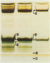
This study investigates the physical basis of color effects in the detection of proteins in polyacrylamide gels by silver staining. Specifically, the hypothesis that different colors may correlate with the development of silver grains of characteristic sizes was investigated by electron microscopy. Protein bands that stained brown, yellow, and blue were excised from stained gels and prepared for electron microscopy by thin-sectioning. In each case, the size distributions of globular silver grains were determined directly from the electron micrographs. We found that blue bands have larger silver grains (with diameters of 40-100 nm) than yellow (21-39 nm) or brown bands (17-35 nm). On the basis of these and other observations, a general mechanism is proposed whereby chemical specificity of electrophoretically separated proteins is expressed in color-specific silver staining.
Full text
PDF


Images in this article


Selected References
These references are in PubMed. This may not be the complete list of references from this article.
- Dzandu J. K., Deh M. E., Barratt D. L., Wise G. E. Detection of erythrocyte membrane proteins, sialoglycoproteins, and lipids in the same polyacrylamide gel using a double-staining technique. Proc Natl Acad Sci U S A. 1984 Mar;81(6):1733–1737. doi: 10.1073/pnas.81.6.1733. [DOI] [PMC free article] [PubMed] [Google Scholar]
- Goldman D., Merril C. R., Ebert M. H. Two-dimensional gel electrophoresis of cerebrospinal fluid proteins. Clin Chem. 1980 Aug;26(9):1317–1322. [PubMed] [Google Scholar]
- Knott G. D. Mlab--a mathematical modeling tool. Comput Programs Biomed. 1979 Dec;10(3):271–280. doi: 10.1016/0010-468x(79)90075-8. [DOI] [PubMed] [Google Scholar]
- Labaw L. W., Padlan E. A., Segal D. M., Davies D. R. An em study of phosphorylcholine-binding fab immunoglobulin fragment crystals. J Ultrastruct Res. 1975 Jun;51(3):326–339. doi: 10.1016/s0022-5320(75)80097-9. [DOI] [PubMed] [Google Scholar]
- Laemmli U. K. Cleavage of structural proteins during the assembly of the head of bacteriophage T4. Nature. 1970 Aug 15;227(5259):680–685. doi: 10.1038/227680a0. [DOI] [PubMed] [Google Scholar]
- Merril C. R., Pratt M. E. A silver stain for the rapid quantitative detection of proteins or nucleic acids on membranes or thin layer plates. Anal Biochem. 1986 Jul;156(1):96–110. doi: 10.1016/0003-2697(86)90160-0. [DOI] [PubMed] [Google Scholar]
- Merril C. R., Switzer R. C., Van Keuren M. L. Trace polypeptides in cellular extracts and human body fluids detected by two-dimensional electrophoresis and a highly sensitive silver stain. Proc Natl Acad Sci U S A. 1979 Sep;76(9):4335–4339. doi: 10.1073/pnas.76.9.4335. [DOI] [PMC free article] [PubMed] [Google Scholar]
- Morrissey J. H. Silver stain for proteins in polyacrylamide gels: a modified procedure with enhanced uniform sensitivity. Anal Biochem. 1981 Nov 1;117(2):307–310. doi: 10.1016/0003-2697(81)90783-1. [DOI] [PubMed] [Google Scholar]
- Nielsen B. L., Brown L. R. The basis for colored silver-protein complex formation in stained polyacrylamide gels. Anal Biochem. 1984 Sep;141(2):311–315. doi: 10.1016/0003-2697(84)90047-2. [DOI] [PubMed] [Google Scholar]
- Switzer R. C., 3rd, Merril C. R., Shifrin S. A highly sensitive silver stain for detecting proteins and peptides in polyacrylamide gels. Anal Biochem. 1979 Sep 15;98(1):231–237. doi: 10.1016/0003-2697(79)90732-2. [DOI] [PubMed] [Google Scholar]