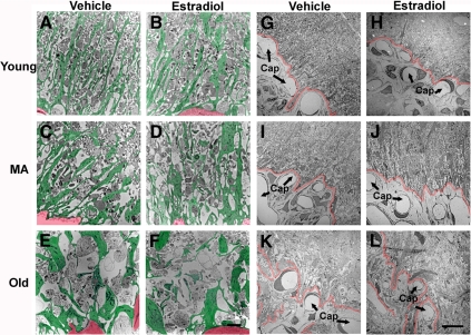
Figure 4.
Electron microscopic images (low magnification) showing the organization of the pericapillary zone of the median eminence. A–F, The pattern of glial processes in the pericapillary zone of the median eminence from one representative rat per group is shown. Glial processes are pseudocolored in green, and a corner of the portal capillary plexus (presented for orientation) is pseudocolored in red. Glial processes of young rats, irrespective of hormone treatment, tended to be narrow and linearly oriented toward the portal capillary system. These glial processes became larger and wider and this radial orientation diminished with age. Scale bar (A–F), 2 μm. G–L, The boundary between the pericapillary zone and the portal capillary region (Cap; arrows) is indicated in red for representative rats of each group. This boundary appeared to become increasingly convoluted and less clearly delineated with age. Scale bar (G–L), 10 μm. MA, Middle-aged.

