Abstract
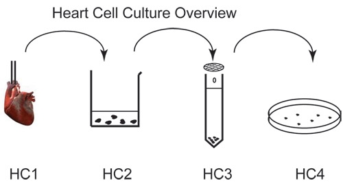
Cultured primary adult rodent heart cells are an important model system for cardiovascular research. Nevertheless, establishment of robust, viable cultured adult myocytes can be a technically challenging, rate-limiting step for many researchers. Here we described a protocol to obtain a high yield of adult rat heart myocytes that remain viable in culture for several days. The heart is isolated and perfused with collagenase and protease under low Ca2+ conditions to recover single myocytes. Ca2+-tolerant cells are obtained by stepwise increases in extracellular Ca2+ concentration in three subsequent wash steps. Cells are filtered, resuspended in culture medium, and plated on laminin coated slips. Cultured myocytes obtained using this protocol are viable for up to four days and are suitable for most experiments including electrophysiology, biochemistry, imaging and molecular biology.
Protocol
Part I. Preparation for surgery
The day before the rat surgery, make certain that all solutions are made, but do not add any enzymes yet. Buffers A, B and the wash buffer should all be filtered for sterility prior to storage. The buffers should all be stored at 4 °C.
6-well tissue culture plates must be plated with 10-20 μg/mL laminin in 2 mL 1XSFM the day before as well, and should be stored at 37 °C in a CO2 incubator.
On the day of the surgery, prepare the surgical instruments by sterilizing the clamp, scissors, and forceps with 70 % ethanol and let them dry on paper towel.
A 1 mL syringe filled with 0.5 mL Heparin Solution (90 U/ml) is made ready.
Now it is good practice to wash the perfusion apparatus by running through 70% ethanol, using the peristaltic pump in reverse. Once finished rinsing with ethanol, rinse the apparatus with double distilled water, and finally rinse with Buffer A. The flow through is collected in a beaker placed in a 37 °C water bath.
To prepare the cleaned perfusion apparatus, fill the right syringe tube with 40 mL of Buffer A and the left syringe tube with 40 mL of Buffer B.
Prime the perfusion tubing to the tip of the catheter by allowing Buffer A to flow through the apparatus. Refill to the buffer A syringe to the 40 mL level. Make sure there are no air bubbles trapped in the tubing after this step.
Now, close the buffer A syringe stopcock valve and repeat the process with Buffer B such that the perfusion tubing is now primed with Buffer B.
Finally, the perfusion apparatus is left with the Buffer B stopcock valve open, but with the flow stopped using the regulator on the IV line.
Discard the buffer flow-though in the collecting beaker.
The last thing to do before the surgery is to add the required enzymes to your stock solution, and then filter the enzyme solution just as the other solutions were filtered the day before. Once filtered, this solution can be left at room temperature.
Part II. The Rat heart dissection
Following standard protocol euthanize the rat with Isoflurane in a gas chamber.
Sterilize the incision area on the thorax with 70% ethanol.
Open chest using scissors and expose heart.
Inject left ventricle with 0.5 mL Heparin solution (90 U/mL).
Remove heart with large portion of aorta intact, place in ice cold Buffer B.
Part III. Myocytes isolation
A typical rat heart should yield a high fraction of rod-shaped myocytes (as opposed to dead rounded cells) if the following procedure is properly adhered to.
To begin, clamp aorta to the catheter. The tip of the catheter should not be pushed too far into the heart to ensure good perfusion of the heart through the coronary artery.
Once clamped, tie the aorta to the catheter with a suture.
Next, open the IV line regulator to allow a fast drip rate (~60 drops/min) and allow Buffer B to wash out the heart.
During Buffer B wash, use the small scissors and forceps to remove the atria and any fatty or lung tissue clinging to the heart.
After Buffer B is finished, switch the flow to Buffer A.
While Buffer A is allowed to flow for 5 minutes, several small tasks should be quickly performed.
First, warm the Enzyme Solution in a 37 °C water bath.
Finally, when Buffer A is depleted from the syringe (but still present in the tubing), load 50 mL of the Enzyme Solution in syringe A of the perfusion apparatus
It is now necessary to discard all the flow-through from the collection beaker in the water bath (by pipetting), until the Enzyme Solution perfuses heart.
As soon as the Enzyme Solution perfuses the heart, activate the peristaltic pump which is set up to transfer the enzyme solution from the collecting beaker to replenish syringe A.
Allow the enzyme solution to flow through the heart for 10 min. As the heart digests, it begins to look bloated.
Add 37.5 μL of 0.1 M CaCl2 to the enzyme solution in syringe A to give an effective concentration of 0.1 mM Ca2+.
After 10 minutes increase the concentration of calcium to 0.2 mM by adding an additional 50 μL of 0.1 M CaCl2 to syringe A and let the perfusion proceed for a further 10 minutes.
Cut off the ventricles and transfer to a small sterile beaker containing 20 mL Enzyme Solution.
In the beaker increase the Calcium concentration to 0.4 mM by adding 40 more μL of 0.1 M CaCl2.
Gently mince the heart into 10 or more pieces with a pair of small sterile scissors.
Incubate the beaker for 5 minutes at 37 °C with gentle rocking.
Add 40 mL of 0.1 M CaCl2 to give effective concentration of 0.6 mM Ca2+ and gently triturate the heart pieces with a plastic transfer pipet 3-5 times.
After incubating for an additional 5 minutes at 37 °C, add 40 mL of 0.1 M CaCl2 and gently triturate as before.
Separate digested single myocytes from non-digested connective tissue using filtration through a sterile 500 μm mesh.
Allow the cells to settle in a 50 ml tube for 10 minutes at room temperature.
Discard supernatant with a transfer pipet.
Gently resuspend cells in Wash Buffer #1 and allow the cells to settle for 10-20 min at room temperature.
While the cells are settling, take a small aliquot, and use this time as an opportunity to assess the quality and viability of the cells in suspension under an inverted microscope. A good preparation will have a high proportion (>80%) of rod like cells with crisp striations.
Once the cells have settled, discard the supernatant and gently resuspend cells in Wash Buffer #2. Let the cells settle at room temperature, which should again take 10-20 minutes, and discard the supernatant.
Repeat the washing process once more with Wash Buffer #3, and the cells will be ready for culture.
Part IV. Myocyte cell culture
Gently resuspend cells in 5% Serum Media. Before transferring the cells to tissue culture plates, wash the plates with 1x SFM. Transfer the cells at a desired density and incubate in a CO2 tissue culture incubator for 3-5 hours.
Switch the media to 1x SFM.
Cells can be cultured for up to four days and used as required for experiments.
Part V. Representative Results/Outcome
There should be more than 70% live heart myocytes under inverted microscope when the protocol is done correctly.

Table 1: Solution recipe for 1 L 10X Ca2+-free KH Buffer:
| mass | Final Concentration | |
| NaCl | 68.96 g | 1180 mM |
| KCl | 3.58 g | 48 mM |
| HEPES | 59.58 g | 250 mM |
| K2HPO4 | 2.85 g | 12.5 mM |
| MgSO4 | 3.08 g | 12.5 mM |
Table 2: Solution recipe for 1 L Buffer A:Add 1.98 g glucose to 100 mL 10X KH buffer and bring to a final volume of 1 L. Adjust osmolarity to near physiology condition (~300) with glucose. Bring to pH 7.4 with NaOH.
| - | Mass | Final Concentration |
| 10X KH Buffer | 100 mL | 100 ml |
| Glucose | 1.98 g | 11 mM |
| Bring to 1 L | ||
| Bring to pH 7.4 with NaOH | ||
| Adjust osmolarity to near physiological condition(~300) with glucose. |
Table 3: Solution recipe for EnzymeBuffers
| mass | Final Concentration | |
| Collagenase Type II | 25 mg | 0.05% (w/v) |
| Protease XIV | 10 mg | 0.02% (w/v) |
| BDM | 0.025 g | 5 mM |
| Carnitine | 0.020 g | 2 mM |
| Taurine | 0.031 g | 5 mM |
| Glutamic Acid | 0.020 g | 2 mM |
| 0.1 M CaCl2 | 12.5 uL | 25 uM |
| Buffer A | Fill to 50 ml |
Table 4: Solution recipe for 25X BDM/Taurine/BSA (B/T/B)
| mass | Final Concentration | |
| BDM | 0.076 g | 125 mM |
| Taurine | 0.094 g | 125 mM |
| BSA | 0.1% (w/v) | |
| Buffer A | Fill to 6 mL |
Table 5: Solution recipe for Buffer B and Wash Buffers
| Buffer: | B | #1 | #2 | #3 |
| Add: | ||||
| 0.5 M CaCl2 | 0.4 mL | 0.1 mL | 0.125 mL | 0.15 mL |
| Final concentration of CaCl2 | 1 mM | 1 mM | 1.25 mM | 1.5 mM |
| 25X B/T/B | --- | 2 mL | 2 mL | 2 mL |
| Buffer A to final volume | 200 mL | 50 mL | 50 mL | 50 mL |
Table 6: Solution Recipe for Culturing Media
| 2X Serum-free Media (SFM) | ||
| Final Concentration | ||
| (after dilution) | ||
| Carnitine | 0.099 g | 10 mM (5 mM) |
| Taurine | 0.063 g | 10 mM (5 mM) |
| Creatine | 0.075 g | 10 mM (5 mM) |
| Antibiotic/Antimycotic | 0.5 ml | 1% (0.5%) (v/v) |
| Media 199 | 45 mL | Fill to 50 mL |
| 1X SFM | ||
| Final Concentration | ||
| 2X SFM | 25 mL | 1X |
| Media 199 | Fill to 50 mL | |
| 1X 5% Serum Media | ||
| Final Concentration | ||
| 2X SFM | 25 mL | 1X |
| FBS | 2.5 mL | 5% (v/v) |
| Media 199 | Fill to 50 mL |
Discussion
A critical step is the speed with which the isolated heart is hung up on the perfusion system. The length of the enzymatic digestion period may be a little different for each rat. The adjustment depends on how soft the heart becomes after the regular period of digestion. The slowly recovery of Ca2+ after enzyme digestion is essential for obtaining Ca2+-tolerant healthy cells.
For guinea pig, the protocol is the same except hyaluronidase is used instead of Protease XIV. Typically, we find the that initial percentage of living cells is lower for guinea pig compared to rat, although they survive just as long in culture.
References
- Miriyala J, Nguyen T, Yue DT, Colecraft HM. Role of CaVbeta subunits, and lack of functional reserve, in protein kinase A modulation of cardiac CaV1.2 channels. Circ Res. 2008;102(7):e54–e64. doi: 10.1161/CIRCRESAHA.108.171736. [DOI] [PubMed] [Google Scholar]
- SX Takahashi, Mittman S, Colecraft HM. Distinctive modulatory effects of five human auxiliary beta2 subunit splice variants on L-type calcium channel gating. Biophys. 2003:84–845. doi: 10.1016/S0006-3495(03)70027-7. [DOI] [PMC free article] [PubMed] [Google Scholar]


