Abstract
Recurrent hepatitis C infection (HCV) following liver transplantation causes accelerated allograft cirrhosis. Here we characterized HCV-specific immunity in adult liver transplant recipients (n=74) with and without allograft cirrhosis. Patients were divided into hepatic inflammation/ no cirrhosis (METAVIR scores 0–2, HIN) and hepatic cirrhosis (score 3–4, HFC). As control, 20 normal subjects and 10 non-HCV liver transplant patients were included. Twenty-five different serum cytokines were analyzed using LUMINEX. Frequency of T-cells specific to HCV-derived proteins (NS3, NS4, NS5, Core) was characterized using ELISPOT immunoassays. There was no difference in clinical characteristics between HIN (n=49) and HFC (n=25) groups. HIN group had high serum IFN-γ and IL-12 while HFC demonstrated elevated IL-4, IL-5, and IL-10 (p<0.01). HCV (NS3, NS4, NS5, Core) -specific IFN-γ producing CD4+ T-cells were elevated in the HIN group whereas the HFC patients showed predominance of HCV-specific IL-5 and IL-10 producing CD4+ T-cells.
Conclusions
Lack of HCV-specific Th1-type T-cell immunity is observed in liver transplant recipients with advanced allograft cirrhosis.
Keywords: HCV, immunity, and liver allograft cirrhosis
Introduction
Chronic hepatitis C infection has emerged as the leading indication for liver transplantation. Recent data reveal that end-stage liver disease secondary to HCV accounts for about 40–45% of all transplants (1). However, HCV recurrence following transplantation is a universal phenomenon. HCV RNA can be detected in virtually all patients post-transplant (2, 3) with at least 50% of recipients demonstrating histological evidence of HCV induced hepatitis at 1-year (4). Post-transplant hepatitis C viral load at 1-year is typically about 10–20 fold greater than the pre-transplant levels (5). The progression to liver allograft cirrhosis following HCV recurrence is significantly enhanced, with a median time to cirrhosis of 10 years, compared to 30–40 years in HCV-infected immunocompetent patients (6, 7). Liver allograft failure and mortality rates are higher in patients with post-transplant HCV recurrence than non-HCV infected recipients. Previous reports have demonstrated that over 25% of patients with HCV recurrence may die or require re-transplantation within 5 yrs post-transplantation (8). Whether there is a subset of patients with HCV recurrence that is pre-disposed to early onset cirrhosis and liver allograft failure remains unknown.
Studies performed in non-transplant patients with chronic HCV infection have indicated that T cell-mediated immunity is important in determining the outcome of HCV infection (9–11). Patients that clear the infection have a broader and more potent HCV specific immune response than those with chronic infection. CD4+ T lymphocytes of patients that clear HCV have been shown to produce high levels of IFN-γ in response to HCV stimulation compared to patients with chronic infection. In contrast, chronic infection is favored by a Th2 cytokine profile (IL-4, IL-10) (12, 13).
In the transplant setting, Schirren et al demonstrated the presence of HCV-specific immunity in recipients with recurrent HCV by developing T cell lines both from the peripheral blood as well as allograft infiltrating lymphocytes (14). However, the nature of these HCV-specific T cells and their role in the progression of HCV induced allograft cirrhosis needs to be defined. Zekry et al measured intrahepatic mRNA content of cytokines and demonstrated that cholestatic hepatitis, a severe form of HCV-induced hepatitis, is associated with IL-10 and IL-4 (Th2 cytokines) (15). However, mRNA may not translate into proteins. Furthermore, the source of these cytokines is unknown since the analysis was done using total extracts from hepatic biopsy specimens.
The aim of the present study was to characterize the peripheral blood cytokine profile and HCV-specific T cell response in patients with chronic HCV recurrence and determine their correlation with development of allograft cirrhosis. Our premise was that the OLT recipients with hepatic inflammation alone and no fibrosis would have HCV-specific Th1-immunity to HCV while those with allograft fibrosis/cirrhosis would develop Th2-predominant immunity.
Materials and Methods
Study Subjects
Study subjects included adult OLT recipients transplanted for end-stage liver disease secondary to HCV infection and subsequently developed HCV recurrence. Patients with co-existing hepatitis B infection were excluded. The transplant recipients had undergone a sequential immunosuppressive protocol consisting of methylprednisolone, 1000 mg preoperatively and subsequent tapering to 20 mg daily, 5 days after transplantation. The steroids were eventually weaned off at 6 months. Tacrolimus or cyclosporine were given at a dose of 0.5 mg/kg and 4 mg/kg starting on day 1 after transplantation, to obtain a serum trough level of 6–10 ng/ml and 300–400 mg/ml respectively. In addition, patients also received mycophenolic acid (either Cellcept 1 gm twice a day or myfortic 720 mg twice a day) following transplantation. Patients that developed renal insufficiency were transitioned from calcineurin inhibitors to sirolimus (5 mg bolus and then 3 mg once daily to maintain a levels between 6–10 ng/ml). HCV recurrence was confirmed by the presence of anti–HCV antibody by second-generation enzyme-linked immunoabsorbent assay (ELISA) (Abbot Laboratories, Chicago, IL, USA), positive semi quantitative reverse-transcriptase polymerase chain reaction specific for HCV RNA in peripheral blood (Roche Diagnostics, Branchburg, NJ, USA), and by histological changes on liver biopsy. No other cause of hepatitis other than HCV infection was identified in these individuals. To avoid any confounding effect of the early post-transplant ischemia-reperfusion injuries and other technical issues, patients were only included in the study if they were at least one year post-transplant. Twenty HCV negative, healthy control individuals and ten non-HCV liver transplant recipients were also included in this study. The study was performed according to the guidelines of the Declaration of Helsinki and approved by the Institutional Review Board of Washington University School of Medicine. Informed written consent was obtained from all recipients and controls.
Liver Histology
Pathology of the liver in chronic HCV infection was classified by the standard METAVIR scoring system for necro-inflammatory grade of activity and stage of fibrosis (16). Liver biopsies were reviewed by a panel of pathologists to determine the presence or absence of bridging fibrosis or cirrhosis, and subjects were categorized accordingly. The necro-inflammatory histology of the included study subjects was consistent with HCV recurrence but none had cholestatic hepatitis. The following scores were used as the different stages of fibrosis: 0, no fibrosis; 1, minimal fibrosis without any septa; 2, portal fibrosis with rare septa; 3, numerous septa with bridging fibrosis without cirrhosis; and 4, cirrhosis. Recipients with METAVIR stage of 0, 1, and 2 were classified into a group with predominant histology of inflammation (HIN) alone without any bridging fibrosis or cirrhosis while those with stage of 3 and 4 were classified into a group with predominant histology of fibrosis/cirrhosis (HFC) (Table 1). This nomenclature does not imply that patients in the HFC did not have any inflammation.
Table 1.
Demographic and clinical profile of study subjects
| HIN* (n=49) | HFC* (n=25) | p value | |
|---|---|---|---|
| Age | 51.7 ± 6.77 | 52.5 ± 5.98 | 0.9 |
| Gender (M:F) | 26 : 23 | 13 : 12 | 0.7 |
| Fibrosis stage | 0–2 | 3–4 | |
| HCV genotype | α | β | |
| 1 | 29 | 14 | |
| Non-1 | 6 | 4 | |
| HCV load (log IU/ml) | |||
| Pre-Transplant | 13.57 ± 12.46 | 13.67 ± 12.27 | 0.7 |
| HCV load (IU/ml) | |||
| Post-transplant | 15.78 ± 13.68 | 15.75 ± 13.42 | 0.7 |
| Pre-transplant CTP Score | |||
| 5–6 | 10 | 6 | |
| 7–9 | 28 | 12 | |
| 10–15 | 11 | 7 | 0.6 |
| CMV Status | γ | δ | |
| D+R− | 7 (14.3%) | 4 (16%) | |
| D−R+ | 7 (14.3%) | 3 (12%) | 0.7 |
| D+R+ | 21 (42.9%) | 8 (32%) | |
| D−R− | 6 (12.2%) | 5 (20%) | |
| Coexistent Alcohol | 17 | 10 | 0.8 |
| Immunosuppression | |||
| Tacrolimus | 16 (64%) | 0.48 | |
| Cyclosporine | 32 (65.3%) | 7 (24%) | |
| Sirolimus | 8 (16.3%) | 3 (12%) | |
| MMF | 9 (18.4%) | 12 (48%) | 0.9 |
| Myfortic | 16 (32.7%) | 2 (8%) | |
| AZA | 2 (4.1%) | 0 | |
| Prednisone | 0 | 5 (20%) | |
| 12 (24.5%) | |||
| No. acute rejection | 15 (30.1%) | 9 (36%) | 0.9 |
|
Mean cumulative solumedrol (gms) |
0.18 | 0.15 | 0.8 |
HIN= Hepatic Inflammation, HFC= Hepatic Fibrosis/ cirrhosis, D=Donor, R=Recipient
α, β, γ, δ not available in 14, 7, 8, and 5 cases, respectively.
Antigens
Recombinant HCV core, NS3, NS4, NS5 and mumps antigen was obtained from Viral Therapeutics, Inc (Ithaca, NY, USA). All the four proteins were endotoxin-free as tested by LAL assay kit (Charles Rivers, Charleston, SC, USA).
Isolation of peripheral blood mononuclear cells from blood
Peripheral blood mononuclear cells (PBMC) were isolated from whole blood obtained from HCV infected OLT recipients and normal healthy control individuals by density gradient centrifugation using Ficoll-Paque (Amersham Pharmacia, Uppsala, Sweden) and resuspended in RPMI-1640 medium (Gibco BRL, Grand Island, NY, USA) supplemented with 10% FBS (Hyclone, Logan, UT, USA).
Enzyme-LinkedImmunospot Assay
Enzyme-Linked immunospot assay (ELISPOT) was carried out as described previously (12). Briefly, Immunospot plates (Pharmingen, San Diego, CA, USA) were coated with a monoclonal antibody to IFN-γ, IL-5 and IL-10 (5 µg/ml, Pharmingen) at 4°C overnight. Then the plates were blocked with bovine serum albumin (1%) for 1 hour and washed three times with phosphate-buffered saline. The PBMCs were then cultured in triplicate wells of the ImmunoSpot plates (3 × 10⁵/200 µL) in the presence of recombinant HCV proteins core, NS3, NS4 and NS5 (5 µg/ml) for 48 h. After the incubation times, the plates were washed with PBS three times and then with PBS/ Tween-20 (0.5%) three times. Then biotinylated monoclonal antibodies specific for IFN-γ (3 µg/ml, Pharmingen) or IL-5 (3 µg/ml, Pharmingen) or IL-10 (3 µg/ml, Pharmingen) resuspended in phosphate-buffered saline/bovine serum albumin/Tween-20 were added to the plates. After 12 hours at 4°C, the plates were washed (three times) and horseradish peroxidase–labeled streptavidin (1:2000, Pharmingen) was added for 2 hours at room temperature. The spots were developed using -amino-9-ethylcarbazole solution (Pharmingen). Then the plates were washed and spots were analyzed in an ImmunoSpot Image Analyzer (Cellular Technology Ltd., Cleveland, OH, USA). Cells cultured in medium alone were used as negative control. Cells cultured in the presence of the PHA (10 µg/ml) and mumps antigen (5 µg/ml) were used as positive controls for CD4+ T cell responses. The number of spots observed in the negative control wells was subtracted from the number of spots observed in the experimental wells.
Luminex Assay
Plasma levels of cytokines (IL-1β, IL-1ra, IL-2, IL-2R, IL-4, IL-5, IL-6, IL-7, IL-8, IL-10, IL-12p40, IL-13, IL-15, IL-17, TNF-α, IFN-α, IFN-γ, GM-CSF), and chemokines (MIP-1γ, MIP-1β, IP-10, MIG, Eotaxin, RANTES, MCP-1) in HCV infected OLT recipients were analyzed using the solid phase sandwich Multiplex Bead immunoassays (Invitrogen, Carlsbad, CA, USA) as previously described (17). Briefly, primary antibody coated beads and incubation buffer were pipetted into 96-well filter plates (Fisher, Pittsburgh, PA, USA). The standards and samples were incubated in the presence of the primary antibody beads for 2 hours at room temperature on an orbital shaker. Following this, the wells were washed twice with the wash buffer provided using a vacuum manifold and then biotinylated detector antibodies were added to the wells. After further incubation for 30 minutes at room temperature on an orbital shaker, the wells were washed twice and streptavidin-RPE solution was added for 15 minutes. Finally, the wells were washed thoroughly and read using the Luminex xMAPTM system. The cytokine levels were calculated against the mean fluorescence intensity (MFI) of the standards and expressed as pg/ml.
Statistical Analysis
The Mann-Whitney U test was used to determine the differences in CD4+ T-cell responses between the patients (HIN group vs. HFC group). Correlations were calculated using the Spearman rank test. The alpha was set a priori at p < 0.05.
Results
Clinical characteristics of HCV infected OLT recipients
In this study that included 74 subjects, 49 exhibited hepatic inflammation and no cirrhosis (HIN group) on hepatic biopsy while 25 showed fibrosis/cirrhosis (HFC). The clinico-demographic profile of these patients is summarized in Table 1. There was no significant difference in the mean age, CTP score, HCV RNA load, HCV genotype, concomitant alcohol use, and CMV co-infection. In addition, there was no significant difference in the immunosuppressive regimen, number of acute rejection episodes, and mean pulse steroid dose used for acute rejection, in the two groups (Table 1). The study excluded patients with concomitant Hepatitis B infection. Further, to avoid any confounding effect of early post-transplant ischemia-reperfusion injury and other technical issues, patients were only included in the study if they were at least one year post-transplant. For the HIN and HFC groups the mean sampling interval was 47.23 ± 24.80 and 52.65 ± 20.52 months, respectively (p=0.78). We also analyzed serum cytokines in ten non-HCV transplant recipients for control. The mean age of these patients was 49 ± 9.8 years, male : female ratio 7:3, etiology of liver disease was ethanol (6), primary biliary cirrhosis (2), and cryptogenic (2). The samples were obtained at comparable time intervals as the study group counterparts (44.21 ± 21.54 months).
Analysis of serum cytokines in patients with HCV recurrence
All study subjects had evidence of chronic hepatitis associated with HCV recurrence on hepatic biopsy. In order to test our hypothesis that HCV induced cirrhosis is associated with lack of Th1 response, we first analyzed 25 different cytokines and chemokines relevant in the transplant setting. These included: IL-1β, IL-1ra, IL-2, IL-2R, IL-4, IL-5, IL-6, IL-7, IL-8, IL-10, IL-12p40, IL-13, IL-15, IL-17, TNF-α, IFN-α, IFN-γ, GM-CSF, MIP-1γ, MIP-1β, IP-10, MIG, Eotaxin, RANTES, MCP-1. The analysis was done using the LUMINEX immunoassays. As shown in Table 2, patients with HIN revealed significantly increased levels of Th1-cytokines compared to the HFC and control groups (IFN-γ: HIN 910 ± 190, HFC 54 ± 15, Controls 51 ± 31 pg/ml, p=0.019; IL-12: HIN 987 ± 97, HFC 89 ± 28, Controls 98 ± 39 pg/ml, p=0.020). In contrast, HFC patients had predominance of the typical Th2-cytokines (IL-4: HFC 473 ± 66, HIN 40 ± 15, Controls 54 ± 37 pg/ml, p=0.029; IL-5: HFC 235 ± 61, HIN 17 ± 9, Controls 21 ± 11 pg/ml, p=0.021; IL-10: HFC 980 ± 113, HIN 193 ± 29, Controls 36 ± 16 pg/ml, p=0.001). There was no significant difference in the levels of the other cytokines (IL-1, IL-1ra, IL-2, IL-2R, IL-6, IL-7, IL-8, IL-13, IL-15, IL-17, TNF-α, IFN-α, GM-CSF) and chemokines (MIP-1 γ, MIP-1 β, IP-10, MIG, Eotaxin, RANTES, MCP-1) that were also analyzed (data not shown). These data suggested that while OLT recipients with hepatic fibrosis following HCV recurrence develop a Th2-predominant response to HCV, patients more resistant to hepatic fibrosis following HCV recurrence mount a Th1-predominant response.
Table 2.
Plasma levels of cytokines in OLT patients
| HIN n=49 |
HFC n=25 |
Healthy controls n=20 |
Non-HCV Transplant n=10 |
p (HIN vs HFC) |
|
|---|---|---|---|---|---|
| IFN-γ | 910 ± 190 | 54 ± 15 | 51 ± 31 | 47 ± 44 | 0.019 |
| IL-4 | 40 ± 15 | 473 ± 66 | 54 ± 37 | 94 ± 32 | 0.029 |
| IL-5 | 17 ± 9 | 235 ± 61 | 21 ± 11 | 78 ± 29 | 0.021 |
| IL-10 | 193 ± 29 | 980 ± 113 | 36 ± 16 | 143 ± 45 | 0.001 |
| IL-12 | 987 ± 97 | 89 ± 28 | 98 ± 39 | 77 ± 33 | 0.02 |
HIN= Hepatic Inflammation
HFC= Hepatic Fibrosis/ cirrhosis
All values expressed as pg/ml
Expansion of HCV-specific Th1-type CD4+ T cells is associated with absence of hepatic cirrhosis following HCV recurrence
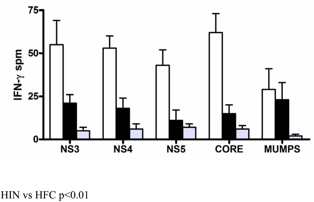
The above results suggested that Th2-predominant immunity was associated with hepatic cirrhosis following HCV recurrence. However, serum cytokines are non-specific and can vary due to the natural state of the recipients. Therefore, we next analyzed the HCV specific T cell response in these subjects. First, the frequency of peripheral IFN-γ producing Th1-type T cells reactive to HCV derived proteins NS3, NS4, NS5 and core was determined by ELISPOT assay. The frequency of these IFN-γ producing T cells was expressed as IFN-γ spots per 106 PBMCs (spm). As shown in Figure 1, HIN group exhibited a high frequency of IFN-γ producing T cells to all the four viral proteins: NS3 (55 ± 14 spm), NS4 (53 ± 7 spm), NS5 (43 ± 9 spm) and core (62 ± 11 spm), when compared to the HFC group: NS3 (21 ± 5 spm, p=0.025), NS4 (18 ± 6 spm, p=0.031), NS5 (11 ± 6 spm, p=0.011) and core (15 ± 5 spm, p=0.018). In contrast, the frequency of IFN-γ producing T cells specific to a common third-party mumps antigen was not statistically different between the two groups (HIN: 29 ± 12 spm Vs HFC: 23 ± 10 spm, p=0.45). PBMCs isolated from the healthy control did not show a significant response to any of the four HCV derived proteins (NS3: 5 ± 2 spm, NS4: 6 ± 3 spm, NS5: 7 ± 2 spm, Core: 6 ± 2 spm). The IFN-γ producing T cells were predominantly CD4+ since depletion of CD4+ T cells from the PBMCs lead to >70% loss of IFN-γ production in both the HIN and HFC groups (n=5 each, data not shown). Taken together, this indicates that OLT recipients belonging to HIN group with no hepatic cirrhosis develop an expansion of Th1-type CD4+ T cells specific to HCV antigens.
Figure 1. Lack of HCV-specific IFN-γ producing T lymphocytes in OLT recipients with HCV-induced allograft cirrhosis.
The frequency of peripheral blood T lymphocytes specific to HCV derived proteins, NS3, NS4, NS5 and core was determined by means of IFN-γ ELISPOT assay. The data is representative of all experiments done in triplicate and the standard error bars are demonstrated. Patients with hepatic inflammation (HIN, n=49, white bars) demonstrated significantly higher frequency of IFN-γ producing HCV-specific T lymphocytes compared to patients with hepatic fibrosis (HFC, n=25, black bars) and healthy control individuals (n=20, grey bars, p<0.05). The frequency of T lymphocytes reactive to the mumps antigen was comparable.
Hepatic cirrhosis following HCV recurrence in OLT recipients is associated with development of Th2-type HCV specific CD4+ T cells
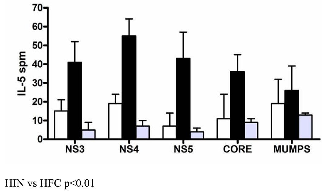
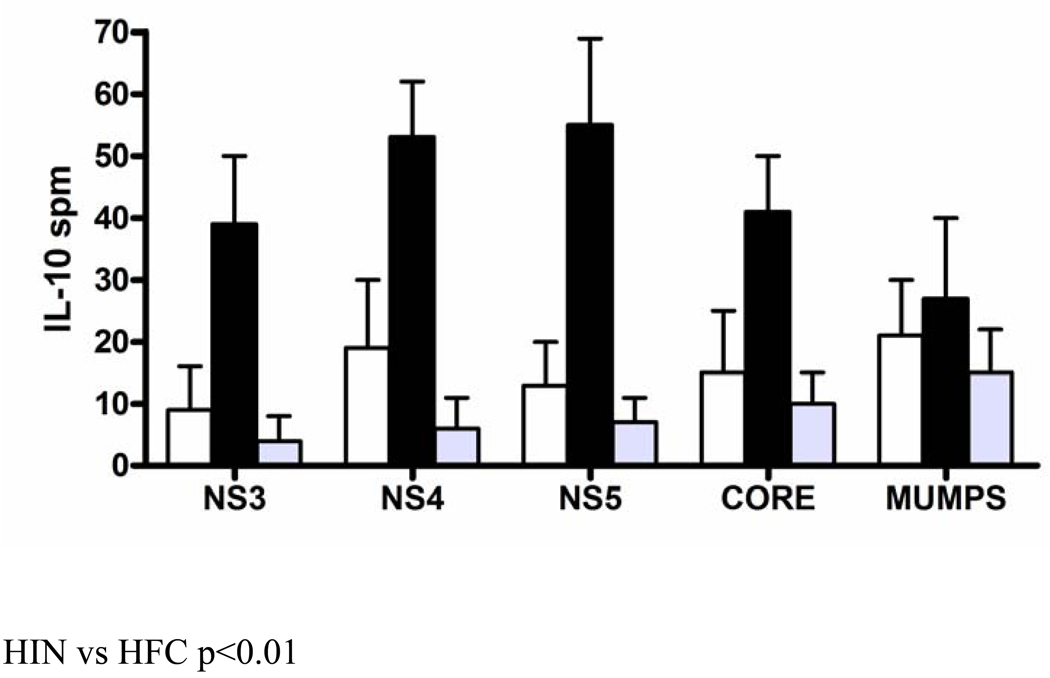
IL-5 and IL-10 belong to the Th2-cytokines and were found to be elevated in the HFC group. We further hypothesized that the Th2-predominant immunity observed in the HFC group would be associated with an expansion of Th2-type HCV-specific CD4+ T cells. The frequency of IL-5 and IL-10 reactive T cells specific to HCV derived proteins NS3, NS4, NS5 and core was determined by IL-5 and IL-10 ELISPOT assays, respectively. As shown in Figure 2, HFC group demonstrated an increased frequency of IL-5 producing Th2-type T cells to all four viral proteins: NS3 (41 ± 11 spm), NS4 (55 ± 9 spm), NS5 (43 ± 14 spm) and core (36 ± 9 spm) when compared to the HIN group: NS3 (15 ± 6 spm, p=0.011), NS4 (19 ± 5 spm, p=0.028), NS5 (7 ± 7 spm, p=0.015) and core (11 ± 13 spm, p=0.023). The normal subjects did not produce IL-5 upon stimulation with the HCV derived proteins (NS3: 5 ± 4 spm, NS4: 7 ± 3 spm, NS5: 4 ± 2 spm, Core: 9 ± 2 spm). Similarly, as demonstrated in Figure 3, the HFC group exhibited an expansion of Th2-type IL-10 producing T cells specific to all four viral proteins: NS3 (39 ± 11 spm), NS4 (53 ± 9 spm), NS5 (55 ± 14 spm) and core (41 ± 9 spm) when compared to the HIN group: NS3 (9 ± 7 spm, p=0.031), NS4 (19 ± 11 spm, p=0.033), NS5 (13 ± 7 spm, p=0.017) and core (15 ± 10 spm, p=0.027). The normal subjects did not produce IL-10 in response to the HCV derived proteins (NS3: 4 ± 4 spm, NS4: 6 ± 5 spm, NS5: 7 ± 4 spm, Core: 10 ± 5 spm). The frequency of IL-5 and IL-10 producing T cells specific to the common third-party mumps antigens was comparable between the two groups (Figure 2 and Figure 3). Depletion of CD4+ T cells again lead to almost complete loss of both IL-5 and IL-10 production in either group (n=5 each, data not shown). Taken together, these data indicate that hepatic cirrhosis following HCV recurrence in patients with OLT is associated with HCV-specific Th2-type immunity.
Figure 2. High frequency of HCV-specific IL-5 producing T lymphocytes in OLT recipients with HCV-induced allograft cirrhosis.
The frequency of peripheral blood T lymphocytes specific to HCV derived proteins, NS3, NS4, NS5 and core was determined by means of IL-5 ELISPOT assay. The data is representative of all experiments done in triplicate and the standard error bars are demonstrated. Patients with hepatic fibrosis (HFC, n=25, black bars) demonstrated significantly higher frequency of IL-5 producing HCV-specific T lymphocytes compared to patients with hepatic inflammation (HIN, n=49, white bars) and healthy control individuals (n=20, grey bars, p<0.05). The frequency of T lymphocytes reactive to the mumps antigen was comparable.
Figure 3. High frequency of HCV-specific IL-10 producing T lymphocytes in OLT recipients with HCV-induced allograft cirrhosis.
The frequency of peripheral blood T lymphocytes specific to HCV derived proteins, NS3, NS4, NS5 and core was determined by means of IL-10 ELISPOT assay. The data is representative of all experiments done in triplicate and the standard error bars are demonstrated. Patients with hepatic fibrosis (HFC, n=25, black bars) demonstrated significantly higher frequency of IL-10 producing HCV-specific T lymphocytes compared to patients with hepatic inflammation (HIN, n=49, white bars) and healthy control individuals (n=20, grey bars, p<0.05). The frequency of T lymphocytes reactive to the mumps antigen was comparable.
Discussion
The International Liver Transplant Society Consensus Panel meeting recently identified several factors that promote progression to allograft failure following HCV recurrence in liver allograft recipients (18). These can be classified as: A) Recipient- increasing age, female sex, severity of disease pre-transplant, and race; B) Donor- increasing age, warm ischemia time, and allograft steatosis; C) Virological- high pre-transplant HCV load; and D) Other-immunosuppression, high steroid use, concomitant CMV infection, and early onset HCV recurrence. In addition, the host anti-viral immune response is also crucial. In general, CD8+ T cells constitute an important effector arm of the anti-viral immunity. Interestingly, HCV can persist despite the presence of CD8+ cytotoxic T cells (19–21). Therefore, it has been suggested that CD8+ T cell response is less effective in clearing HCV compared to other viruses like influenza and Hepatitis B. CD4+ T cells, on the other hand, have a dominant role both in the initiation and propagation of anti-viral immunity. The balance between Th1/Th2 CD4+ T cells may also influence the expansion of CD8+ T cells (22, 23). While Th1 predominance has been associated with HCV resolution, Th2 response is frequently observed in non-transplant patients with chronic infection and cirrhosis (13).
The immune response in transplant recipients with chronic HCV infection is poorly understood. Although HCV recurs in almost all patients following transplantation, the progression to cirrhosis is variable. Bridging fibrosis is one of the principal prognostic factors in patients with chronic HCV. Khan et al demonstrate that stage 3 and 4 cirrhosis, characterized by evidence of bridging fibrosis, is an independent risk factor for HCV related morbidity and mortality (24). Therefore, we considered patients with bridging fibrosis (stage 3) as hepatic cirrhosis (HFC) and those with no bridging fibrosis (stages 0, 1, and 2) as hepatic inflammation (HIN). There was no significant difference in the clinical and demographic risk factors outlined by the international liver transplant society consensus panel between the two groups (Table 1).
We performed a large analysis of 25 clinically relevant cytokines and chemokines in the study subjects and found that patients with HFC had a predominance of Th2 cytokines in the serum including IL-4, IL-5, and IL-10. In contrast, the HIN group demonstrated higher levels of Th1 cytokines including IL-12, and IFN-γ. IL-12 is involved in the differentiation of naive T cells into Th1 cells which are important for resistance against pathogens. IL-12 is also known to stimulate IFN-γ from T cells and reduce IL-4 mediated suppression of IFN-γ. IFN-γ further plays a pivotal role in the development of cellular and humoral immunity against the pathogen.
Since cytokines are non-specific markers, the HCV-specific response was characterized using PBMC from the OLT recipients. The HFC group demonstrated predominance of Th2-lymphocytes specific to the HCV-derived proteins in contrast to the HIN group with a Th1-predominace response. These results are also in close agreement with previous studies carried out on non-transplant chronic HCV patients (12, 13). It has been reported that peripheral blood CD4+ T cells from patients with chronic HCV infection have a strong Th2-type response to HCV derived antigens (13). We have also previously demonstrated that non-transplant patients with advanced HCV-induced cirrhosis demonstrate a Th2 predominance response (12). The results obtained in this study indicate that transplant recipients with and without HCV induced cirrhosis demonstrate a similar pattern of HCV-specific immunity as the non-transplant HCV-infected patients.
Why some recipients develop a Th2 response still remains unclear. According to the previous reports, non-transplant patients with advanced HCV-indcuced cirrhosis have Th2 immunity. It is unknown when the switch from Th1- to Th2-immunity occurs in the non-transplant patients with chronic HCV infection. Since these patients ultimately go on to receive a transplant, it may be possible that patients with early onset HCV induced allograft cirrhosis had Th2 immunity pre-transplant. Casanovas-Taltavull et al earlier demonstrated that patients with a sustained viral response after antiviral therapy or those with spontaneous clearance of the virus post-transplant have a stronger proliferation index to HCV derived proteins and produce increased IFN-γ upon mitogenic stimulation compared to pre-transplant patients (25). Therefore, there may be a role in prospectively testing the HCV-specific immunity in potential transplant candidates to determine whether recipients with Th1-immunity pre-transplant have better HCV-related outcomes. It would also be important to investigate whether this cytokine profile is stable in the patients over time. Our present (cross-sectional) study forms the basis for such a prospective human trial. Since the present report was planned as a cross-sectional study, serial samples were not collected on the study patients and therefore could not be performed.
OLT recipients receive strong induction and immunosuppressive therapy during the peri-transplant period. This significantly decreases the T cells counts. HCV RNA can be detected very early following transplantation. In fact, Garcia et al showed that the first wave of HCV re-infection can occur as early as allograft reperfusion (26). This raises the possibility that the immediate and universal HCV recurrence could be a result of the initial T cell depletion. We hypothesize that patients that ultimately develop a Th1-predominant response to HCV are more resistant to allograft cirrhosis compared to patients with Th2 response. Under natural circumstances, post-transplant inflammation and the high allo-antigen load from the donor liver would promote development of Th1 immunity. Interestingly, we found that patients in the HFC group had a trend towards increased (leukocyte-rich) packed red blood (PRBC) transfusion within 3 months prior to transplantation (Bharat A and Mohanakumar T unpublished observation). Pre-operative blood transfusion has been shown to have a tolerogenic (or immunosuppressive) effect that can lead to reduced incidence of allograft rejection (27, 28). Quigley et al further demonstrated that pre-operative blood transfusion induces “suppressor T cells” (29). Recently, Demirkiran et al found that patients with HCV re-infection have high levels of Foxp3 in the allograft. Foxp3 is a transcriptional factor associated with CD4+CD25+ regulatory T cells that suppress Th1 response (30). These regulatory T cells in OLT recipients can suppress Th1- and lead to Th2-immunity. The association between blood transfusion and regulatory T cells is under investigation. In addition, immunosuppressive agents can also promote Th2 immunity. Although patients in both study groups were given similar agents, the cumulative doses can still vary in these patients. Furthermore, patients with acute rejection are likely to get increased immunosuppression and steroids and therefore are predisposed to early HCV-induced allograft cirrhosis. We found that patients in the HFC group had a higher incidence of acute rejection (36% Vs 30.1%, p=0.9). Although this difference was not statistically significant, there is a possibility of a type 2 error. Alternatively, we cannot exclude the possibility that the development of Th2-immunity is a direct result of factors produced by HCV. Further studies are required to address these questions and better characterize the pathogenesis of HCV induced allograft failure.
In summary, we characterized the cytokine profile and HCV specific T cell immunity in the peripheral blood of OLT recipients with chronic HCV infection. To our knowledge, this is the first study demonstrating variations in T cell immunity in hepatitis C positive transplant recipients with and without hepatic cirrhosis. Specifically, OLT recipients with no fibrosis exhibited a multispecific CD4+ Th1 T-cell response whereas OLT recipients with advanced fibrosis demonstrated CD4+ Th2 T-cell predominance.
Acknowledgements
We would like to thank Ms. Billie Glasscock for her assistance in preparing this manuscript.
This work was supported by NIH/NIDDK DK065982 (TM).
References
- 1.Wong JB, McQuillan GM, McHutchison JG, Poynard T. Estimating future hepatitis C morbidity, mortality, and costs in the United States. Am J Public Health. 2000;90(10):1562–1569. doi: 10.2105/ajph.90.10.1562. [DOI] [PMC free article] [PubMed] [Google Scholar]
- 2.Feray C, Samuel D, Thiers V, Gigou M, Pichon F, Bismuth A, et al. Reinfection of liver graft by hepatitis C virus after liver transplantation. J Clin Invest. 1992;89(4):1361–1365. doi: 10.1172/JCI115723. [DOI] [PMC free article] [PubMed] [Google Scholar]
- 3.Wright TL, Donegan E, Hsu HH, Ferrell L, Lake JR, Kim M, et al. Recurrent and acquired hepatitis C viral infection in liver transplant recipients. Gastroenterology. 1992;103(1):317–322. doi: 10.1016/0016-5085(92)91129-r. [DOI] [PubMed] [Google Scholar]
- 4.Feray C, Gigou M, Samuel D, Paradis V, Wilber J, David MF, et al. The course of hepatitis C virus infection after liver transplantation. Hepatology. 1994;20(5):1137–1143. doi: 10.1002/hep.1840200506. [DOI] [PubMed] [Google Scholar]
- 5.Gane EJ, Naoumov NV, Qian KP, Mondelli MU, Maertens G, Portmann BC, et al. A longitudinal analysis of hepatitis C virus replication following liver transplantation. Gastroenterology. 1996;110(1):167–177. doi: 10.1053/gast.1996.v110.pm8536853. [DOI] [PubMed] [Google Scholar]
- 6.Pelletier SJ, Iezzoni JC, Crabtree TD, Hahn YS, Sawyer RG, Pruett TL. Prediction of liver allograft fibrosis after transplantation for hepatitis C virus: persistent elevation of serum transaminase levels versus necroinflammatory activity. Liver Transpl. 2000;6(1):44–53. doi: 10.1002/lt.500060111. [DOI] [PubMed] [Google Scholar]
- 7.Berenguer M, Ferrell L, Watson J, Prieto M, Kim M, Rayon M, et al. HCV-related fibrosis progression following liver transplantation: increase in recent years. J Hepatol. 2000;32(4):673–684. doi: 10.1016/s0168-8278(00)80231-7. [DOI] [PubMed] [Google Scholar]
- 8.Charlton M, Seaberg E, Wiesner R, Everhart J, Zetterman R, Lake J, et al. Predictors of patient and graft survival following liver transplantation for hepatitis C. Hepatology. 1998;28(3):823–830. doi: 10.1002/hep.510280333. [DOI] [PubMed] [Google Scholar]
- 9.Ferrari C, Valli A, Galati L, Penna A, Scaccaglia P, Giuberti T, et al. T-cell response to structural and nonstructural hepatitis C virus antigens in persistent and self-limited hepatitis C virus infections. Hepatology. 1994;19(2):286–295. [PubMed] [Google Scholar]
- 10.Lechmann M, Ihlenfeldt HG, Braunschweiger I, Giers G, Jung G, Matz B, et al. T- and B-cell responses to different hepatitis C virus antigens in patients with chronic hepatitis C infection and in healthy anti-hepatitis C virus--positive blood donors without viremia. Hepatology. 1996;24(4):790–795. doi: 10.1002/hep.510240406. [DOI] [PubMed] [Google Scholar]
- 11.Missale G, Bertoni R, Lamonaca V, Valli A, Massari M, Mori C, et al. Different clinical behaviors of acute hepatitis C virus infection are associated with different vigor of the anti-viral cell-mediated immune response. J Clin Invest. 1996;98(3):706–714. doi: 10.1172/JCI118842. [DOI] [PMC free article] [PubMed] [Google Scholar]
- 12.Sreenarasimhaiah J, Jaramillo A, Crippin J, Lisker-Melman M, Chapman WC, Mohanakumar T. Lack of optimal T-cell reactivity against the hepatitis C virus is associated with the development of fibrosis/cirrhosis during chronic hepatitis. Hum Immunol. 2003;64(2):224–230. doi: 10.1016/s0198-8859(02)00781-4. [DOI] [PubMed] [Google Scholar]
- 13.Tsai SL, Liaw YF, Chen MH, Huang CY, Kuo GC. Detection of type 2-like T-helper cells in hepatitis C virus infection: implications for hepatitis C virus chronicity. Hepatology. 1997;25(2):449–458. doi: 10.1002/hep.510250233. [DOI] [PubMed] [Google Scholar]
- 14.Schirren CA, Jung MC, Gerlach JT, Worzfeld T, Baretton G, Mamin M, et al. Liver-derived hepatitis C virus (HCV)-specific CD4(+) T cells recognize multiple HCV epitopes and produce interferon gamma. Hepatology. 2000;32(3):597–603. doi: 10.1053/jhep.2000.9635. [DOI] [PubMed] [Google Scholar]
- 15.Zekry A, Bishop GA, Bowen DG, Gleeson MM, Guney S, Painter DM, et al. Intrahepatic cytokine profiles associated with posttransplantation hepatitis C virus-related liver injury. Liver Transpl. 2002;8(3):292–301. doi: 10.1053/jlts.2002.31655. [DOI] [PubMed] [Google Scholar]
- 16.Bedossa P, Poynard T The METAVIR Cooperative Study Group. An algorithm for the grading of activity in chronic hepatitis C. Hepatology. 1996;24(2):289–293. doi: 10.1002/hep.510240201. [DOI] [PubMed] [Google Scholar]
- 17.Bharat A, Narayanan K, Street T, Fields RC, Steward N, Aloush A, et al. Early posttransplant inflammation promotes the development of alloimmunity and chronic human lung allograft rejection. Transplantation. 2007;83(2):150–158. doi: 10.1097/01.tp.0000250579.08042.b6. [DOI] [PubMed] [Google Scholar]
- 18.Wiesner RH, Sorrell M, Villamil F. Report of the first International Liver Transplantation Society expert panel consensus conference on liver transplantation and hepatitis C. Liver Transpl. 2003;9(11):S1–S9. doi: 10.1053/jlts.2003.50268. [DOI] [PubMed] [Google Scholar]
- 19.Chang KM, Gruener NH, Southwood S, Sidney J, Pape GR, Chisari FV, et al. Identification of HLA-A3 and -B7-restricted CTL response to hepatitis C virus in patients with acute and chronic hepatitis C. J Immunol. 1999;162(2):1156–1164. [PubMed] [Google Scholar]
- 20.Chang KM, Thimme R, Melpolder JJ, Oldach D, Pemberton J, Moorhead-Loudis J, et al. Differential CD4(+) and CD8(+) T-cell responsiveness in hepatitis C virus infection. Hepatology. 2001;33(1):267–276. doi: 10.1053/jhep.2001.21162. [DOI] [PubMed] [Google Scholar]
- 21.Rice CM, Walker CM. Hepatitis C virus-specific T lymphocyte responses. Curr Opin Immunol. 1995;7(4):532–538. doi: 10.1016/0952-7915(95)80099-9. [DOI] [PubMed] [Google Scholar]
- 22.Eckels DD, Wang H, Bian TH, Tabatabai N, Gill JC. Immunobiology of hepatitis C virus (HCV) infection: the role of CD4 T cells in HCV infection. Immunol Rev. 2000;174:90–97. doi: 10.1034/j.1600-0528.2002.017403.x. [DOI] [PubMed] [Google Scholar]
- 23.Jacobson Brown PM, Neuman MG. Immunopathogenesis of hepatitis C viral infection: Th1/Th2 responses and the role of cytokines. Clin Biochem. 2001;34(3):167–171. doi: 10.1016/s0009-9120(01)00210-7. [DOI] [PubMed] [Google Scholar]
- 24.Khan MH, Farrell GC, Byth K, Lin R, Weltman M, George J, et al. Which patients with hepatitis C develop liver complications? Hepatology. 2000;31(2):513–520. doi: 10.1002/hep.510310236. [DOI] [PubMed] [Google Scholar]
- 25.Casanovas-Taltavull T, Ercilla MG, Gonzalez CP, Gil E, Vinas O, Canas C, et al. Long-term immune response after liver transplantation in patients with spontaneous or post-treatment HCV-RNA clearance. Liver Transpl. 2004;10(5):584–594. doi: 10.1002/lt.20105. [DOI] [PubMed] [Google Scholar]
- 26.Garcia-Retortillo M, Forns X, Feliu A, Moitinho E, Costa J, Navasa M, et al. Hepatitis C virus kinetics during and immediately after liver transplantation. Hepatology. 2002;35(3):680–687. doi: 10.1053/jhep.2002.31773. [DOI] [PubMed] [Google Scholar]
- 27.Opelz G, Sengar DP, Mickey MR, Terasaki PI. Effect of blood transfusions on subsequent kidney transplants. Transplant Proc. 1973;5(1):253–259. [PubMed] [Google Scholar]
- 28.Flye MW, Burton K, Mohanakumar T, Brennan D, Keller C, Goss JA, et al. Donor-specific transfusions have long-term beneficial effects for human renal allografts. Transplantation. 1995;60(12):1395–1401. doi: 10.1097/00007890-199560120-00004. [DOI] [PubMed] [Google Scholar]
- 29.Quigley RL, Wood KJ, Morris PJ. Transfusion induces blood donor-specific suppressor cells. J Immunol. 1989;142(2):463–470. [PubMed] [Google Scholar]
- 30.Bharat A, Fields RC, Mohanakumar T. Regulatory T cell-mediated transplantation tolerance. Immunol Res. 2005;33(3):195–212. doi: 10.1385/IR:33:3:195. [DOI] [PubMed] [Google Scholar]


