Abstract
Lung cancer is the leading cause of cancer-related mortality in the world, resulting in over a million deaths each year. Non-small cell lung cancers (NSCLCs) are characterized by a poor immunogenic response, which may be the result of immunosuppressive factors such as prostaglandin E2 (PGE2) present in the tumor environment. The effect of PGE2 in the suppression of anti-tumor immunity and its promotion of tumor survival has been established for over three decades, but with limited mechanistic understanding. We have previously reported that PGE2 activates hematopoietic progenitor kinase 1 (HPK1), a hematopoietic-specific kinase known to negatively regulate T-cell receptor signaling. Here, we report that mice genetically lacking HPK1 resist the growth of PGE2-producing Lewis lung carcinoma (LLC). The presence of tumor-infiltrating lymphocytes (TILs) and T-cell transfer into T cell-deficient mice revealed that tumor rejection is T cell mediated. Further analysis demonstrated that this may be significantly due to the ability of HPK1 −/− T cells to withstand PGE2-mediated suppression of T-cell proliferation, IL-2 production, and apoptosis. We conclude that PGE2 utilizes HPK1 to suppress T cell-mediated anti-tumor responses.
Electronic supplementary material
The online version of this article (doi:10.1007/s00262-009-0761-0) contains supplementary material, which is available to authorized users.
Keywords: T cell, Tumor immunology, Immunosuppression, Prostaglandin E2, Lung cancer
Introduction
Tumor escape, the last phase of the cancer immunoediting model, is characterized by the emergence of somatic mutants that have acquired sufficient gain/loss of function defects, which allows these cancer cells to resist eradication by the immune system [9]. Of these evasive mechanisms, production of immunosuppressive factors by tumor cells is recognized as one of the most effective means to disarm anti-tumor immunity.
One of the characteristic aberrations found in cancers, such as non-small lung cancers (NSLCs), is the upregulation of cyclooxygenase 2 (COX-2) [13, 15, 43], the rate-limiting enzyme that catalyzes the conversion of arachidonic acid into five classes of prostanoids, of which prostaglandin E2 (PGE2) is the predominant by-product [6]. PGE2 is a multifunctional ligand whose biological actions are determined in part by the type of E prostanoid receptors (EP) it binds [26, 27]. In many transformed cells, PGE2 engagement initiates signals that block apoptosis, promote angiogenesis and stimulate tumor growth [12, 15, 16, 20, 24, 42]. Paradoxically, the binding of tumor-derived PGE2 to EP receptors expressed on T cells initiates a cascade of immunoinhibitory signals that block T-cell proliferation, IL-2 production and tumor necrosis factor (TNF) secretion [8, 10, 28, 31, 35]. In addition, PGE2 can inhibit T-cell functions indirectly by inducing the synthesis of immunosuppressive cytokines such as IL-10 [40]. Therefore, the ability of PGE2 to promote tumor growth while suppressing the functions of immune cells makes it an ideal mechanism for cancer cells to utilize for their growth and survival.
The pro-growth properties of PGE2 on epithelial cells and its anti-proliferative effects on hematopoietic cells led us to hypothesize that a hematopoietic cell-restricted negative regulator may contribute to the PGE2-mediated immune suppression. Our discovery that PGE2 stimulation activated hematopoietic progenitor kinase 1 (HPK1) [34] led us to investigate whether this hematopoietic cell-restricted member of the Ste20 family of serine/threonine kinases plays a role in the PGE2-mediated suppression of the anti-tumor immune response. HPK1 was considered as a candidate because it is also a known negative regulator of T-cell receptor signaling pathways [17, 19, 38]. In this report, we assess the impact of the loss of HPK1 on PGE2-mediated functions. We use a genetic approach to study the role of HPK1 in mounting an effective immune response against a PGE2-secreting murine tumor, the Lewis lung carcinoma (LLC).
Materials and methods
Animals
HPK1 −/− mice were generated as follows: a 4.7 kb genomic BamHI–XhoI fragment covering the first three exons of HPK1 was inserted into a pMC1-neo vector, which served as the long arm of the targeting construct. The second half of exon 1 and the adjacent intron were replaced with a PGK1-neo selection cassette in antisense orientation. A 466 bp fragment starting with exon 2 was generated by PCR amplification and served as the short arm of the targeting construct. The linearized construct was electroporated into E14 embryonic stem cells. Positive clones were identified by PCR with the primers: 5′-GGG AGC CAA GAA ATT TGA GAG CTC-3′ (common primer), 5′-CCG GTG GAT GTG GAA TGT GTG-3′ (targeted allele) and CCC TTC TGT CTC CTC CAC CAC (wild-type allele), and injected into C57BL/6 blastocysts. Mice lacking RAG2 on a C57BL/6 background and wild-type controls were from Taconic (Hudson, NY).
Antibodies, media and reagents
The following antibodies were used for T-cell stimulation and FACS staining of intracellular and cell surface markers. All were from Becton-Dickinson (BD) Pharmingen (San Jose, CA): anti-murine CD3ε, CD28, CD4-FITC, IL-2-PE, and unlabeled and biotinylated anti-murine IL-2. Annexin V-PE, 7-AAD (7-Amino-Actinomycin D), and GolgiStop were also from BD Pharmingen. Anti CD3ε,CD4, and CD8 mAbs used for immunohistological studies were purchased from Ventana Medical Systems (Tucson, AZ). Polyclonal rabbit anti-EP receptor antibodies were purchased from Cayman Chemicals (Ann Arbor, MI). Polyclonal rabbit anti-β-actin was purchased from Rockland Immunochemicals (Gilbertsville, PA). RPMI 1640 media (Cellgro, Herndon, VA), supplemented with 10% bovine calf serum (Gemini Bio-products, West Sacramento, CA), β-mercaptoethanol (50 µM) from Gibco (Carlsbad, CA), and l-glutamine (2 mM)/penicillin (100 U/mL)/streptomycin (100 µg/mL) also from Gemini Bio-Products were used as components for complete medium. Quillaja Bark Saponin was from Sigma-Aldrich (St. Louis, MO). Prostaglandin E2 was from Calbiochem (San Diego, CA).
Proliferation assay
Negatively selected, purified T cells were prepared using the Pan T-cell isolation kit from Miltenyi Biotech, Inc. (Auburn, CA). 2 × 10⁵ T cells were seeded in a 96-well plate and incubated with various dilutions of anti-CD3ε and 0.5 µg/mL of anti-CD28 for 72 h in the presence or absence of 1 nM PGE2. Cells were pulsed with 1 µCi/well 3H-thymidine (MP Biomedicals, Irvine, CA) for 18 h before harvest.
Enzyme-linked immunosorbent assay (ELISA)
Supernatants from the proliferation assay were collected for ELISA prior to addition of 3H-thymidine. Supernatants from CTL assay were collected after 18 h of culture in an E:T ratio of 40:1. Protocol used for ELISA followed BD Pharmingen instructions (for IL-2) and R&D instructions (for IFN-γ).
Intracellular staining, apoptosis, and FACS analysis
RBC-lysed wild-type or HPK1 −/− splenocytes were stimulated with or without 1 nM PGE2 and 1 µg/mL anti-CD3ε plus 0.5 µg/mL anti-CD28 for 72 h in the presence of GolgiStop for the last 5 h of culture. Cells were then fixed with 2% paraformaldehyde, followed by permeabilization with a buffer containing 0.1% saponin and 0.1% BSA. Cells were then incubated with PE-conjugated anti-IL-2 mAb for 30 min. IL-2 producing cells were determined by flow cytometry using FACSCalibur and analyzed using FlowJo software (FlowJo, LLC, Ashland, OR). For measurement of apoptosis, HPK1 −/− and wild-type T cells were stimulated as above, with or without PGE2 and then stained with annexin V-PE and 7AAD after 72 h of culture.
LLC tumor model
Lewis lung carcinoma tumor cell line (clone 3LL) was obtained from ATCC (Manassas, VA). 3LL cells (5 × 10⁵) were injected intravenously into the retro-orbital sinus. Mice were killed 14 days post-cell injection using carbon dioxide asphyxiation, and lung tissue was collected and fixed in 10% buffered formalin for histological analysis. In mice that received drug treatment, 2.5 mg/kg of COX-2 inhibitor II (SC-791) from Calbiochem (San Diego, CA) in PBS was administered to mice intraperitonially three times per week for the duration of the study. Tumor burden was quantified by both the number of foci in total lung tissue and the total tumor area in the lungs; assessed by microscopic examination with a calibrated grid using Sigma Scan software (Systat Software, San Jose, CA).
Histology
Lung tissue was stained with hematoxylin and eosin (H&E), as well as anti-CD3ε, CD4, and CD8 antibodies for immunohistochemistry analysis. Tumor burden was assessed by microscopic examination of H&E stained sections that show visible foci.
Immunohistochemistry
Anti CD3ε, CD4, and CD8 antibodies require antigen retrieval in 0.01 M citrate of pH 6.0 for 10, 20, and 20 min, respectively. The primary antibodies were detected using biotinylated goat anti-mouse secondary antibody followed by streptavidin–biotin peroxidase conjugate. The complex containing peroxidase was visualized by the addition of diaminobenzidine (DAB). Tissue sections were counter-stained with hematoxylin and samples were mounted using “permount”, a permanent mounting media (Fisher Scientific, Pittsburgh, PA). All other reagents were from Ventana Nexus and the detection was carried out on a Ventana Nexus instrument.
CTL and IFN-γ assay
3LL cells were irradiated and labeled with 200 µCi/mL 51Cr. Splenocytes from tumor-bearing animals were depleted of red blood cells by treating them with RBC lysis buffer (Sigma-Aldrich). Cytotoxicity against 3LL was measured at variable effector to target (E:T) ratios after 6 h of co-culture in V-bottom 96-well plates, in the presence of 10 U/mL of IL-2. Percent lysis was calculated using the following formula: (%cytotoxicity = 100 × E − min/max − min) where “min” is the minimum (target cells alone) and “max” is maximum (100%) lysis. For IFN-γ analysis, RBC-lysed splenocytes from 3LL-injected mice were stimulated ex vivo at a 40:1 ratio of T cells to irradiated 3LL cells for 18 h, and IFN-γ levels were determined by ELISA according to the manufacturer’s instructions. All reagents for IFN-γ ELISA were from R&D Systems, Inc. (Minneapolis, MN).
T-cell transfer
Cells were purified from the spleen and lymph nodes of HPK1 −/− and wild-type littermates by negative selection as described above. In all experiments, T-cell purity was between 94 and 98% as assessed by anti CD3-FITC staining. Purified T cells (20 × 106) were injected intravenously into the retro-orbital sinus of RAG2−/− mice at the same time as the intravenous injection of 3LL (as above).
Statistical analysis
Error bars represent the standard error of the mean, as indicated in the figure legends. Differences were analyzed using Student’s t test. P values of <0.05 were regarded as significant.
Results
Generation of HPK1 knockout mice
We have previously demonstrated that PGE2 activates hematopoietic progenitor kinase 1 (HPK1) [34], a known negative regulator of T-cell receptor signaling [19, 32, 38]. We, therefore, investigated whether HPK1 may be responsible for the PGE2-induced suppression of T cell-mediated responses. To address this, we generated mice lacking HPK1 using standard homologous recombination techniques (Fig. 1a–d). HPK1 −/− mice were healthy, reproduced with Mendelian ratios and had a normal life span (data not shown). Analysis revealed that CD4+ to CD8+ T-cell ratios in the thymus, lymph nodes, and spleen were normal in HPK1 −/− mice when compared to T cells from wild-type animals (Fig. 1e). These results suggested that the lack of HPK1 does not affect T-cell development, in agreement with a recent report from Tan et al. who independently generated an HPK1-deficient mouse [38].
Fig. 1.
Generation of HPK1-deficient mice. a Gene-targeting strategy. Top a portion of the wild-type murine HPK1 locus showing relevant restriction sites: BamHI, EcoRI, XhoI, and XbaI. Exons are shown as filled rectangles and the position of the 3′ flanking probe is indicated. The structure of the targeting vector (middle) and the mutant locus (bottom) are also shown. b PCR-based genomic analysis for HPK1 −/− mice. Tail genomic DNA was subjected to PCR analysis using primer pairs specific for the wild-type HPK1 allele and neo-specific primer. A 726-nucleotide fragment is expected for the wild-type allele and a 670-nucleotide fragment for the HPK1-disrupted allele. c Southern blot analysis of tail DNA isolated from wild-type, heterozygous (HPK1 +/−), and HPK1 −/− mice. Genomic DNA was digested with EcoRI and hybridized with the 3′ flanking probe. A 2.4-kb fragment is expected for the wild-type allele and a 3.8-kb fragment would indicate the presence of the neomycin cassette. d Western blot analysis of protein lysates from spleens of wild-type and knockout mice using antibodies against HPK1. e CD4+ and CD8+ ratios in the thymus (thy), lymph nodes (ln) and spleen (sp) of wild-type and HPK1 −/− mice
HPK1−/− T cells are resistant to PGE2-mediated suppression of IL-2 production
The best-characterized effects that PGE2 exerts on T cells are its negative impact on TCR-induced IL-2 production and proliferation [8, 22, 28]. Therefore, we investigated whether the absence of HPK1, a known negative regulator of TCR-induced IL-2 production [19], would alter the ability of PGE2 to inhibit IL-2 production. T cells from the spleens of HPK1 −/− or wild-type mice were co-stimulated with plate-bound anti-CD3 and soluble anti-CD28 mAbs (CD3 + CD28) for 72 h, in the presence or absence of PGE2. The amount of IL-2 in the supernatants of these stimulated T cells was determined by ELISA (Fig. 2a, left panel). HPK1 −/− T cells exposed to PGE2 were markedly resistant to the inhibitory effect of PGE2 on IL-2 production when compared to identically treated wild-type T cells. Specifically, HPK1 −/− T cells stimulated with PGE2 produced 26% less IL-2 than those left untreated, whereas an 88% reduction was observed in identically stimulated wild-type T cells (Fig. 2a, left panel). Anti-IL-2 intracellular staining confirmed that HPK1 −/− T cells were more resistant to PGE2-mediated inhibition of IL-2 production (Fig. 2a, right panel), as indicated by the 9% reduction of IL-2+ CD4+ HPK1 −/− T cells, compared to a 75% reduction of IL-2+ CD4+ T cells from wild-type mice. Because the resistance to PGE2-mediated suppression could have been caused by the diminished expression of the prostaglandin E2 receptors (EP1–EP4) in HPK1 −/− T cells, we performed Western blot analyses on lymphocyte whole cell lysates prepared from wild-type and HPK1 −/− lymph nodes, using antibodies that recognize specific EP receptors. Analysis of the EP Western blot data revealed that the absence of HPK1 did not reduce the expression of the EP receptors, when the EP expression levels were compared to the amounts of proteins in the loading control lanes (see Supplemental Fig. 1). These findings support the conclusion that the lack of HPK1 renders T cells significantly resistant to PGE2-mediated inhibition of IL-2 production.
Fig. 2.
Resistance of HPK1 −/− T cells to PGE2 inhibition of IL-2 production and proliferation. T cells stimulated with 1 µg/mL anti-CD3 and 0.5 µg/mL anti-CD28 in the presence or absence of 1 nM PGE2. a IL-2 levels of supernatants measured by ELISA (left panel) or intracellular staining (right panel). b T-cell proliferation by 3H thymidine incorporation. 3H thymidine was added in the last 18 h of stimulation. c Line graphs representing proliferation (left panel) or IL-2 levels (right panel) at varying anti-CD3 concentrations, as shown on the X-axis. Cells were stimulated in 96-well plates for 3 days with the varying anti-CD3 concentrations, and a fixed amount of CD28 (0.5 µg/mL) in the presence or absence of 1nM PGE2. As shown, filled squares are HPK1 −/− and filled circles are wild-type T cells. Solid symbols indicate conditions without PGE2 and open symbols are with PGE2. d The degree of inhibition of proliferation on the addition of PGE2 was compared when HPK1 −/− or wild-type T cells produced comparable levels of IL-2 at 0.25 or 2 µg/mL of anti-CD3 used for stimulation, respectively. Error bars represent the standard error of the mean of an experiment done in triplicate (*P < 0.05)
HPK1−/− T cells are resistant to PGE2-mediated suppression of proliferation
Given that PGE2 stimulation also inhibits TCR-induced T-cell proliferation [22], we investigated whether the loss of HPK1 would have an impact on the anti-proliferative effect induced by PGE2. We compared the levels of 3H-thymidine incorporation by wild-type and HPK1 −/− T cells that were stimulated with CD3 + CD28 for 72 h and found that proliferation of HPK1 −/− T cells was only inhibited by 24% in the presence of PGE2 (Fig. 2b). This degree of inhibition of HPK1 −/− T cells by PGE2 was in contrast to the 77% reduction in the proliferation of wild-type T cells treated with PGE2. Since we observed an increase in the levels of proliferation and IL-2 production from HPK1 −/− when compared to wild-type conditions in the absence of PGE2, the amount of anti-CD3 used for stimulation was titrated to address whether the resistance to PGE2-mediated inhibition may be dispensable at a lower threshold levels of stimulation. At all degrees of IL-2 production (Fig. 2c, right panel) and proliferation (Fig. 2c left panel) examined, there remained a significant resistance to the addition of PGE2 in the absence of HPK1 (Fig. 2c). Furthermore, we recognized that the amount of IL-2 produced might affect the degree of proliferation. Therefore, we compared two points at which HPK1 −/− and wild-type T cells produced similar amounts of IL-2, but at different anti-CD3 concentrations. The proliferation of HPK1 −/− T cells at 0.25 µg/mL of anti-CD3 used for stimulation was inhibited by 28% when compared to the 88% inhibition observed by wild-type T cells that produced similar amounts of IL-2 at 2 µg/mL of anti-CD3 (Fig. 2d). Taken together, our results confirm that the absence of HPK1 confers T cells with a significant resistance to PGE2-mediated inhibition of both proliferation and IL-2 production.
HPK1−/− T cells are resistant to PGE2-mediated induction of apoptosis
In addition to suppressing T-cell functions, PGE2 stimulation has been shown to promote apoptosis in thymocytes and mature peripheral T cells [25, 29, 41]. Since HPK1 is known to act as a pro-apoptotic molecule in T cells [36], it is possible that HPK1 −/− T cells are less susceptible to apoptosis-inducing stimuli, such as PGE2. Indeed, in the absence of HPK1, T cells stimulated by CD3 + CD28 for 72 h underwent less apoptosis as evidenced by Annexin V staining when compared to identically treated wild-type T cells (Fig. 3). Not only are HPK1 −/− T cells resistant to TCR-induced apoptosis, but they are also resistant to apoptosis triggered by PGE2.
Fig. 3.
Resistance to apoptosis by HPK1 −/− T cells. HPK1 −/− or wild-type T cells, in the absence or presence of PGE2, were stained with 7AAD and Annexin V. The 7AAD negative population, which represents living cells, was assessed for Annexin V staining
Prevention of Lewis lung carcinoma development by the ablation of HPK1
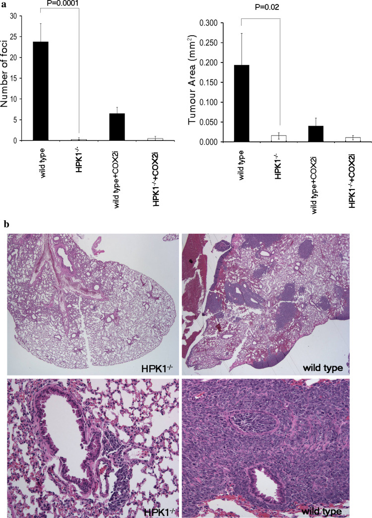
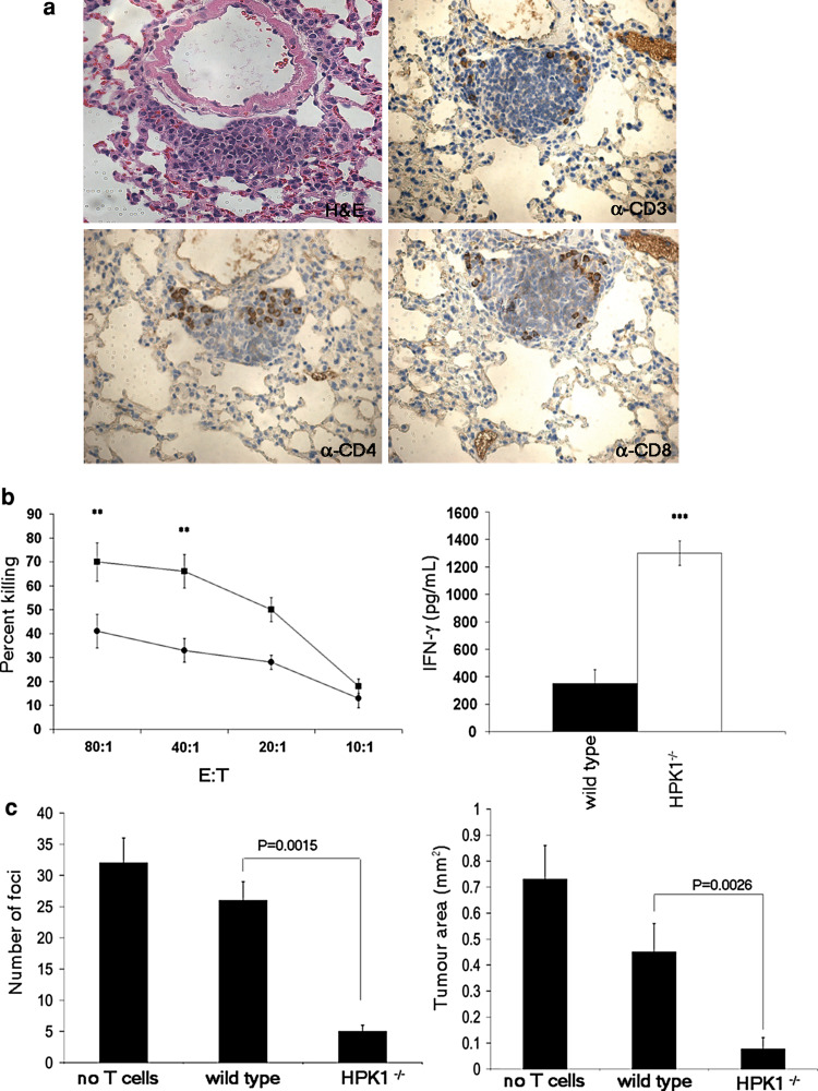
The ability of HPK1 −/− T cells to resist the immunosuppressive and pro-apoptotic action of PGE2 suggests that HPK1 −/− tumor-infiltrating lymphocytes (TILs) may continue to carry out their effector function in a PGE2-rich tumor microenvironment. The PGE2-producing Lewis lung carcinoma cell line possesses immunosuppressive properties and is widely used as a murine model of lung cancer with a strong T-cell involvement [23, 37, 39]. To test whether the loss of HPK1 would result in a resistance to the growth of 3LL, we injected 5 × 10⁵ 3LL cells intravenously into wild-type or HPK1 −/− mice. The presence of tumor foci in the lungs was assessed 14 days after 3LL injection by hematoxylin and eosin (H&E) staining. No foci were detected in 75% of the HPK1 −/− mice examined, and the remaining 25% only developed one or two tumor foci compared to an average of 22 foci in wild-type mice (Fig. 4a, b). Furthermore, when compared to wild-type controls, the size of individual foci in the lungs of HPK1 −/− mice was significantly smaller. Immunohistochemistry revealed that CD3+ lymphocytic infiltrates were present in those solitary tumor foci, whereas none was detected in the foci of wild-type mice (Fig. 5a). Further analysis revealed both CD4+ and CD8+ T-cell infiltrates. When PGE2 production was blocked in wild-type mice by intraperitoneal administration of a COX-2 inhibitor (SC-791), the number of foci was markedly reduced compared to that from untreated wild-type mice (Fig. 4a). Furthermore, these foci were now infiltrated by lymphocytes (Fig. 5a), in agreement with prior reports [39]. COX-2 inhibitors reduced the number of lung foci, but not as much as the reduction in foci seen in the HPK1 −/− mice. These results suggest that besides resistance to PGE2, removing HPK1 results in an enhanced anti-tumor response by these T cells. This interpretation would be consistent with the enhanced T-cell response seen in the HPK1 −/− mice, reported by Tan et al. Taken together, our results indicate that HPK1 −/− mice have a robust anti-tumor response against PGE2 producing 3LL that may consist of an enhanced T-cell response to the tumor, as well as a resistance to PGE2 suppression.
Fig. 4.
HPK1 −/− mice are resistant to the growth of Lewis lung carcinoma. 3LL cells were injected intravenously into HPK1 −/− or wild-type mice, in the presence or absence of a COX-2 inhibitor (COX2i), and tumor burden in the lungs was assessed after 14 days. a Total number of lung foci and total tumor area in the lungs with H&E staining. White and black bars represent HPK1 −/− and wild-type conditions, respectively. b With H&E staining, a 20× magnification (top panels) of a single lobe from the lungs of HPK1 −/− or wild-type mice, respectively. A single tumor focus of 60× magnification from HPK1 −/− or wild-type lungs (bottom panels). P values shown represent the standard error of the mean of an experiment (n = 6)
Fig. 5.
Enhanced anti-tumor T-cell response in HPK1 −/− mice. a 40× magnification of H&E staining with superimposed IHC of anti-CD3, CD4, and CD8 staining, respectively, of a tumor focus from HPK1 −/− lungs. b Varying ratios of splenocytes, from 3LL-injected HPK1 −/− (filled squares) or wild-type (filled circles) mice were co-cultured with irradiated, 51Cr-labeled 3LL cells. Irradiated 3LL cells were co-cultured with splenocytes from tumor-bearing HPK1 −/− (white bar) or wild-type (black bar) mice, and supernatants were collected for IFN-γ ELISA (n = 3, **P < 0.01, ***P < 0.001). c Purified T cells from wild-type or HPK1 −/− mice were injected intravenously with 3LL cells into RAG2 −/− mice. The number of tumor lung foci and the tumor area was assessed as in Fig. 4 (n = 5). Error bars in all experiments represent the standard error of the mean of an experiment
T cell-dependent resistance of HPK1−/− mice to tumor growth
T-cell infiltration in tumors that developed in HPK1-deficient mice implicates T cells in their resistance to the growth of Lewis lung carcinoma. To compare the anti-tumor T-cell response of wild-type and HPK1 −/− mice, splenocytes were isolated from mice that received 3LL cells 14 days earlier, and their ability to lyse 3LL cells was determined ex vivo by a cytotoxicity assay. When HPK1 −/− splenocytes were co-cultured with irradiated 3LL cells at varying effectors to targets (E:T) ratios, they were five times more effective in killing 3LL cells than wild-type splenocytes (Fig. 5b). Furthermore, when supernatants from this assay were collected and analyzed for the production of IFN-γ by ELISA, significantly more IFN-γ was detected in the supernatants collected from HPK1 −/− T-cell co-cultures versus those from wild-type T cells (Fig. 5b).
To verify the role of HPK1 −/− T cells in the rejection of 3LL cells, we purified and intravenously transferred wild-type or HPK1 −/− T cells along with 3LL cells into T cell-deficient, RAG2 −/− hosts. After 14 days, there was a profound reduction in the number and size of tumor foci in the lungs of mice that received HPK1 −/− T cells when compared to the number and size of tumors from mice that received wild-type T cells or to controls that received no T cells (Fig. 5c). These findings confirm that the resistance to 3LL tumor development in HPK1 −/− mice is T cell-dependent.
Discussion
The primary objective of cancer immunotherapy is to manipulate a non-responsive immune system to effectively participate in tumor rejection. Our finding that the lack of HPK1 resulted in an efficient rejection of Lewis lung carcinoma suggests that HPK1 is involved in the control of anti-tumor immune responses. The involvement of HPK1 in the control of anti-tumor activity is consistent with its role as a negative regulator of TCR-induced signals [19, 38]. Similar enhanced anti-tumor responses in mice that are deficient in known negative regulators have been documented. For instance, the loss of the ubiquitin ligase activity of Cbl-b correlates with an enhanced ability of Cbl-b −/− mice to reject transplanted, as well as spontaneously arising, tumors [7, 21]. Administering neutralizing mAbs capable of blocking negative signaling pathways downstream of CTLA-4 and PD-1 also resulted in enhanced anti-tumor activity [2, 11, 14, 18, 30]. Because HPK1 and the aforementioned negative regulators may utilize unique signaling mechanisms to inhibit T-cell activation, they represent possible targets for the development of cancer therapies.
Our initial observation that the kinase activity of HPK1 is responsive to PGE2 stimulation led us to propose that HPK1 plays a functional role downstream of PGE2-induced signals. Through genetic ablation of HPK1, we have generated mice whose hematopoietic cells are resistant to immunosuppression mediated by PGE2 stimulation, but whose T cells also demonstrate enhanced functions, such as proliferation, IL-2 production, and cytotoxicity. In aggregate, the resistance to PGE2, in conjunction with the resistance to apoptosis and the increased T-cell responsiveness to TCR-generated signal, may contribute toward the enhanced anti-tumor activity observed in HPK1 −/− mice.
Lewis lung carcinoma is a PGE2-dependent murine model of lung cancer. The importance of PGE2 in this model system has been demonstrated through the use of COX-2 inhibitors and anti-PGE2 neutralizing antibodies, resulting in an increased anti-tumor activity and subsequent tumor rejection by PGE2-manipulated mice [39]. The inability of COX-2 inhibitor-treated wild-type mice to eradicate 3LL tumors as efficiently as HPK1 −/− mice does suggest that an HPK1-regulated, PGE2-independent anti-tumor mechanism(s) may also exist. Our results here demonstrate that HPK1 −/− T cells are also more resistant to PGE2-induced apoptosis, a finding that is consistent with the existing data that implicates HPK1 as a pro-apoptotic molecule in TCR signaling pathways [3–5]. Not only did we observe resistance to PGE2-induced apoptosis in HPK1 −/− T cells, but fewer Annexin V+ T cells were found in CD3 + CD28 expanded HPK1 −/− cultures in the absence of PGE2. These findings strengthened the conclusion that HPK1 functions as a pro-apoptotic molecule downstream of both the TCR and the EP receptors.
The concept that HPK1 function is regulated by both PGE2 and the TCR pathways is consistent with our finding that TCR and PGE2 stimulation utilize distinct signal transduction mechanisms to activate HPK1 [33]. Protein tyrosine kinase-dependent HPK1 activation via TCR engagement also requires the interaction between HPK1 and Src homology domain-containing adapter proteins as well as PKD activation [1]. This signaling mechanism contrasts dramatically with the G-protein-coupled EP receptor signaling pathway used by PGE2 stimulation to activate HPK1. PGE2-induced HPK1 activity requires PKA activation and does not need protein tyrosine kinase activation or adapter protein interactions with HPK1 [33]. Taken together, these data suggest that both TCR and EP receptors may activate HPK1 via different signaling mechanisms to achieve the same goal. Therefore, the role of HPK1 in T-cell-mediated signaling events in response to PGE2-producing tumors can be clearly differentiated from its role as a negative regulator of TCR-mediated events.
In conclusion, understanding the mechanism whereby HPK1 is regulated by, and, in turn, regulates PGE2 signal transduction pathways could provide a novel approach that will facilitate the development of HPK1-based immunotherapy for cancer and perhaps inflammatory diseases that also utilize PGE2.
Electronic supplementary material
Below is the link to the electronic supplementary material.
Prostaglandin E2 receptor levels in wild type and HPK1-/- lymphocytes prepared from lymph nodes. Whole cell lysates prepared from wild type (WT) and HPK1-/- (KO) lymph node T cells were resolved by SDS-PAGE and were subjected to Western blot analyses using rabbit polyclonal antibodies that recognize specific EP receptors. Anti-β-actin blotting (bottom panel) was used as loading control. (TIF 619 kb)
Acknowledgments
This work was supported by an NIH grant RO1 CA 70758 (SJB) and by a grant from the V Foundation for Cancer Research awarded to S.S. We would like to thank Dr. Richard Williams (Imperial College, London) for review of the manuscript.
Open Access
This article is distributed under the terms of the Creative Commons Attribution Noncommercial License which permits any noncommercial use, distribution, and reproduction in any medium, provided the original author(s) and source are credited.
References
- 1.Arnold R, Patzak IM, Neuhaus B, Vancauwenbergh S, Veillette A, Van Lint J, Kiefer F. Activation of hematopoietic progenitor kinase 1 involves relocation, autophosphorylation, and transphosphorylation by protein kinase D1. Mol Cell Biol. 2005;25:2364–2383. doi: 10.1128/MCB.25.6.2364-2383.2005. [DOI] [PMC free article] [PubMed] [Google Scholar]
- 2.Azuma M, Ito D, Yagita H, Okumura K, Phillips JH, Lanier LL, Somoza C. B70 antigen is a second ligand for CTLA-4 and CD28. Nature. 1993;366:76–79. doi: 10.1038/366076a0. [DOI] [PubMed] [Google Scholar]
- 3.Bode JG, Nimmesgern A, Schmitz J, Schaper F, Schmitt M, Frisch W, Haussinger D, Heinrich PC, Graeve L. LPS and TNFalpha induce SOCS3 mRNA and inhibit IL-6-induced activation of STAT3 in macrophages. FEBS Lett. 1999;463:365–370. doi: 10.1016/S0014-5793(99)01662-2. [DOI] [PubMed] [Google Scholar]
- 4.Brenner D, Golks A, Becker M, Muller W, Frey CR, Novak R, Melamed D, Kiefer F, Krammer PH, Arnold R. Caspase-cleaved HPK1 induces CD95L-independent activation-induced cell death in T and B lymphocytes. Blood. 2007;110:3968–3977. doi: 10.1182/blood-2007-01-071167. [DOI] [PubMed] [Google Scholar]
- 5.Brenner D, Golks A, Kiefer F, Krammer PH, Arnold R. Activation or suppression of NFkappaB by HPK1 determines sensitivity to activation-induced cell death. EMBO J. 2005;24:4279–4290. doi: 10.1038/sj.emboj.7600894. [DOI] [PMC free article] [PubMed] [Google Scholar]
- 6.Chell S, Kaidi A, Williams AC, Paraskeva C. Mediators of PGE2 synthesis and signalling downstream of COX-2 represent potential targets for the prevention/treatment of colorectal cancer. Biochim Biophys Acta. 2006;1766:104–119. doi: 10.1016/j.bbcan.2006.05.002. [DOI] [PubMed] [Google Scholar]
- 7.Chiang JY, Jang IK, Hodes R, Gu H. Ablation of Cbl-b provides protection against transplanted and spontaneous tumors. J Clin Invest. 2007;117:1029–1036. doi: 10.1172/JCI29472. [DOI] [PMC free article] [PubMed] [Google Scholar]
- 8.Chouaib S, Welte K, Mertelsmann R, Dupont B. Prostaglandin E2 acts at two distinct pathways of T-lymphocyte activation: inhibition of interleukin 2 production and down-regulation of transferrin receptor expression. J Immunol. 1985;135:1172–1179. [PubMed] [Google Scholar]
- 9.Dunn GP, Old LJ, Schreiber RD. The immunobiology of cancer immunosurveillance and immunoediting. Immunity. 2004;21:137–148. doi: 10.1016/j.immuni.2004.07.017. [DOI] [PubMed] [Google Scholar]
- 10.Goodwin JS, Bankhurst AD, Messner RP. Suppression of human T-cell mitogenesis by prostaglandin. Existence of a prostaglandin-producing suppressor cell. J Exp Med. 1977;146:1719–1734. doi: 10.1084/jem.146.6.1719. [DOI] [PMC free article] [PubMed] [Google Scholar]
- 11.Hirano F, Kaneko K, Tamura H, Dong H, Wang S, Ichikawa M, Rietz C, Flies DB, Lau JS, Zhu G, Tamada K, Chen L. Blockade of B7-H1 and PD-1 by monoclonal antibodies potentiates cancer therapeutic immunity. Cancer Res. 2005;65:1089–1096. [PubMed] [Google Scholar]
- 12.Hsu AL, Ching TT, Wang DS, Song X, Rangnekar VM, Chen CS. The cyclooxygenase-2 inhibitor celecoxib induces apoptosis by blocking Akt activation in human prostate cancer cells independently of Bcl-2. J Biol Chem. 2000;275:11397–11403. doi: 10.1074/jbc.275.15.11397. [DOI] [PubMed] [Google Scholar]
- 13.Huang M, Stolina M, Sharma S, Mao JT, Zhu L, Miller PW, Wollman J, Herschman H, Dubinett SM. Non-small cell lung cancer cyclooxygenase-2-dependent regulation of cytokine balance in lymphocytes and macrophages: up-regulation of interleukin 10 and down-regulation of interleukin 12 production. Cancer Res. 1998;58:1208–1216. [PubMed] [Google Scholar]
- 14.Iwai Y, Ishida M, Tanaka Y, Okazaki T, Honjo T, Minato N. Involvement of PD-L1 on tumor cells in the escape from host immune system and tumor immunotherapy by PD-L1 blockade. Proc Natl Acad Sci USA. 2002;99:12293–12297. doi: 10.1073/pnas.192461099. [DOI] [PMC free article] [PubMed] [Google Scholar]
- 15.Krysan K, Merchant FH, Zhu L, Dohadwala M, Luo J, Lin Y, Heuze-Vourc’h N, Pold M, Seligson D, Chia D, Goodglick L, Wang H, Strieter R, Sharma S, Dubinett S (2003) COX-2-dependent stabilization of survivin in non-small cell lung cancer. FASEB J, 03-0369fje [DOI] [PubMed]
- 16.Krysan K, Reckamp KL, Sharma S, Dubinett SM. The potential and rationale for COX-2 inhibitors in lung cancer. Anticancer Agents Med Chem. 2006;6:209–220. doi: 10.2174/187152006776930882. [DOI] [PubMed] [Google Scholar]
- 17.Le Bras S, Foucault I, Foussat A, Brignone C, Acuto O, Deckert M. Recruitment of the actin-binding protein HIP-55 to the immunological synapse regulates T cell receptor signaling and endocytosis. J Biol Chem. 2004;279:15550–15560. doi: 10.1074/jbc.M312659200. [DOI] [PubMed] [Google Scholar]
- 18.Leach DR, Krummel MF, Allison JP. Enhancement of antitumor immunity by CTLA-4 blockade. Science. 1996;271:1734–1736. doi: 10.1126/science.271.5256.1734. [DOI] [PubMed] [Google Scholar]
- 19.Liou J, Kiefer F, Dang A, Hashimoto A, Cobb MH, Kurosaki T, Weiss A. HPK1 is activated by lymphocyte antigen receptors and negatively regulates AP-1. Immunity. 2000;12:399–408. doi: 10.1016/S1074-7613(00)80192-2. [DOI] [PubMed] [Google Scholar]
- 20.Liu CH, Chang SH, Narko K, Trifan OC, Wu MT, Smith E, Haudenschild C, Lane TF, Hla T. Overexpression of cyclooxygenase-2 is sufficient to induce tumorigenesis in transgenic mice. J Biol Chem. 2001;276:18563–18569. doi: 10.1074/jbc.M010787200. [DOI] [PubMed] [Google Scholar]
- 21.Loeser S, Loser K, Bijker MS, Rangachari M, van der Burg SH, Wada T, Beissert S, Melief CJ, Penninger JM. Spontaneous tumor rejection by cbl-b-deficient CD8+ T cells. J Exp Med. 2007;204:879–891. doi: 10.1084/jem.20061699. [DOI] [PMC free article] [PubMed] [Google Scholar]
- 22.Makoul GT, Robinson DR, Bhalla AK, Glimcher LH. Prostaglandin E2 inhibits the activation of cloned T cell hybridomas. J Immunol. 1985;134:2645–2650. [PubMed] [Google Scholar]
- 23.Mandelboim O, Berke G, Fridkin M, Feldman M, Eisenstein M, Eisenbach L. CTL induction by a tumour-associated antigen octapeptide derived from a murine lung carcinoma. Nature. 1994;369:67–71. doi: 10.1038/369067a0. [DOI] [PubMed] [Google Scholar]
- 24.Masferrer JL, Leahy KM, Koki AT, Zweifel BS, Settle SL, Woerner BM, Edwards DA, Flickinger AG, Moore RJ, Seibert K. Antiangiogenic and antitumor activities of cyclooxygenase-2 inhibitors. Cancer Res. 2000;60:1306–1311. [PubMed] [Google Scholar]
- 25.McConkey DJ, Orrenius S, Jondal M. Agents that elevate cAMP stimulate DNA fragmentation in thymocytes. J Immunol. 1990;145:1227–1230. [PubMed] [Google Scholar]
- 26.Nataraj C, Thomas DW, Tilley SL, Nguyen M, Mannon R, Koller BH, Coffman TM. Receptors for prostaglandin E2 that regulate cellular immune responses in the mouse. J Clin Invest. 2001;108:1229–1235. doi: 10.1172/JCI13640. [DOI] [PMC free article] [PubMed] [Google Scholar]
- 27.Negishi M, Sugimoto Y, Ichikawa A. Molecular mechanisms of diverse actions of prostanoid receptors. Biochim Biophys Acta (BBA)—Lipids Lipid Metab. 1995;1259:109–119. doi: 10.1016/0005-2760(95)00146-4. [DOI] [PubMed] [Google Scholar]
- 28.Paliogianni F, Kincaid RL, Boumpas DT. Prostaglandin E2 and other cyclic AMP elevating agents inhibit interleukin 2 gene transcription by counteracting calcineurin-dependent pathways. J Exp Med. 1993;178:1813–1817. doi: 10.1084/jem.178.5.1813. [DOI] [PMC free article] [PubMed] [Google Scholar]
- 29.Pica F, Franzese O, D’Onofrio C, Bonmassar E, Favalli C, Garaci E. Prostaglandin E2 induces apoptosis in resting immature and mature human lymphocytes: a c-Myc-dependent and Bcl-2-independent associated pathway. J Pharmacol Exp Ther. 1996;277:1793–1800. [PubMed] [Google Scholar]
- 30.Quezada SA, Peggs KS, Curran MA, Allison JP. CTLA4 blockade and GM-CSF combination immunotherapy alters the intratumor balance of effector and regulatory T cells. J Clin Invest. 2006;116:1935–1945. doi: 10.1172/JCI27745. [DOI] [PMC free article] [PubMed] [Google Scholar]
- 31.Rappaport RS, Dodge GR. Prostaglandin E inhibits the production of human interleukin 2. J Exp Med. 1982;155:943–948. doi: 10.1084/jem.155.3.943. [DOI] [PMC free article] [PubMed] [Google Scholar]
- 32.Sauer K, Liou J, Singh SB, Yablonski D, Weiss A, Perlmutter RM. Hematopoietic progenitor kinase 1 associates physically and functionally with the adaptor proteins B cell linker protein and SLP-76 in lymphocytes. J Biol Chem. 2001;276:45207–45216. doi: 10.1074/jbc.M106811200. [DOI] [PubMed] [Google Scholar]
- 33.Sawasdikosol S, Pyarajan S, Alzabin S, Matejovic G, Burakoff SJ. Prostaglandin E2 activates HPK1 kinase activity via a PKA-dependent pathway. J Biol Chem. 2007;282:34693–34699. doi: 10.1074/jbc.M707425200. [DOI] [PubMed] [Google Scholar]
- 34.Sawasdikosol S, Russo KM, Burakoff SJ. Hematopoietic progenitor kinase 1 (HPK1) negatively regulates prostaglandin E2-induced fos gene transcription. Blood. 2003;101:3687–3689. doi: 10.1182/blood-2002-07-2316. [DOI] [PubMed] [Google Scholar]
- 35.Scales WE, Chensue SW, Otterness I, Kunkel SL. Regulation of monokine gene expression: prostaglandin E2 suppresses tumor necrosis factor but not interleukin-1 alpha or beta-mRNA and cell-associated bioactivity. J Leukoc Biol. 1989;45:416–421. [PubMed] [Google Scholar]
- 36.Schulze-Luehrmann J, Santner-Nanan B, Jha MK, Schimpl A, Avots A, Serfling E. Hematopoietic progenitor kinase 1 supports apoptosis of T lymphocytes. Blood. 2002;100:954–960. doi: 10.1182/blood-2002-01-0089. [DOI] [PubMed] [Google Scholar]
- 37.Sharma S, Huang M, Dohadwala M, Pold M, Batra RK, Dubinett SM. Cyclooxygenase 2-dependent regulation of antitumor immunity in lung cancer. Methods Mol Med. 2003;75:723–736. doi: 10.1385/1-59259-324-0:723. [DOI] [PubMed] [Google Scholar]
- 38.Shui JW, Boomer JS, Han J, Xu J, Dement GA, Zhou G, Tan TH. Hematopoietic progenitor kinase 1 negatively regulates T cell receptor signaling and T cell-mediated immune responses. Nat Immunol. 2007;8:84–91. doi: 10.1038/ni1416. [DOI] [PubMed] [Google Scholar]
- 39.Stolina M, Sharma S, Lin Y, Dohadwala M, Gardner B, Luo J, Zhu L, Kronenberg M, Miller PW, Portanova J, Lee JC, Dubinett SM. Specific inhibition of cyclooxygenase 2 restores antitumor reactivity by altering the balance of IL-10 and IL-12 synthesis. J Immunol. 2000;164:361–370. doi: 10.4049/jimmunol.164.1.361. [DOI] [PubMed] [Google Scholar]
- 40.Strassmann G, Patil-Koota V, Finkelman F, Fong M, Kambayashi T. Evidence for the involvement of interleukin 10 in the differential deactivation of murine peritoneal macrophages by prostaglandin E2. J Exp Med. 1994;180:2365–2370. doi: 10.1084/jem.180.6.2365. [DOI] [PMC free article] [PubMed] [Google Scholar]
- 41.Suzuki K, Tadakuma T, Kizaki H. Modulation of thymocyte apoptosis by isoproterenol and prostaglandin E2. Cell Immunol. 1991;134:235–240. doi: 10.1016/0008-8749(91)90346-D. [DOI] [PubMed] [Google Scholar]
- 42.Tsujii M, DuBois RN. Alterations in cellular adhesion and apoptosis in epithelial cells overexpressing prostaglandin endoperoxide synthase 2. Cell. 1995;83:493–501. doi: 10.1016/0092-8674(95)90127-2. [DOI] [PubMed] [Google Scholar]
- 43.Tsujii M, Kawano S, Tsuji S, Sawaoka H, Hori M, DuBois RN. Cyclooxygenase regulates angiogenesis induced by colon cancer cells. Cell. 1998;93:705–716. doi: 10.1016/S0092-8674(00)81433-6. [DOI] [PubMed] [Google Scholar]
Associated Data
This section collects any data citations, data availability statements, or supplementary materials included in this article.
Supplementary Materials
Prostaglandin E2 receptor levels in wild type and HPK1-/- lymphocytes prepared from lymph nodes. Whole cell lysates prepared from wild type (WT) and HPK1-/- (KO) lymph node T cells were resolved by SDS-PAGE and were subjected to Western blot analyses using rabbit polyclonal antibodies that recognize specific EP receptors. Anti-β-actin blotting (bottom panel) was used as loading control. (TIF 619 kb)


