Abstract
Clade B of the New World arenaviruses contains both pathogenic and nonpathogenic members, whose surface glycoproteins (GPs) are characterized by different abilities to use the human transferrin receptor type 1 (hTfR1) protein as a receptor. Using closely related pairs of pathogenic and nonpathogenic viruses, we investigated the determinants of the GP1 subunit that confer these different characteristics. We identified a central region (residues 85 to 221) in the Guanarito virus GP1 that was sufficient to interact with hTfR1, with residues 159 to 221 being essential. The recently solved structure of part of the Machupo virus GP1 suggests an explanation for these requirements.
Arenaviruses are bisegmented, single-stranded RNA viruses that use an ambisense coding strategy to express four proteins: NP (nucleoprotein), Z (matrix protein), L (polymerase), and GP (glycoprotein). The viral GP is sufficient to direct entry into host cells, and retroviral vectors pseudotyped with GP recapitulate the entry pathway of these viruses (5, 13, 24, 31). GP is a class I fusion protein comprising two subunits, GP1 and GP2, cleaved from the precursor protein GPC (4, 14, 16, 18, 21). GP1 contains the receptor binding domain (19, 28), while GP2 contains structural elements characteristic of viral membrane fusion proteins (8, 18, 20, 38). The N-terminal stable signal peptide (SSP) remains associated with the mature glycoprotein after cleavage (2, 39) and plays a role in transport, maturation, and pH-dependent fusion (17, 35, 36, 37).
The New World arenaviruses are divided into clades A, B, and C based on phylogenetic relatedness (7, 9, 11). Clade B contains the human pathogenic viruses Junin (JUNV), Machupo (MACV), Guanarito (GTOV), Sabia, and Chapare, which cause severe hemorrhagic fevers in South America (1, 10, 15, 26, 34). Clade B also contains the nonpathogenic viruses Amapari (AMAV), Cupixi, and Tacaribe (TCRV), although mild disease has been reported for a laboratory worker infected with TCRV (29).
Studies with both viruses and GP-pseudotyped retroviral vectors have shown that the pathogenic clade B arenaviruses use the human transferrin receptor type 1 (hTfR1) to gain entry into human cells (19, 30). In contrast, GPs from nonpathogenic viruses, although capable of using TfR1 orthologs from other species (1), cannot use hTfR1 (1, 19) and instead enter human cells through as-yet-uncharacterized hTfR1-independent pathways (19). In addition, human T-cell lines serve as useful tools to distinguish these GPs, since JUNV, GTOV, and MACV pseudotyped vectors readily transduce CEM cells, while TCRV and AMAV GP vectors do not (27; also unpublished data). These properties of the GPs do not necessarily reflect a tropism of the pathogenic viruses for human T cells, since viral tropism is influenced by many factors and T cells are not a target for JUNV replication in vivo (3, 22, 25).
Generation of chimeric GPs.
At present, there is limited information about the determinants of receptor binding within the arenavirus GP1. Previous studies using GP1 immunoadhesins (IMs) have suggested that the N-terminal 22 amino acids and C-terminal 10 amino acids of clade B GP1s are dispensable for receptor binding (1, 19, 27, 30). To further map the regions involved in the interactions that are characteristic of the pathogenic clade B viruses, we generated a series of chimeric GPs based on the exchange of GP1 protein segments between closely related pathogenic and nonpathogenic viruses from the same sublineage, specifically the B1 viruses JUNV and TCRV and the B2 viruses GTOV and AMAV (9, 11).
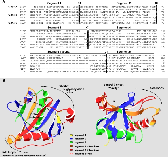
Sequence alignment of representative New World GP1 proteins using the Clustal X software (12) revealed four completely conserved cysteine residues (Fig. 1A) that are also conserved in the Old World arenaviruses. The cysteines were used to delineate 5 segments within GP1, which were swapped to generate the chimera series T(J), comprising JUNV sequences inserted into a TCRV GP backbone, and the GTOV/AMAV chimeras A(G) and G(A). The crystal structure of a truncated GP1 from MACV (residues 87 to 239) has recently been solved (6), revealing that conserved cysteines 1 and 4 and conserved cysteines 2 and 3 form disulfide bonds (Fig. 1B). Given the close evolutionary relatedness between the GPs of the clade B arenaviruses and the similarity in the patterns of predicted secondary structures across GP1 (data not shown), the structural information available for the MACV GP1 is likely to be a useful blueprint for the JUNV and GTOV GP1 structures, although as noted by Bowden and coworkers (6), there is considerable variation in the location of predicted N-glycosylation sites, which suggests that structural variation will exist in the carbohydrate-rich regions of the protein.
FIG. 1.
(A) Sequence alignment of GP1 sequences from representative arenaviruses in New World clades A, B, and C. The positions of four completely conserved cysteines (C1 to C4) residues are shown, which were used to delineate five distinct segments in GP1. The sequences used were Pichinde virus (PICV) (GenBank accession no. P31840), GTOV (GenBank accession no. Q8AYW1), AMAV (GenBank accession no. YP_001649208), and Oliveros virus (OLVV) (GenBank accession no. Q84168). Gaps introduced to maximize alignment are indicated by dashes. (B) Locations of segments within MACV GP1 crystal structure. Schematic of MACV GP1 crystal structure (6), generated using structural coordinates pdb code 2WFO, generated using the Swiss-PdbViewer 4.0.1 software (23; http://www.expasy.org/spdbv/). Specific segments are color coded as indicated. The boundary between the N and C termini of segment 4 corresponds to the boundary between the A(Gn4) and A(Gc4) chimeras. The structure contains a central β-sheet, a series of side loops containing conserved residues predicted to be solvent accessible, and an opposite region of side loops that contain most of the N-glycosylation sites. The locations of disulfide links between conserved cysteines 1 and 4 and conserved cysteines 2 and 3 are indicated on the left-hand panel.
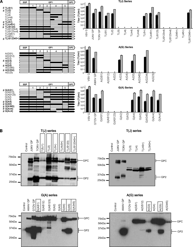
The functionality of each chimeric GP was assessed by generating pseudotyped retroviral vectors and transducing HEK293A cells as described previously (27, 31, 32) (Fig. 2A). In the T(J) series, only chimeras generated by single-segment swaps of segments 1, 2, and 5 or the combined substitution of segments 1 to 3 or the C terminus of segment 4 plus segment 5 were functional. No chimeras containing either complete or partial substitutions in segment 4 alone were functional. The A(G) series tolerated single-segment swaps of segments 3 or 5, while segments 2 or 4 could be accommodated only in the context of a larger swap of the central region spanning segments 2 to 4. The G(A) series revealed a different pattern again, with single-segment swaps of 1, 4, or 5 being tolerated, as well as the larger swap of segments 3 to 5.
FIG. 2.
(A) Chimeric GPs. Schematic representations of chimeric GPs and titers of the corresponding pseudotyped retroviral vectors on HEK293A cells. Functional chimeras are indicated by a # symbol before the chimera. The boundaries used to generate the chimeras are based on the signal peptide SSP (S) and conserved cysteine residues in GP1 that delineate segments 1 through 5. TCRV and AMAV sequences (white) and JUNV and GTOV sequences (black) are indicated. Only the N-terminal part of the GP2 sequence is represented. In addition, the boundaries of the following partial segments are as indicated: Jm4 extends to JUNV residue 207, Jn4 extends to JUNV residue 184, Jc4 extends from JUNV residue 208, + indicates extension into JUNV GP2 to residue 280, An4 extends to AMAV residue 183, and Ac4 extends from AMAV residue 184. Titers were determined as previously described (27, 31), and values shown are the means plus standard deviations (SD) (error bars) for three to six independent experiments. The titers are shown for both unconcentrated vector stocks (black) and 10× concentrated stocks (gray); an asterisk indicates no detectable titer. VSV, vesicular stomatitis virus. (B) Western blotting of GP-pseudotyped vector particles. Vectors were generated in HEK293T cells, pelleted by ultracentrifugation, deglycosylated, and resolved by sodium dodecyl sulfate-polyacrylamide gel electrophoresis (SDS-PAGE) as previously described (31). Functional chimeric GPs are boxed. Depending on PAGE conditions, GPC could be seen as both the full-length GPC, as well as the SSP cleaved form comprising only GP1-GP2. TCRV and JUNV proteins were detected using anti-GP2 antiserum, while GTOV and AMAV proteins were constructed as C-terminal Flag-tagged proteins and detected using anti-Flag antibody.
To address the basis for the nonfunctional chimeras, we analyzed GP incorporation into vector particles by Western blotting (Fig. 2B). GPs of the T(J) series were detected using an anti-GP2 rabbit polyclonal antibody raised against peptide CDMLRLFYNKNAIKTLN (Covance, San Diego, CA), while G(A) and A(G) chimeras were constructed with C-terminal Flag tag epitopes and detected using the M2 anti-Flag antibody (Sigma-Aldrich, Saint Louis, MO). GP2 cleavage products of the correct size were detected for the parental GPs and all functional chimeras, except G(A345), whereas all nonfunctional chimeras revealed absent or aberrantly cleaved products. We also failed to detect cleavage of the untagged G(A345) protein using monoclonal antibody QD04-AF03 that recognizes JUNV GP1 (31, 33, 39) and is cross-reactive against GTOV and AMAV GP1 (data not shown), so that it is possible that only a very low level of cleavage of this chimera, undetectable by Western blotting, is sufficient to direct entry. The crystal structure of MACV GP1 (residues 87 to 239) reveals that the residues contained within linear segments 2, 3, and 4 interact extensively, with each contributing one or more β-strands to a large central β-sheet (Fig. 1B). The complexity of these interactions may explain why so many chimeras were not tolerated, despite the sequence similarity between the parental GP1 sequences.
Evaluation of CEM tropism and hTfR1 use by GP chimeras.
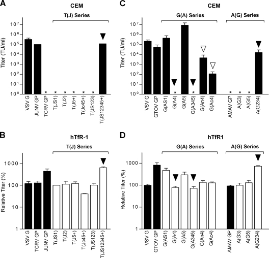
We and others have previously shown that the ability to transduce human CEM cells and the ability to respond to the presence of hTfR1 transfected into CHO-K1 cells [CHO-K1(hTfR1)] are properties of the GPs from pathogenic viruses JUNV, MACV, and GTOV, but not nonpathogenic viruses AMAV and TCRV (1, 19, 27, 30). Using these features as a probe, we analyzed the properties of the functional chimeric GPs. For the T(J) series, we found that only the complete JUNV GP1 swapped into the TCRV backbone in chimera T(JS12345+) was able to direct entry into CEM cells or increase the titer on CHO cells expressing hTfR1 (CHO-hTfR1) (Fig. 3A and B). Substitution of the N-terminal part of the JUNV GP1 in construct T(J123) or the C-terminal substitution in T(Jc45+) was not sufficient to alter tropism, suggesting that either the receptor binding domain in JUNV GP specifically requires the N terminus of segment 4 and/or that generation of a functional domain requires residues located throughout the larger fold of the GP1 subunit.
FIG. 3.
Characterization of chimeric GPs. (A and C) The titers of the vectors pseudotyped with parental GPs or indicated chimeras were determined on CEM cells as previously described (27). Values shown are the means plus SD (error bars) for three to six independent experiments. An asterisk indicates no detectable titer, even for 10× concentrated vector stocks. Chimeric GPs with altered characteristics that no longer reflect the parental GP backbone are indicated with a black arrowhead, and partial effects are indicated with a white arrowhead. (B and D) Relative titers of the indicated vectors on CHO-K1 cells transiently transfected with hTfR1 versus mock-transfected CHO-K1 cells. Values are the means plus SD for more than five independent experiments. Chimeric GPs with altered characteristics that no longer reflect the parental GP backbone are indicated with a black arrowhead. VSV, vesicular stomatitis virus.
For the functional G(A) chimeras, both the single substitution of segment 4 and the more extensive substitution of segments 3 to 5 altered tropism and prevented transduction of CEM cells or the use of hTfR1 (Fig. 3C and D). Replacement of just the N or C termini of segment 4 in chimeras G(An4) and G(Ac4) resulted in only a partial ability to transduce CEM cells, with the G(Ac4) chimera being more severely affected, and both chimeras displayed only a slight enhancement in titers on the CHO-K1(hTfR1) cells that was not statistically significant (P > 0.05). Overall, this suggests that residues within both the N and C termini of segment 4 are involved in receptor use by the GTOV GP, with residues in the C terminus being especially important. In agreement with an important role for segment 4, studies with the reciprocal A(G) chimeras revealed that substituting segment 3 or 5 alone did not alter tropism, while the combined substitution of segments 2 to 4 in chimera A(G234) allowed the efficient transduction of CEM cells and the use of hTfR1. Unfortunately, the lack of functionality of chimera A(G4) prevented us from directly examining whether substitution of the GTOV segment 4 alone into the AMAV GP was sufficient to switch tropism and receptor use.
Interestingly, we observed enhanced transduction of CEM cells by chimera G(A5) compared to either the GTOV or AMAV GP parents, and this was also apparent on HEK293A cells. This increased titer did not appear to be due to greater incorporation of GP proteins into the pseudotyped vectors or differences in GPC processing (Fig. 2B), and the change did not enhance the ability of the protein to recognize hTfR1, since the ratio of titers on CHO-K1(hTfR1) cells versus CHO-K1 cells was actually less than observed with the GTOV protein. It is possible that this modification of the C terminus of GP1 resulted in structural changes and/or the introduction of more-permissive contact residues that increased the protein's affinity for the receptors present on CEM and HEK293A cells or, alternatively, enhanced more downstream aspects of the fusion process.
GP1 immunoadhesin binding activity.
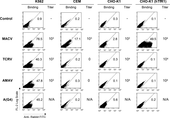
The lack of functionality of the T(J) or A(G) GP chimeras containing full or partial substitutions of segment 4 frustrated our ability to further characterize receptor binding determinants in this region. Segment 4 constitutes an extensive region within GP1 (Fig. 1B) that could be involved in both intra- and intermolecular interactions, including those with the GP2 subunit or within a GP multimer. To reduce the influence of such interactions, we next evaluated immunoadhesins (IMs) comprising GP1 fused to the hinge and Fc regions of an IgG heavy chain that we and others have previously used to characterize arenavirus GP1 binding (1, 19, 27, 30). Functional (binding-competent) IMs were generated for full-length MACV, AMAV, and TCRV GPs, as well as the chimera A(G4), and used to bind to human K562 and CEM cells, as well as to CHO-K1 and CHO-K1(hTfR1) cells (Fig. 4). At the same time, matched stocks of GP-pseudotyped retroviral vectors were analyzed for their ability to transduce the same panel of cells.
FIG. 4.
IM binding and transduction efficiencies of parental and chimeric GPs. The values in the Binding columns show IM binding measured on the indicated cell lines as previously described (19, 27). The percentage of cells showing a positive shift in each representative experiment is indicated in the right-hand part of the graph. Control, secondary antibody only. GP-pseudotyped retroviral vectors were generated as previously described (27) and used to transduce indicated cell types. The titers were determined by fluorescence-activated cell sorting (FACS) as transducing units per ml (TU/ml) and are shown approximated to the nearest factor of 10 (N/A, not available due to lack of activity in A(G4) GP; -, not applicable). FITC, fluorescein isothiocyanate.
The three parental GP constructs gave robust titers on K562 cells, as we have previously observed (19, 27), and their corresponding IMs resulted in good levels of binding to the same cells. On CEM cells, only the MACV GP vectors gave titers and only the MACV IM produced detectable binding activity. Comparing CHO-K1 cells to CHO-K1(hTfR1) cells, MACV IM binding and GP titers both showed a significant increase. In contrast, although the AMAV and TCRV GP vectors were able to efficiently transduce CHO-K1 cells (and were unaffected by the presence of hTfR1), we did not observe binding by their corresponding IMs. A similar lack of binding to HEK293T cells has been reported for both TCRV and AMAV IMs, despite titers from the equivalent GP pseudoviruses (1). These findings suggest that the interaction between GP1 and the receptor that is used by TCRV or AMAV on CHO-K1 or HEK293T cells is of lower affinity than is necessary to observe IM binding or, alternatively, that stable binding by these GP1 proteins requires additional interactions that are available only in the mature GP complex.
Interestingly, although the A(G4) chimera was nonfunctional as a GP, we found that this substitution of segment 4 was tolerated as an IM, resulting in readily detectable binding on K562 cells. However, A(G4) IM binding was less robust than the wild-type IMs, requiring twice as much of the concentrated IM preparation to achieve binding levels on K562 cells similar to those observed with either the AMAV or GTOV IMs (Fig. 4). The functionality of the A(G4) IM allowed us to address whether segment 4 of GTOV in an AMAV backbone was sufficient to switch tropism by analyzing binding on the full panel of cell lines, where we were unable to detect any binding to either CEM or CHO-K1(hTfR1) cells. These results could indicate that segment 4 of GTOV alone is not sufficient to allow hTfR1 use or since the A(G4) IM had lower overall activity than the wild-type AMAV or GTOV IMs, could simply reflect a technical problem with the A(G4) IM preparations.
The results presented here confirm and further refine features of the clade B receptor binding domain, beyond those previously determined by the analysis of GP1 IMs (1, 19, 27, 30). Notably, we found that substitution of segments 2 to 4 of AMAV GP1 (residues 85 to 221) with the equivalent region from GTOV was sufficient to switch the properties of the protein, allowing entry into CEM cells and increased titer on CHO-K1 cells transfected with hTfR1. We also observed that the substitution of GTOV segment 4 (residues 159 to 221) with the corresponding AMAV sequence produced a chimeric GP that was now unable to transduce CEM cells or utilize hTfR1 and that the replacement of either the N- or C-terminal half of segment 4 caused partial effects. These data suggest that segment 4 contains residues required for the specific interaction of the GTOV GP with hTfR1 and that both halves of segment 4 contribute important residues.
The recently solved structure of MACV GP1 (residues 87 to 239) has revealed an unusual structure, with a large central flat β-sheet area that appears to be within an open cavity (6). The sheet is constructed from antiparallel β-strands from segments 2 and 4 at its center, with β-strands from segments 1 and 3 at the edges (Fig. 1B). It is bordered by loop structures predicted to contain conserved, solvent-accessible residues on one side, with the majority of N-glycosylation sites on the opposite side. Both the N and C termini of segment 4 contribute adjacent β-strands (β-strands 6 and 7 in reference 6) to the central β-sheet, as well as contributing residues to the predicted exposed loop regions, and our finding of an important role for segment 4 suggests that either structure could constitute a contact face with hTfR1 or alternate receptors. Similarly, the observed requirement for GTOV segments 2, 3, and 4 to generate a fully functional chimeric GP with acquired hTfR1 usage suggests that all three regions influence the proper structural conformation of a receptor binding domain. Segments 2, 3, and 4 each contribute one or more strands to the central β-sheet, and they each also contain predicted solvent-exposed residues. However, these conserved exposed residues are present in very different geometric planes in the side loops, making it unlikely that they are all involved in receptor binding. We note that no information is yet available about GP1-GP2 interactions or interactions that occur within the complete multimeric GP complex, so it is possible that such interactions could engage some or all of these conserved residues. An alternative explanation is that the contributions of segments 2, 3, and 4 are essential for the structural integrity of the central β-sheet and that this unusual structural feature either represents the receptor binding domain itself or is important for the correct presentation of such a domain.
Although in the present study we focused on the use of hTfR1 and the characteristic tropism of the pathogenic clade B GPs, our data do not rule out the possibility that the regions of GP1 we have highlighted also play roles in receptor interactions for the nonpathogenic viruses or the larger family of arenaviruses. The GPs from AMAV and TCRV cannot bind to human TfR1 (1, 19), but they enter cells using the TfR1 orthologs of several mammalian species (1). Moreover, a small number of mutations in the apical domain of hTfR1, in a loop located between β-strands 1 and 2, were sufficient to convert hTfR1 to a receptor that could now be used by TCRV and AMAV GP-pseudotyped vectors (1). As argued by the authors, this in turn could mean that only minor changes within the TCRV and AMAV GP1 would be required to allow these viruses to use the human form of TfR1 (1). Indeed, it is possible that only a few contact residues, distributed across segments 2 to 4, are important for robust hTfR1 binding.
Studies of receptor binding using either truncation mutants or chimeric proteins always face the challenge of maintaining a suitable structure to obtain viable proteins. This in turn makes it harder to distinguish mutations that affect actual receptor contact residues and those that alter the overall structural scaffold of a receptor binding domain. The structure of the MACV GP1 (6) clearly shows that, regardless of the precise location of the receptor binding site, the different segments that we have identified as being important for receptor interactions are inextricably linked, so that it is likely that only further structural studies of GP1 complexed with hTfR1 will allow the correct assignment of the receptor contact residues. Arguably, this will allow a better understanding of the threat posed by arenaviruses with regard to the potential mutation of nonpathogenic strains into more dangerous viruses.
Acknowledgments
We thank David Stuart and Thomas Bowden (University of Oxford) for providing us with the MACV GP1 crystal structure coordinates, Stefan Kunz (University of Lausanne) for Flag-tagged GTOV and AMAV GP constructs, and Kevin Haworth (University of Southern California) for bioinformatics analysis.
This work was supported by PHS grant 1U54 AI065359 from NIAID to the Pacific Southwest Regional Center of Excellence for Biodefense and Emerging Infectious Diseases (P.M.C.) and a Saban Research Institute Career Development Award (T.R.).
Footnotes
Published ahead of print on 4 November 2009.
REFERENCES
- 1.Abraham, J., J. A. Kwong, C. G. Albariño, J. G. Lu, S. R. Radoshitzky, J. Salazar-Bravo, M. Farzan, C. F. Spiropoulou, and H. Choe. 2009. Host-species transferrin receptor 1 orthologs are cellular receptors for nonpathogenic New World clade B arenaviruses. PLoS Pathog. 5:e1000358. [DOI] [PMC free article] [PubMed] [Google Scholar]
- 2.Agnihothram, S. S., J. York, and J. H. Nunberg. 2006. Role of the stable signal peptide and cytoplasmic domain of G2 in regulating intracellular transport of the Junin virus envelope glycoprotein complex. J. Virol. 80:5189-5198. [DOI] [PMC free article] [PubMed] [Google Scholar]
- 3.Avila, M. M., R. M. Laguens, R. P. Laguens, and M. C. Weissenbacher. 1981. Tissue selectivity and virulence indicators of 3 strains of Junin virus. Medicina (Bueno Aires) 41:157-166. (In Spanish.) [PubMed] [Google Scholar]
- 4.Beyer, W. R., D. Pöpplau, W. Garten, D. von Laer, and O. Lenz. 2003. Endoproteolytic processing of the lymphocytic choriomeningitis virus glycoprotein by the subtilase SKI-1/S1P. J. Virol. 77:2866-2872. [DOI] [PMC free article] [PubMed] [Google Scholar]
- 5.Beyer, W. R., M. Westphal, W. Ostertag, and D. von Laer. 2002. Oncoretrovirus and lentivirus vectors pseudotyped with lymphocytic choriomeningitis virus glycoprotein: generation, concentration, and broad host range. J. Virol. 76:1488-1495. [DOI] [PMC free article] [PubMed] [Google Scholar]
- 6.Bowden, T. A., M. Crispin, S. C. Graham, D. J. Harvey, J. M. Grimes, E. I. Jones, and D. I. Stuart. 2009. Unusual molecular architecture of Machupo virus attachment glycoprotein. J. Virol. 83:8259-8265. [DOI] [PMC free article] [PubMed] [Google Scholar]
- 7.Bowen, M. D., C. J. Peters, and S. T. Nichol. 1996. The phylogeny of New World (Tacaribe complex) arenaviruses. Virology 219:285-290. [DOI] [PubMed] [Google Scholar]
- 8.Buchmeier, M. J. 2002. Arenaviruses: protein structure and function. Curr. Top. Microbiol. Immunol. 262:159-173. [DOI] [PubMed] [Google Scholar]
- 9.Charrel, R. N., X. de Lamballerie, and S. Emonet. 2008. Phylogeny of the genus Arenavirus. Curr. Opin. Microbiol. 11:362-368. [DOI] [PubMed] [Google Scholar]
- 10.Charrel, R. N., and X. de Lamballerie. 2003. Arenaviruses other than Lassa virus. Antiviral Res. 57:89-100. [DOI] [PubMed] [Google Scholar]
- 11.Charrel, R. N., H. Feldmann, C. F. Fulhorst, R. Khelifa, R. de Chesse, and X. de Lamballerie. 2002. Phylogeny of New World arenaviruses based on the complete coding sequences of the small genomic segment identified an evolutionary lineage produced by intrasegmental recombination. Biochem. Biophys. Res. Commun. 296:1118-1124. [DOI] [PubMed] [Google Scholar]
- 12.Chenna, R., H. Sugawara, T. Koike, R. Lopez, T. J. Gibson, D. G. Higgins, and J. D. Thompson. 2003. Multiple sequence alignment with the Clustal series of programs. Nucleic Acids Res. 31:3497-3500. [DOI] [PMC free article] [PubMed] [Google Scholar]
- 13.Christodoulopoulos, I., and P. M. Cannon. 2001. Sequences in the cytoplasmic tail of the gibbon ape leukemia virus envelope protein that prevents its incorporation into lentivirus vectors. J. Virol. 75:4129-4138. [DOI] [PMC free article] [PubMed] [Google Scholar]
- 14.Colman, P. M., and M. C. Lawrence. 2003. The structural biology of type I viral membrane fusion. Nat. Rev. Mol. Cell Biol. 4:309-319. [DOI] [PubMed] [Google Scholar]
- 15.Delgado, S., B. R. Erickson, R. Agudo, P. J. Blair, E. Vallejo, C. G. Albariño, J. Vargas, J. A. Comer, P. E. Rollin, T. G. Ksiazek, J. G. Olson, and S. T. Nichol. 2008. Chapare virus, a newly discovered arenavirus isolated from a fatal hemorrhagic fever case in Bolivia. PLoS Pathog. 4:e1000047. [DOI] [PMC free article] [PubMed] [Google Scholar]
- 16.Eckert, D. M., and P. S. Kim. 2001. Mechanisms of viral membrane fusion and its inhibition. Annu. Rev. Biochem. 70:777-810. [DOI] [PubMed] [Google Scholar]
- 17.Eichler, R., O. Lenz, T. Strecker, M. Eickmann, H. D. Klenk, and W. Garten. 2003. Identification of Lassa virus glycoprotein signal peptide as a trans-acting maturation factor. EMBO Rep. 4:1084-1088. [DOI] [PMC free article] [PubMed] [Google Scholar]
- 18.Eschli, B., K. Quirin, A. Wepf, J. Weber, R. Zinkernagel, and H. Hengartner. 2006. Identification of an N-terminal trimeric coiled-coil core within arenavirus glycoprotein 2 permits assignment to class I viral fusion proteins. J. Virol. 80:5897-5907. [DOI] [PMC free article] [PubMed] [Google Scholar]
- 19.Flanagan, M. L., J. Oldenburg, T. Reignier, N. Holt, G. A. Hamilton, V. K. Martin, and P. M. Cannon. 2008. New World clade B arenaviruses can use transferrin receptor 1 (TfR1)-dependent and -independent entry pathways, and glycoproteins from human pathogenic strains are associated with the use of TfR1. J. Virol. 82:938-948. [DOI] [PMC free article] [PubMed] [Google Scholar]
- 20.Gallaher, W. R., C. Di Simone, and M. J. Buchmeier. 2001. The viral transmembrane superfamily: possible divergence of Arenavirus and Filovirus glycoproteins from a common RNA virus ancestor. BMC Microbiol. 1:1. [DOI] [PMC free article] [PubMed] [Google Scholar]
- 21.Glushakova, S. E., I. S. Lukashevich, and L. A. Baratova. 1990. Prediction of arenavirus fusion peptides on the basis of computer analysis of envelope protein sequences. FEBS Lett. 269:145-147. [DOI] [PubMed] [Google Scholar]
- 22.González, P. H., P. M. Cossio, R. Arana, J. I. Maiztegui, and R. P. Laguens. 1980. Lymphatic tissues in Argentine hemorrhagic fever. Pathologic features. Arch. Pathol. Lab. Med. 104:250-254. [PubMed] [Google Scholar]
- 23.Guex, N., and M. C. Peitsch. 1997. Swiss-Model and the Swiss-Pdb Viewer: an environment for comparative protein modeling. Electrophoresis 18:2714-2723. [DOI] [PubMed] [Google Scholar]
- 24.Kunz, S., K. H. Edelmann, J. C. de la Torre, R.Gorney, and M. B. Oldstone. 2003. Mechanisms for lymphocytic choriomeningitis virus glycoprotein cleavage, transport, and incorporation into virions. Virology 314:168-178. [DOI] [PubMed] [Google Scholar]
- 25.Laguens, M., J. G. Chambó, and R. P. Laguens. 1983. In vivo replication of pathogenic and attenuated strains of Junin virus in different cell populations of lymphatic tissue. Infect. Immun. 41:1279-1283. [DOI] [PMC free article] [PubMed] [Google Scholar]
- 26.Lisieux, T., M. Coimbra, E. S. Nassar, M. N. Burattini, L. T. de Souza, I. Ferreira, I. M. Rocco, A. P. da Rosa, P. F. Vasconcelos, F. P. Pinheiro, et al. 1994. New arenavirus isolated in Brazil. Lancet 343:391-392. [DOI] [PMC free article] [PubMed] [Google Scholar]
- 27.Oldenburg, J., T. Reignier, M. L. Flanagan, G. A. Hamilton, and P. M. Cannon. 2007. Differences in tropism and pH dependence for glycoproteins from the clade B1 arenaviruses: implications for receptor usage and pathogenicity. Virology 364:132-139. [DOI] [PMC free article] [PubMed] [Google Scholar]
- 28.Parekh, B. S., and M. J. Buchmeier. 1986. Proteins of lymphocytic choriomeningitis virus: antigenic topography of the viral glycoproteins. Virology 153:168-178. [DOI] [PubMed] [Google Scholar]
- 29.Peters, C. J. 1995. Arenavirus diseases, p. 227-246. In J. Portfield (ed.), Exotic viral infections. Chapman & Hall Medical, London, United Kingdom.
- 30.Radoshitzky, S. R., J. Abraham, C. F. Spiropoulou, J. H. Kuhn, D. Nguyen, W. Li, J. Nagel, P. J. Schmidt, J. H. Nunberg, N. C. Andrews, M. Farzan, and H. Choe. 2007. Transferrin receptor 1 is a cellular receptor for New World hemorrhagic fever arenaviruses. Nature 446:92-96. [DOI] [PMC free article] [PubMed] [Google Scholar]
- 31.Reignier, T., J. Oldenburg, B. Noble, E. Lamb, V. Romanowski, M. J. Buchmeier, and P. M. Cannon. 2006. Receptor use by pathogenic arenaviruses. Virology 353:111-120. [DOI] [PubMed] [Google Scholar]
- 32.Rojek, J. M., C. F. Spiropoulou, and S. Kunz. 2006. Characterization of the cellular receptors for the South American hemorrhagic fever viruses Junin, Guanarito, and Machupo. Virology 349:476-491. [DOI] [PubMed] [Google Scholar]
- 33.Sanchez, A., D. Y. Pifat, R. H. Kenyon, C. J. Peters, J. B. McCormick, and M. P. Kiley. 1989. Junin virus monoclonal antibodies: characterization and cross-reactivity with other arenaviruses. J. Gen. Virol. 70:1125-1132. [DOI] [PubMed] [Google Scholar]
- 34.Tesh, R. B., P. B. Jahrling, R. Salas, and R. E. Shope. 1994. Description of Guanarito virus (Arenaviridae: Arenavirus), the etiologic agent of Venezuelan hemorrhagic fever. Am. J. Trop. Med. Hyg. 50:452-459. [DOI] [PubMed] [Google Scholar]
- 35.York, J., and J. H. Nunberg. 2009. Intersubunit interactions modulate pH-induced activation of membrane fusion by the Junin virus envelope glycoprotein GPC. J. Virol. 83:4121-4126. [DOI] [PMC free article] [PubMed] [Google Scholar]
- 36.York, J., and J. H. Nunberg. 2007. Distinct requirements for signal peptidase processing and function in the stable signal peptide subunit of the Junín virus envelope glycoprotein. Virology 359:72-81. [DOI] [PubMed] [Google Scholar]
- 37.York, J., and J. H. Nunberg. 2006. Role of the stable signal peptide of the Junin arenavirus envelope glycoprotein in pH-dependent membrane fusion. J. Virol. 80:7775-7780. [DOI] [PMC free article] [PubMed] [Google Scholar]
- 38.York, J., S. S. Agnihothram, V. Romanowski, and J. H. Nunberg. 2005. Genetic analysis of heptad-repeat regions in the G2 fusion subunit of the Junin arenavirus envelope glycoprotein. Virology 343:267-274. [DOI] [PMC free article] [PubMed] [Google Scholar]
- 39.York, J., V. Romanowski, M. Lu, and J. H. Nunberg. 2004. The signal peptide of the Junín arenavirus envelope glycoprotein is myristoylated and forms an essential subunit of the mature G1-G2 complex. J. Virol. 78:10783-10792. [DOI] [PMC free article] [PubMed] [Google Scholar]


