Abstract
Because bone reconstruction in irradiated sites is less than ideal, we applied a regenerative gene therapy method in which a cell-signaling virus was localized to biomaterial scaffolds to regenerate wounds compromised by radiation therapy. Critical-sized defects were created in rat calvariae previously treated with radiation. Gelatin scaffolds containing lyophilized adenovirus encoding BMP-2 (AdBMP-2) or freely suspended AdBMP-2 were transplanted. Lyophilized AdBMP-2 significantly improved bone quality and quantity over free AdBMP-2. Bone mineral density was reduced after radiotherapy. Histological analyses demonstrated that radiation damage led to less bone regeneration. The woven bone and immature marrow formed in the radiated defects indicated that irradiation retarded normal bone development. Finally, we stored the scaffolds with lyophilized AdBMP-2 at –80°C to determine adenovirus stability. Micro-CT quantification demonstrated no significant differences between bone regeneration treated with lyophilized AdBMP-2 before and after storage, suggesting that virus-loaded scaffolds may be convenient for application as pre-made constructs.
Keywords: tissue engineering, gene delivery, lyophilization, radiotherapy, bone regeneration, gene therapy
Introduction
Radiotherapy is commonly used as primary treatment and as an adjuvant to the surgical excision of oral squamous cell cancer in the head and neck. While radiotherapy is effective in destroying potential residual cancer cells, the side effects to adjacent tissue are well-documented and include damage to normal epithelial, dermal, and endothelial cells (Reuther et al., 2003). The ensuing hypocellularity and hypoxic environment leads to scarring and fibrosis that make secondary reconstruction of the surgical site difficult. In addition, reconstruction of recurrent tumors and the treatment of extensive osteoradionecrosis, as a side-effect of radiotherapy, often require the removal of large amounts of bone in a previously irradiated field. Free tissue transfer with vascularized flaps from distant sites, including the fibula, radius, iliac crest, and scapula (Disa and Cordeiro, 2000; Emerick and Teknos, 2007), is the current state-of-the-art reconstruction method for large maxillofacial defects that include bone. While these procedures have been proven effective, they require extended hospitalization for flap monitoring and recovery, generally compromise normal anatomy, and require a secondary donor site, with associated morbidity and complications.
As an alternative to the use of free tissue transfer, tissue-engineering approaches are being developed to regenerate large skeletal defects. Potential advantages to these approaches include the possibility of mirroring normal form and function without donor site morbidity. We chose to model the most difficult clinical reconstructive setting, utilizing previously irradiated sites for reconstruction. This model is most applicable to the treatment of recurrent disease and the post-radiotherapy complication of osteoradionecrosis. Technology successfully applied to this stringent environment would also be likely to succeed in a clinical setting where radiotherapy is delivered within 6 wks following definitive surgical treatment, the most common approach for the treatment of primary squamous cell carcinomas within the oral cavity.
One tissue-engineering approach to the regeneration of bone involves the delivery of cell-signaling factors such as growth factors or genes from biomaterial scaffolds. Growth factors such as VEGF, TGF-β1, and BMPs have been delivered to defects and have significantly improved bone repair in irradiated sites (Khouri et al., 1996; Howard et al., 1998; Würzler et al., 1998; Ehrhart et al., 2005; Kaigler et al., 2006). However, these studies did not demonstrate complete regeneration of critical-sized defects with only protein delivery. Furthermore, milligram doses are often necessary in protein-based therapies, which are extremely expensive and thus may be impractical for universal clinical application. Therefore, regenerative gene therapy may be an effective alternative method of addressing these difficulties by sustainably expressing osteoinductive factors through cells that are transduced in vivo (Franceschi, 2005). The effectiveness of BMP-transduced cells has been studied for the regeneration of critical-sized bone defects (Krebsbach et al., 2000; Lee et al., 2001; Gysin et al., 2002; Chang et al., 2003; Koh et al., 2008), and this ex vivo gene therapy has been shown to be capable of regenerating bone in defects compromised by radiotherapy (Nussenbaum et al., 2003, 2005). While ex vivo gene therapy improves regeneration in bone defects compromised by irradiation, this method requires the harvest of autologous cells from patients for in vitro transduction followed by in vivo transplantation. This sequence is very laborious and expensive and increases the risk of infection.
An in vivo regenerative gene therapy approach in which genes are directly delivered from biomaterials would circumvent the need for the harvest and transplantation of autologous cells. We have developed methods to deliver adenovirus-encoding bone morphogenetic proteins (BMPs) from biomaterials and demonstrated that such viral delivery is effective in regenerating bone in vivo (Hu et al., 2007, 2008). With this gene delivery platform, host cells grown on scaffolds were transduced in situ to induce bone formation in critical-sized defects (Hu et al., 2007). Therefore, we hypothesized that this in vivo gene therapy strategy would be able to regenerate craniofacial defects compromised by pre- operative radiotherapy, an environment that is ordinarily refractory to most tissue-engineering approaches. In addition, due to the high stability of lyophilized viral vectors, we investigated the feasibility of this gene delivery in preparing adenovirus-loaded scaffolds as pre-made constructs for surgical convenience.
Materials & Methods
Generation of Recombinant Adenoviral Vectors
An adenovirus-encoding BMP-2 gene and CMV promoter sequence (AdBMP-2) was constructed by Cre/lox recombination with E1 and E3 deletion as previously described (Yang et al., 2003). Briefly, a full length of BMP-2 cDNA was cloned into pAdlox, and then was co-transfected with Ψ5 into CRE8 cells (Hardy et al., 1997). Viral vectors were amplified in 293 cells and purified by gradient ultracentrifugation.
Animal Irradiation
Surgical sites in Fisher rats were irradiated as previously described (Nussenbaum et al., 2005). Briefly, rats were anesthetized, and a single 12-Gy dose was delivered to Dmax at a source-to-skin distance of 80 cm in an 11.47-minute exposure to the surgical site. The rest of the body was shielded (Fields et al., 2000). The radiation dose was administered 2 wks before the surgical procedure, to mimic a clinical pre-surgical radiation protocol.
Polymer Matrix Loaded with Adenovirus for BMP-2 Gene Delivery
Gelatin sponges (Pfizer, New York, NY, USA) were used as scaffolds to deliver AdBMP-2. For the virus lyophilization groups, 108 PFU adenoviral vectors were loaded in scaffolds before being freeze-dried for 24 hrs (Hu et al., 2007). A free AdBMP-2 group was also prepared with the same virus concentration in PBS. To investigate the stability of freeze-dried adenovirus, we stored one group of AdBMP2 lyophilized in scaffolds at -80°C for 1 mo prior to implantation.
Calvarial Defect Model and Specimen Harvest
Critical-sized defects were created in calvariae as previously described (Hu et al., 2007). Briefly, a defect was created with the use of an 8-mm-diameter trephine bur. The calvarial disks were removed, and viral-loaded scaffolds or controls were placed in the defects. Five animals per group were implanted for 5 wks. All specimens were fixed in 10% buffered formalin phosphate and stored in 70% alcohol prior to analysis. All animal protocols followed the guidelines established by the University of Michigan Committee for the Use and Care of Animals.
Micro-CT 3D Reconstruction
Specimens were scanned by μ-CT on a MS8-CMR-100 μ-CT scanner (EVS Corp, London, ON, Canada). Reconstructed images were analyzed with Microview v2.1.0 software (GE, Waukesha, WI, USA). Circular regions of 8 mm in diameter were cropped as the region of interest (ROI). The bone volume fraction (BVF) and bone mineral density (BMD) of the regenerated bone were evaluated in the ROI, respectively. The superficial views were captured, and the bone coverage in defects was evaluated as bone area fraction (BAF).
Results
3-D Images Reconstructed by μ-CT Scanning
To facilitate bone regeneration, we delivered adenovirus-encoding BMP-2 either as a free suspension or lyophilized within gelatin scaffolds before being implanted into calvarial defects compromised by pre-operative irradiation. Adenovirus-encoding LacZ was used as a negative control, to illustrate the effects of AdBMP2 on bone formation. The cranial specimens were harvested for μ-CT scanning to reconstruct 3-D images and processed for histological analysis.
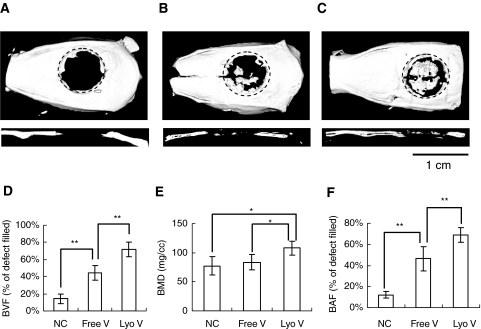
There was almost no regenerated bone in the defects except in the immediate vicinity of the surgical margins in the negative controls, demonstrating that AdLacZ was incapable of inducing new bone formation (Fig. 1A). On scaffolds with AdBMP2 suspensions, a few bony islands were distributed throughout the defects, but newly formed bone did not significantly cover the defects (Fig. 1B). In contrast, when AdBMP2 was delivered in a lyophilized formulation, significant regeneration was achieved, nearly spanning the entire defect (Fig. 1C).
Figure 1.
Bone formation in critical-sized calvarial defects compromised by radiation therapy. We performed µ-CT analysis to visualize and quantify bone regeneration. The 3-D images were reconstructed to illustrate the top and sagittal section views of (A) AdLacZ lyophilized in gelatin scaffolds, (B) AdBMP-2 freely suspended in gelatin scaffolds, and (C) AdBMP-2 lyophilized in gelatin scaffolds. The newly formed bone was evaluated by (D) bone volume fraction (BVF) in defects, by (E) bone mineral density (BMD) of newly formed bone, and by (F) bone area fraction (BAF) in defects assessed from the projected area ratio of the µ-CT image. The data were expressed as mean ± standard deviation, and the statistical differences between groups were analyzed by Student's t test (*p < 0.05; **p < 0.01). The dotted line represents the surgical margin of the cranial defect. N = 5.
Quantitative Assessment of Regenerated Bone
To quantify newly formed bone, we evaluated both BVF and BMD by μ-CT scanning (Figs. 1D, 1E). Compared with the negative control group, suspended AdBMP2 enhanced the BVF from 14.2 ± 5.3% to 44.6 ± 8.5%. However, there was no significant difference in BMD between these two groups (77 ± 16 mg/cc vs. 88 ± 13 mg/cc). In contrast, AdBMP2 lyophilized within scaffolds significantly improved both BVF and BMD over the AdLacZ and free AdBMP2 groups.
To judge the extent of regenerated bone in the cranial defects, we determined the BAF of newly formed bone based on 3-D projection images captured by μ-CT (Fig. 1F). Compared with the free AdBMP-2 group, in which bone covered only a modest region of the defects (46.4 ± 11.3%), lyophilized AdBMP2 treatment significantly increased bone formation, and most of the area of the defects was covered by mineralized tissue (68.7 ± 7.1%). These results suggest that the therapeutic effects of AdBMP2 were greatly improved when AdBMP was locally delivered within scaffolds.
The Effects of Bone Regeneration in Critical-sized Defects Compromised by Radiotherapy
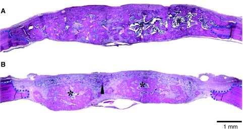
For evaluation of the extent to which pre-operative radiotherapy retarded bone regeneration, rats without radiation treatment (No-XRT) were implanted with gelatin sponges containing lyophilized AdBMP-2. At the time of specimen harvest, scaffolds with lyophilized AdBMP-2 induced new bone growth that nearly filled the entire defect, and no residual scaffold material was observed (Fig. 2A). The regenerated bone was integrated with the native bone at the defect margins (Appendix Figs. B, C) and supported a robust hematopoietic marrow (Appendix Fig. D).
Figure 2.
Histological analysis of critical-sized calvarial defects with or without pre-operative radiation therapy. Sections were prepared from the midline of defects. The defects without (A) and with (B) radiation treatment were examined. The defect margins are depicted by blue dashed lines. Arrowhead, non-degraded gelatin sponges; #mature bone marrow with adipose tissues; *immature bone marrow.
Defects treated with pre-operative irradiation (Pre-OP) were also successfully regenerated with lyophilized AdBMP2-containing scaffolds. Histological sections illustrated defects that were filled with newly formed bone (Fig. 2B). However, the bone morphology was different from that of the non-radiation-treated group. Here, the bone was admixed with connective tissues, with incomplete bridging. Osseointegration at defect edges was not as complete as that observed in the non-radiation-treated group (Appendix Fig. F), and remnants of non-resorbed scaffold material remained within the tissues (Appendix Fig. G). The woven regenerated bone and sparse marrow suggested that normal bone remodeling was negatively affected by irradiation (Appendix Figs. G, H).
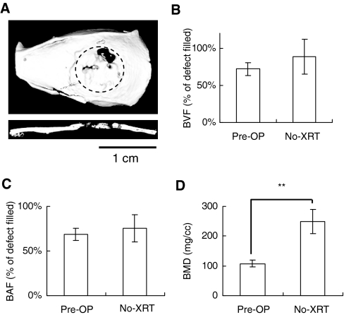
We also compared bone regeneration using μ-CT analysis. Regenerated bone in the Pre-OP group (Fig. 1C) illustrated defect coverage similar to that in the No-XRT group (Fig. 3A). However, compared with the Pre-OP group in which non-union bone gaps existed between newly formed bone and defect margins, bone was well-integrated with defect edges in the No-XRT group.
Figure 3.
Bone formation in critical-sized calvarial defects without radiation therapy (No-XRT). (A) The 3-D images were reconstructed to illustrate the top and sagittal section views. Bone regeneration was compared with that in the irradiated group (Pre-OP) by µ-CT analyses: (B) BVF, (C) BAF, and (D) BMD. The data were expressed as mean ± standard deviation, and the statistical differences between groups were analyzed by Student's t test (*p < 0.05; **p < 0.01). The dotted line represents the surgical margin of the cranial defect. N = 5.
Analysis of the quantified data demonstrated that the BVF and BAF in these two groups had no significant differences (Figs. 3B, 3C). However, the BMD of the Pre-OP group was significantly less than that of the No-XRT group (108 ± 12 mg/cc vs. 250 ± 41mg/cc), suggesting that the level of mineralization in the Pre-OP group was reduced because of the side-effects of radiation (Fig. 3D). Images of the sagittal sections were consistent with this result, demonstrating that the radiographs of defects in the Pre-OP group were less radiopaque than those in the No-XRT group (Figs. 1C, 3A).
Adenoviral Bioactivity is Preserved in Lyophilized Gelatin Scaffolds
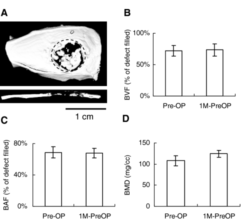
Approximately 80% of adenovirus activity within lyophilized biomaterial scaffolds is maintained for up to 6 mos (Hu et al., 2007). Therefore, we tested the practicality of preparing virus-scaffold complexes as pre-made constructs for potential clinical applications. AdBMP2 lyophilized in gelatin scaffolds was stored at -80°C for 1 mo and implanted into calvarial defects with pre-operative radiotherapy (1M-PreOP). The 3-D images captured by μ-CT scanning illustrated that bone formation in the 1M-PreOP group had bone defect coverage similar to that in the Pre-OP group (Figs. 1C, 4A). The bone volume, area, and density assessments all demonstrated no significant differences between the Pre-OP and 1M-PreOP groups, suggesting that lyophilized AdBMP2 had similar therapeutic effects before and after 1 mo of storage (Figs. 4B-4D). These results suggest that lyophilized AdBMP-2 maintained biologic activity while localized in scaffolds without losing its infectivity after long-term storage.
Figure 4.
Irradiated defects regenerated with AdBMP-2 lyophilized in gelatin scaffolds. To test the stability of viruses in the gelatin scaffolds, we stored pre-made AdBMP-2-lyophilized scaffolds (1M-PreOP) at -80°C for 1 mo prior to surgery. (A) The 3-D images were reconstructed to illustrate the top and sagittal section views. The bone regeneration was compared with that in the radiated group before storage (Pre-OP) by µ-CT analyses: (B) BVF, (C) BAF, and (D) BMD. The data were expressed as mean ± standard deviation, and the statistical differences between groups were analyzed by Student's t test (*p < 0.05; **p < 0.01). The dotted line represents the surgical margin of the cranial defect. N = 5.
Discussion
Compared with the freely suspended virus group, AdBMP-2 lyophilized within gelatin scaffolds demonstrated superior bone formation in bone defects compromised by pre-operative irradiation. Although the BVF and BAF in the suspended AdBMP-2 group were both higher than in the negative control group, there were no significant differences in BMD between these two groups. This suggests that the modest osteogenesis induced by free AdBMP-2 mainly increased bone quantity (BVF and BAF), but not bone quality (BMD). In contrast, lyophilized AdBMP-2 demonstrated BMD improvement over the other two groups, indicating that this local virus delivery enhanced both bone quantity and quality in irradiated defects. Because sufficient mineral density is important for bone homeostasis and function, the lyophilization strategy would likely be more appropriate for treating irradiated bone defects than a conventional bolus gene delivery method.
To determine the effects of radiotherapy on bone regeneration using an in vivo regenerative gene therapy strategy, we implanted gelatin scaffolds containing lyophilized AdBMP-2 into calvarial defects with and without pre-operative irradiation. Such wounds that are subjected to prior radiotherapy are among the most difficult bony reconstructions, in both pre-clinical studies and clinical practice (Deutsch et al., 1999). The newly formed bone in the Pre-OP group had bone volume and defect coverage similar to that in the No-XRT group. However, the sagittal images from μ-CT scanning and the BMD analysis demonstrated that the mineralized tissue in the Pre-OP group was less dense than in the No-XRT group. The histomorphologic assessments also illustrated that woven bone was mainly formed in the Pre-OP defects. In addition, the sparse marrow suggests a sub-optimal environment caused by radiation, and the bone regeneration period may need to be longer for proper recovery.
The long-term storage experiments demonstrated that the biologic activity and therapeutic effects of lyophilized AdBMP-2 were maintained after 1 mo of storage at -80°C. These results suggest that viral vectors encoding cell-signaling genes can be incorporated within biomaterial scaffolds and stored at low temperatures as pre-made constructs, making clinical application convenient for surgeons to apply in an operating room setting.
In this study, we developed an in vivo gene therapy by locally delivering BMP-2 gene within scaffolds, which effectively repaired defects compromised by radiation damage. Compared with ex vivo gene therapy, lyophilized adenovirus transduces cells in situ within the wound sites and simplifies the treatment by avoiding repeated surgeries. Without the use of autologous cells, this method reduces the risk of the contamination that may occur during in vitro cell culture. Furthermore, because virus-scaffold constructs can be prepared and stored in advance of a regenerative procedure, this virus-loaded scaffold could be applied in an operating room setting. From a clinical standpoint, the development of this technology could provide alternative options for bone replacement in irradiated tissue beds without the limitations and side-effects of current reconstructive techniques. In addition, lyophilized adenovirus in situ could have broad applications to non-irradiated tissues where similar reconstruction requirements exist in a tissue environment not compromised by radiotherapy.
Supplementary Material
Acknowledgments
The authors acknowledge and thank Dr. Renny Franceschi for the use of the AdBMP construct.
Footnotes
The research was supported by a grant from the National Institutes of Health (RO1 DE018890) (PHK).
References
- Chang SC, Wei FC, Chuang HL, Chen YR, Chen JK, Lee KC, et al. (2003). Ex vivo gene therapy in autologous critical-size craniofacial bone regeneration. Plast Reconstr Surg 112:1841-1850 [DOI] [PubMed] [Google Scholar]
- Deutsch M, Kroll SS, Ainsle N, Wang B. (1999). Influence of radiation on late complications in patients with free fibular flaps for mandibular reconstruction. Ann Plast Surg 42:662-664 [DOI] [PubMed] [Google Scholar]
- Disa JJ, Cordeiro PG. (2000). Mandible reconstruction with microvascular surgery. Semin Surg Oncol 19:226-234 [DOI] [PubMed] [Google Scholar]
- Ehrhart NP, Hong L, Morgan AL, Eurell JA, Jamison RD. (2005). Effect of transforming growth factor-beta1 on bone regeneration in critical-sized bone defects after irradiation of host tissues. Am J Vet Res 66:1039-1045 [DOI] [PubMed] [Google Scholar]
- Emerick KS, Teknos TN. (2007). State-of-the-art mandible reconstruction using revascularized free-tissue transfer. Expert Rev Anticancer Ther 7:1781-1788 [DOI] [PubMed] [Google Scholar]
- Fields MT, Eisbruch A, Normolle D, Orfali A, Davis MA, Pu AT, et al. (2000). Radiosensitization produced in vivo by once- vs. twice-weekly 2′2′-difluoro-2′-deoxycytidine (gemcitabine). Int J Radiat Oncol Biol Phys 47:785-791 [DOI] [PubMed] [Google Scholar]
- Franceschi RT. (2005). Biological approaches to bone regeneration by gene therapy. J Dent Res 84:1093-1103 [DOI] [PubMed] [Google Scholar]
- Gysin R, Wergedal JE, Sheng MH, Kasukawa Y, Miyakoshi N, Chen ST, et al. (2002). Ex vivo gene therapy with stromal cells transduced with a retroviral vector containing the BMP4 gene completely heals critical size calvarial defect in rats. Gene Ther 9:991-999 [DOI] [PubMed] [Google Scholar]
- Hardy S, Kitamura M, Harris-Stansil T, Dai Y, Phipps ML. (1997). Construction of adenovirus vectors through Cre-lox recombination. J Virol 71:1842-1849 [DOI] [PMC free article] [PubMed] [Google Scholar]
- Howard BK, Brown KR, Leach JL, Chang CH, Rosenthal DI. (1998). Osteoinduction using bone morphogenic protein in irradiated tissue. Arch Otolaryngol Head Neck Surg 124:985-988 [DOI] [PubMed] [Google Scholar]
- Hu WW, Wang Z, Hollister SJ, Krebsbach PH. (2007). Localized viral vector delivery to enhance in situ regenerative gene therapy. Gene Ther 14:891-901 [DOI] [PubMed] [Google Scholar]
- Hu WW, Lang MW, Krebsbach PH. (2008). Development of adenovirus immobilization strategies for in situ gene therapy. J Gene Med 10:1102-1112 [DOI] [PMC free article] [PubMed] [Google Scholar]
- Kaigler D, Wang Z, Horger K, Mooney DJ, Krebsbach PH. (2006). VEGF scaffolds enhance angiogenesis and bone regeneration in irradiated osseous defects. J Bone Miner Res 21:735-744 [DOI] [PubMed] [Google Scholar]
- Khouri RK, Brown DM, Koudsi B, Deune EG, Gilula LA, Cooley BC, et al. (1996). Repair of calvarial defects with flap tissue: role of bone morphogenetic proteins and competent responding tissues. Plast Reconstr Surg 98:103-109 [DOI] [PubMed] [Google Scholar]
- Koh JT, Zhao Z, Wang Z, Lewis IS, Krebsbach PH, Franceschi RT. (2008). Combinatorial gene therapy with BMP2/7 enhances cranial bone regeneration. J Dent Res 87:845-849 [DOI] [PMC free article] [PubMed] [Google Scholar]
- Krebsbach PH, Gu K, Franceschi RT, Rutherford RB. (2000). Gene therapy-directed osteogenesis: BMP-7-transduced human fibroblasts form bone in vivo. Hum Gene Ther 11:1201-1210 [DOI] [PubMed] [Google Scholar]
- Lee JY, Musgrave D, Pelinkovic D, Fukushima K, Cummins J, Usas A, et al. (2001). Effect of bone morphogenetic protein-2-expressing muscle-derived cells on healing of critical-sized bone defects in mice. J Bone Joint Surg-Am 83(A):1032-1039 [DOI] [PubMed] [Google Scholar]
- Nussenbaum B, Rutherford RB, Teknos TN, Dornfeld KJ, Krebsbach PH. (2003). Ex vivo gene therapy for skeletal regeneration in cranial defects compromised by postoperative radiotherapy. Hum Gene Ther 14:1107-1115 [DOI] [PubMed] [Google Scholar]
- Nussenbaum B, Rutherford RB, Krebsbach PH. (2005). Bone regeneration in cranial defects previously treated with radiation. Laryngoscope 115:1170-1177 [DOI] [PubMed] [Google Scholar]
- Reuther T, Schuster T, Mende U, Kubler A. (2003). Osteoradionecrosis of the jaws as a side effect of radiotherapy of head and neck tumour patients—a report of a thirty year retrospective review. Int J Oral Maxillofac Surg 32:289-295 [DOI] [PubMed] [Google Scholar]
- Würzler KK, DeWeese TL, Sebald W, Reddi AH. (1998). Radiation-induced impairment of bone healing can be overcome by recombinant human bone morphogenetic protein-2. J Craniofac Surg 9:131-137 [DOI] [PubMed] [Google Scholar]
- Yang S, Wei D, Wang D, Phimphilai M, Krebsbach PH, Franceschi RT. (2003). In vitro and in vivo synergistic interactions between the Runx2/Cbfa1 transcription factor and bone morphogenetic protein-2 in stimulating osteoblast differentiation. J Bone Miner Res 18: 705-715 [DOI] [PMC free article] [PubMed] [Google Scholar]
Associated Data
This section collects any data citations, data availability statements, or supplementary materials included in this article.


