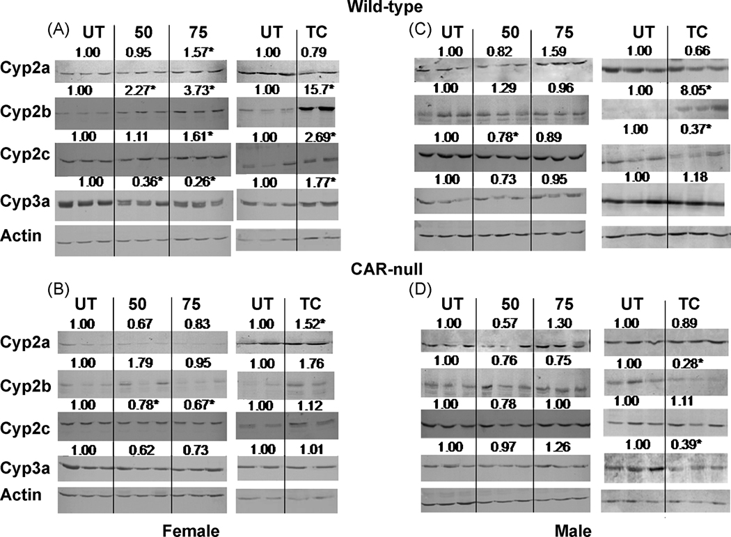
Fig. 5. Western blots of hepatic microsomes from NP and TCPOBOP-treated wild-type and CAR-null male and female mice.
Western blots were performed and visualized as described in the Materials and Methods. Blots were quantified densitometrically and the relative mean differential expression as compared to the controls is reported above the blots. An asterisk indicates a significant difference from the corresponding untreated mice by ANOVA followed by Fisher’s PLSD for NP-treated mice, and Student’s t-test for TC-treated mice (p < 0.05).

