Abstract
Neuronal cell death can be determined by the overall level of reactive oxygen species (ROS) resulting from the combination of extrinsic sources and intrinsic production as a byproduct of oxidative phosphorylation. Key controllers of the intrinsic production of ROS are the mitochondrial uncoupling proteins (UCPs). By allowing a controlled leak of protons across the inner mitochondrial membrane activation of these proteins can decrease ROS and promote cell survival. In both primate models of Parkinson’s disease and mouse models of seizures, increased activity of UCP2 significantly increased neuronal cells survival. In the retina UCP2 is expressed in many neurons and glial cells, but was not detected in rod photoreceptors. Retinal ganglion cell survival following excitotoxic damage was much greater in animals overexpressing UCP2. Traditional Chinese medicines, such as an extract of Cistanche tubulosa, may provide benefit by altering mitochondrial metabolism.
Keywords: Oxidative phosphorylation, Retinal ganglion cell, UCP2, Excitotoxin, Oxidative stress
Introduction
Mitochondria
Mitochondria are essential organelles in all eukaryotic cells and regulate a number of key processes including calcium homeostasis, redox control, and several biosynthetic reactions. Their most important role, however, is in energy production where they serve as the major source of ATP through oxidative phosphorylation. The inner mitochondrial membrane is highly impermeable and maintains a high electrochemical gradient. Embedded in this membrane are a series of transporters and enzymes that make up the respiratory chain. This chain is made up of five separate complexes and has over 80 protein constituents.
Oxidation of nutrient molecules gives rise to the reduced forms of the hydrogen carriers FADH and NADH (reduced cofactors). The reduced cofactors donate electrons to the three of the five complexes of the electron transport chain. The large drop in redox potential across these complexes provides the driving force to pump protons out of the mitochondrial matrix and create a large proton gradient across the membrane. The energy in this gradient is coupled to ATP synthesis by the complex five of the electron transport chain.
An obligatory consequence of these reactions is the generation of reactive oxygen species (ROS) that have the potential of damaging the cells. ROS exist in cells in balance with a variety of antioxidants. Oxidative stress occurs when the balance between ROS and antioxidants is perturbed. The increase in ROS usually comes from extrinsic mechanisms but it is the total ROS load that determines whether a cell is damaged or not. Among the mechanisms by which cells can reduce intrinsic ROS production, and thus be more resistant to extrinsic ROS, is the mild uncoupling that can be achieved through the actions of uncoupling proteins.
Mitochondrial uncoupling proteins
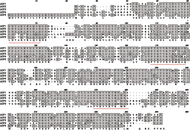
Uncoupling proteins (UCPs) are members of the large gene superfamily of mitochondrial anion carriers (reviewed in [1]). Based on sequence homologies uncoupling protein genes have been identified in plants, protozoa, fungi, and many vertebrates. Five uncoupling proteins have been identified in mammals and all share certain common structural elements (Fig. 1). All five are encoded by nuclear genes and their protein products reside as integral proteins of the inner mitochondrial membrane. The proteins have three approximately 100 amino acid repeats, each with two membrane spanning regions, and each of which has a sequence defined as an energy transfer protein signature.
Fig. 1.
Alignment of mouse UCP protein sequences. The GenBank Accession numbers used were AK002759 (UCP1), NM011671 (UCP2), NM009464 (UCP3), AK014394 (UCP4), and AF155812 (UCP5). The consensus sequence is shown below and identical residues are shaded in gray. The three energy transfer protein signatures are shown underneath in red
The canonical uncoupling protein in mammals is UCP1 which is found exclusively in brown fat, a tissue that uses its energy stores to produce heat. UCP1 is the key molecule in this process because it is able to allow a leak of protons down their potential gradient from the intermembrane space to the matrix of the mitochondria and the energy released by this movement is dissipated as heat [2].
There is still debate about the full range of functions of uncoupling proteins in other tissues. Each of the other UCPs has a characteristic tissue distribution with UCP2 being one of the most prevalent in the nervous system. UCP2 and some of the other uncoupling proteins can, like UCP1, control the leak of protons across the inner mitochondrial membrane [3]. One effect of this will be to reduce the endogenous production of ROS and thus the overall ROS burden. UCP2 may have a much more direct role in regulating ATP production and the coupling of ATP levels to metabolic load [4]. As regulators of energy metabolism in general, and ROS production in particular, UCPs are ideal targets for therapeutic agents. Because UCPs are distributed throughout the body, they are also target for the holistic approaches that characterize many forms of alternative medicine.
Uncoupling proteins and neuronal cell death
UCP2 can protect against oxidative stress
In our first experiments we asked whether overexpression of UCP2 in neuronal cell lines could provide protection against oxidative stress. Transfection of UCP2 plus GFP into several different cell lines made the cells more resistant to H2O2 than cells transfected with GFP alone [5]. Using a low dose of H2O2 that killed about 30% of cells, transfection of UCP2 completely abrogated the toxic effect of the peroxide. Similarly, toxicity of NO was reduced from 40% cell death to less than 15%. We next studied several in vivo models in different species and in different brain regions. For example, activation of UCP2 by its cofactor coenzyme Q protected neurons of the substantia nigra from damage induced by the toxin MPTP [6]. In other experiments we found that mice genetically engineered to overproduce UCP2 were far more resistant to the effects of drug-induced seizures [5]. Our conclusions from these studies were that substances and metabolic conditions that favor mitochondrial uncoupling can preserve neurons and potentially slow the progression of neurodegenerative diseases.
UCP2 is expressed in the retina
Both PCR and immunocytochemical labeling indicated that UCP2 is expressed in the retina [7]. Interestingly, we could not find any evidence that rod photoreceptors express this protein. Rods are one of the most metabolically active cells in the body and it is possible that they cannot function correctly if their mitochondria show any decrease in coupling. Most, if not all, inner retinal cells express UCP2. PCR analysis of cultured Müller cells detected the presence of UCP2 RNA suggesting that both neurons and glia can express this molecule.
To extend our studies in other parts of the CNS we asked whether UCP2 could provide protection against excitotoxic insults. We injected NMDA (160 nmol) or kainic acid (20 nmol) into the eyes of wild-type, UCP2 overexpressing transgenic mice or UCP2 knockout mice. After 24 h we examined the retinas and counted the number of cells in the ganglion cell layer (GCL). The NMDA-injected eyes of wild-type mice and UCP2 knockout mice showed a 36% decrease in cells in the GCL. The UCP2 transgenic mice, however, had lost only 17% of cells. The results with kainate injection were less dramatic but showed a similar trend. Wild-type and UCP2 knockout animals lost 78% of cells in the GCL whereas UCP2 transgenic animals lost 68% of cells.
UCP2 can regulate developmentally programmed cell death
During the first postnatal week mouse retinal ganglion cells (RGC) undergo maturation such that the number of RGCs in various strains of mice decrease from numbers of 131,000 to 224,000 at birth to 45,000 to 76,000 in the adult [8, 9]. The regulation of developmental RGC death is not fully understood but is linked, in part, to appropriate functional connections to target cells. Neurotrophic factors can delay or reduce RGC death and factors derived from postsynaptic cells may play a regulatory role in the process [10–12]. Although the signals responsible for controlling cell survival or cell death at this time are not fully understood, it is well established that the cells die through an apoptotic mechanism. We examined the numbers of apoptotic cells in the C57Bl/6 mouse GCL through the first postnatal week. Wild-type animals showed an increasing number of degenerating cells during the first postnatal days with the peak number at PN4, and this was followed by rapid decline over the next 2 days. UCP2 overexpressing mice on the other hand showed approximately 60% fewer degenerating cells at each time point. As might be expected, the end result of this decrease in cell death were retinas that had a thicker ganglion cell layer in patches across the tissue even in to the adult.
Herbal treatments may act at the level of the mitochondria
Herba Cistanches, the stems of Cistanche species, is a common traditional Chinese medicine and has been named the “Ginseng of the Desert”. Its use was first recorded in about 100 BC in Shen Nong Bencao Jing and is still used to treat a wide variety of ailments. Four major Cistanche species belong to the same genus but the molecular fingerprints of the four are very different [13]. Cistanche tubulosa was first described in an ophthalmology text in 1644 AD. An extract of this plant is produced commercially and has been approved for use as a treatment for dementia by the Chinese FDA. A wide variety of phenylethanoid glycosides have been isolated from Cistanche species and some of these are pharmacologically active [14].
Treatment of mammalian cells with an extract of C. tubulosa led to an increase in mitochondrial membrane potential and to changes in expression of genes related to mitochondria including subunits of ATP synthase, enzymes involved in Krebs cycle and heat shock proteins related to mitochondria (Zhang et al. unpublished results). Further work is needed to dissect this effect and determine how much of the mitochondrial effect is directly on components of the electron transport chain and how much on regulatory proteins such as the uncoupling proteins.
The complexity of the extract used in these experiments makes it difficult to determine whether a single ingredient is responsible for all the effects or multiple ingredients act on different pathways. Nevertheless, these preliminary experiments give support to the idea that traditional Chinese medicine approaches are effective because they target the same pathways as Western medicine.
Discussion
From the above review it is clear that activation of UCP2 can provide protection from a wide variety of insults that can lead to neural cell death. Though we have yet to test the role of UCP2 in any model of retinal degenerative disease, we predict that activation of UCP2 will slow the progression of any disease that involves apoptosis or mitochondrial dysfunction. Another conclusion from these and many other studies is that mitochondria are key determinants of cell death or survival. They act to integrate a wide variety of signals and can precipitate cell death directly through opening of the transition pore and release of cytochrome C, or indirectly through alteration of redox state and cytoplasmic calcium levels.
We currently have only a few compounds that can alter UCP2 activity. These include nucleotides, some fatty acids, and coenzyme Q. As more becomes known about the ways in which these molecules activate UCP2 there will undoubtedly be a search for chemicals that mimic these actions. Given the length of time that traditional Chinese medicine has had to experiment and refine treatments, it should not be surprising that there are medicines that act at key metabolic sites within cells such as the mitochondria. Traditional medicines also represent a more holistic approach and again, we should not be surprised that effective medicines target multiple aspects of cells or organelles that are shared among various tissues.
The challenge now is to decide whether to promote the use of traditional medicines in their present form or to try and dissect their active ingredients and try to formulate mixtures of these that have the same effect.
Acknowledgements
This work was supported by grant EY 013865 from the NIH and by the Macular Vision Research Foundation.
Open Access
This article is distributed under the terms of the Creative Commons Attribution Noncommercial License which permits any noncommercial use, distribution, and reproduction in any medium, provided the original author(s) and source are credited.
References
- 1.Borecký J, Maia IG, Arruda P. Mitochondrial uncoupling proteins in mammals and plants. Biosci Rep. 2001;21:201–12. doi: 10.1023/A:1013604526175. [DOI] [PubMed] [Google Scholar]
- 2.Nicholls DG, Locke RM. Thermogenic mechanisms in brown fat. Physiol Rev. 1984;64:1–64. doi: 10.1152/physrev.1984.64.1.1. [DOI] [PubMed] [Google Scholar]
- 3.Horvath TL, Diano S, Barnstable CJ. Mitochondrial uncoupling protein 2 in the central nervous system. Biochem Pharmacol. 2003;65:1917–21. doi: 10.1016/S0006-2952(03)00143-6. [DOI] [PubMed] [Google Scholar]
- 4.Affourtit C, Brand MD. On the role of uncoupling protein-2 in pancreatic beta cells. Biochim Biophys Acta. 2008;1777:973–9. doi: 10.1016/j.bbabio.2008.03.022. [DOI] [PubMed] [Google Scholar]
- 5.Diano S, Matthews RT, Patrylo P, Warden CH, Bartfai T, Bechmann I, Yang L, Shulman GI, Garcia-Segura LM, Beal MF, Cowley MA, Shanabrough M, Leranth C, Dietrich EH, Eugene Redmond DE D, Jr, Barnstable CJ, Horvath TL. Uncoupling protein 2 prevents neuronal death including that occuring during seizures: a mechanism for pre-conditioning. Endocrinology. 2003;144:5014–21. doi: 10.1210/en.2003-0667. [DOI] [PubMed] [Google Scholar]
- 6.Horvath TL, Diano S, Leranth C, Garcia-Segura LM, Cowley MA, Shanabrough M, Elsworth JD, Sotonyi P, Roth RH, Dietrich EH, Matthews RT, Barnstable CJ, Redmond DE., Jr Coenzyme Q induces mitochondrial uncoupling and prevents dopamine cell loss in a primate model of Parkinson’s disease. Endocrinol. 2003;144:2757–60. doi: 10.1210/en.2003-0163. [DOI] [PubMed] [Google Scholar]
- 7.Barnstable CJ, Li M, Reddy R, Horvath TL. Mitochondrial uncoupling proteins: regulators of retinal cell death. In: LaVail MM, Hollyfield JG, Anderson RE, editors. Retinal Degeneration 2002. New York: Kluwer Academic; 2003. pp. 269–75. [DOI] [PubMed] [Google Scholar]
- 8.Sefton AJ, Horsburgh GM, Lam K. The development of the optic nerve in rodents. Aust NZ J Ophthalmol. 1985;13:135–45. doi: 10.1111/j.1442-9071.1985.tb00414.x. [DOI] [PubMed] [Google Scholar]
- 9.Strom RC, Williams RW. Cell production and cell death in the generation of variation in neuron number. J Neurosci. 1998;18:9948–53. doi: 10.1523/JNEUROSCI.18-23-09948.1998. [DOI] [PMC free article] [PubMed] [Google Scholar]
- 10.McCaffery CA, Bennett MR. Dreher B The survival of neonatal rat retinal ganglion cells in vitro is enhanced in the presence of appropriate parts of the brain. Exp Brain Res. 1982;48:377–86. doi: 10.1007/BF00238614. [DOI] [PubMed] [Google Scholar]
- 11.Spalding KL, Rush RA, Harvey AR. Target-derived and locally derived neurotrophins support retinal ganglion cell survival in the neonatal rat retina. J Neurobiol. 2004;60:319–27. doi: 10.1002/neu.20028. [DOI] [PubMed] [Google Scholar]
- 12.Murphy JA. Clarke DB Target-derived neurotrophins may influence the survival of adult retinal ganglion cells when local neurotrophic support is disrupted: implications for glaucoma. Med Hypotheses. 2006;67:1208–12. doi: 10.1016/j.mehy.2006.04.049. [DOI] [PubMed] [Google Scholar]
- 13.Shi HM, Wang J, Wang MY, Tu PF, Li XB. Identification of Cistanche species by chemical and inter-simple sequence repeat fingerprinting. Biol Pharm Bull. 2009;32:142–6. doi: 10.1248/bpb.32.142. [DOI] [PubMed] [Google Scholar]
- 14.Jiang Y, Tu PF. Analysis of chemical constituents in Cistanche species. J Chromatogr A. 2009;1216:1970–9. doi: 10.1016/j.chroma.2008.07.031. [DOI] [PubMed] [Google Scholar]


