Abstract
Bacteriophage Qβ coat protein forms uniform virus-like particles when expressed recombinantly in a variety of organisms. We have inserted the IgG-binding Z domain at the carboxy terminus of the coat protein and coexpressed this chimeric subunit with native coat protein to create hybrid, IgG-binding virus-like particles. Extracellular osmolytes were found to have an effect on the incorporation efficiency of fusion proteins into VLPs in E. coli when a carbenicillin, but not a kanamycin, selection marker was used. The addition of sucrose to the growth media decreased incorporation efficiency; the osmoprotectant glycine betaine eliminated this effect. The decrease in efficiency was not observed when carbenicillin was omitted from the final expression culture. The addition of sodium chloride instead of sucrose gave rise to particles with a larger number of fusion proteins than the standard conditions. These results illustrate that cellular conditions should be taken into account even in apparently simple systems when natural or engineered protein nanoparticles are made.
Bacteriophage Qβ is a member of the Leviviridae family, forming icosahedral capsids from 180 coat protein subunits around a 4.2 kilobase sense-strand RNA genome. We have exploited the recombinant non-infectious virus-like particle (VLP) as a platform for chemical display, using a short (133 amino acid) wild-type capsid protein sequence as well as several point mutants (1) including those incorporating unnatural amino acids (2). The native infectious Qβ virion displays 3–5 copies of a large (approximately 200 amino acids) domain extension at the C-terminus, termed the A1 protein, which is used to recognize the E. coli host. Pumpens and coworkers previously created mutant recombinant virus-like particles which incorporate C-terminal extensions using fragments of the A1 sequence up to the full-length domain (3). The number of incorporated extensions varied inversely with their length, from a minimum of 10 per particle for a modified version of the full-length A1 protein to a maximum of 81 per particle for extensions of 11–24 amino acids derived from the A1 sequence. We sought to extend this work to the display of folded, functional peptide and protein domains in a manner that allows for as much modularity and control as possible.
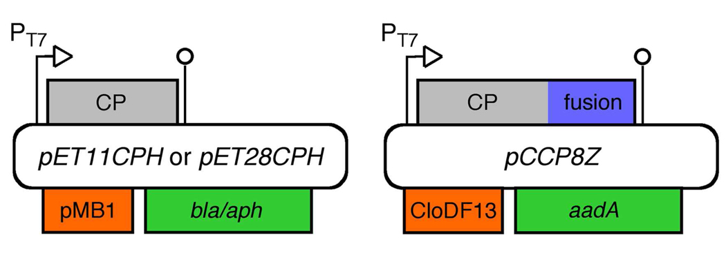
Instead of relying on differential readthrough of a stop codon as in the natural virus, we employed compatible plasmids coding for the wild-type (lacking the A1 extension, designated pET11CPH and pET28CPH) and fused (lacking a stop codon and bearing a C-terminal extension, designated pCCP8Z) capsid proteins (Figure 1). This design allows relative and absolute expression levels to be modulated by the choice of the vector copy number and promoter. For the test case described here, we fused the 58 amino-acid Z domain derived from S. aureus protein A (4) to the C-terminus of the Qβ capsid sequence with an octapeptide spacer (CP8Z). The Z domain binds to the CH2-CH3 hinge region of a group of IgG subtypes, and has been used with other nanoparticle scaffolds (5). IPTG-induced expression in E. coli cells transformed with only the fusion plasmid yielded copious quantities of Qβ-Z protein, but no intact particles, consistent with the expectation that the extended subunit is incapable of assembling into a particle on its own. In contrast, when E. coli cells were transformed with both wild-type and fusion plasmids,(6) hybrid particles were isolated in high yields (approximately 50 mg per liter of culture) after induced expression. Under standard conditions, approximately 20 Z domains were incorporated per particle.
Figure 1.
T7 vectors used to produce hybrid bacteriophage Qβ virus-like particles after co-transformation of E. coli. pMB1 and CloDF13 are non-interacting origins of replication; bla, aph, and aadA are orthogonal selection markers.
Cryoelectron microscopic reconstruction of the icosahedral particles did not show protein density other than that of the standard capsid, presumably due to the sparse and presumably irregular positioning of the Z domains. However, the hybrid VLPs were found to bind strongly to immobilized IgG in an ELISA assay (Supporting Information), whereas native Qβ VLPs showed no interaction. Since Qβ capsids do not disassemble under the conditions used in the ELISA,(7) the result demonstrates that the fused domains are accessible on the exterior surface of the particle.
During the development of this technique, the abundant outer membrane protein OmpF was found to be a frequent contaminant of the Qβ particles, even after several sequential purifications by sucrose gradient sedimentation. Increasing the osmotic pressure of the growth medium with sucrose, which is neither metabolized nor transported into the cells (8), has been found to reduce OmpF expression (9), primarily through the action of the EnvZ-OmpR two-component signaling system (10). Since Eschericia coli exhibits the remarkable ability to grow exponentially across a nearly 100-fold range of environmental osmotic pressures (11), such an approach was judged potentially feasible for Qβ VLP production.
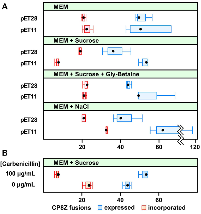
When 5% (w/v) sucrose was added to rich defined media (12) for recombinant Qβ expression, the ratio of OmpF to coat protein in the final post-induction cell culture was indeed diminished, by approximately 60% (Supporting Information). We tested two pairs of PT7 plasmids in the BL21(DE3) strain of E. coli: pET11CPH/pCCP8Z and pET28CPH/pCCP8Z. The plasmids coding for the wild-type short coat protein (CP) sequence differed in their antibiotic resistance marker: the aph gene which confers resistance to the aminoglycoside antibiotic kanamycin (pET28), and the bla gene for resistance to β-lactam antibiotics such as carbenicillin (pET11). The CP8Z sequence was introduced in both cases on a plasmid vector conferring spectinomycin resistance via the aadA gene. After expressing the proteins and purifying the VLPs to homogeneity, the protein content of the particles was analyzed to determine the fraction of coat protein carrying the fused domain (Figure 2). Analysis of post-induction cultures showed both CP and CP8Z protein expression and the yields of isolated particles to be roughly equivalent in all cases.
Figure 2.
(A) Comparison of Z domain fusion incorporation and expression levels with variations in osmolytes and selection markers. (B) The same comparisons in sucrose-supplemented media in the presence or absence of carbenicillin during IPTG-induced protein expression. “Expressed” (blue) and “incorporated” (red) protein levels are given per 180 total protein subunits. The x-axis values represent the number of fusion proteins per 180 total (wild-type + fusion) that were either incorporated into particles (blue) or found in samples of culture isolated 4 hours after induction, including both VLPs and protein not incorporated into particles (blue). These values would be the same if the assembly process was strictly stoichiometric. At least three replicates were performed for each set of data points.
In most experiments, approximately 20 CP8Z subunits were incorporated into each capsid, which is about 40% of the value expected on the basis of expression levels if the fusion and wild-type assembled into particles with equal efficiency. However, the number of incorporated CP8Z fusion proteins dropped to approximately 6 per particle (Figure 2A) in the presence of sucrose when the pET11, but not the pET28, vector was used. This striking observation was made with several batches of rich defined minimal media to which sucrose was added and was also observed for protein inserts other than the Z domain discussed here. We suspected that the increase in osmotic pressure was at least partially responsible for this result.
E. coli rely on organic osmolyte transporters (ProP, ProU, BetU) to rapidly boost the intracellular concentration of betaines under hyperosmotic stress (13). The addition of 1 mM of the potent osmoprotectant glycine betaine to the expression media reversed the deleterious effect of sucrose on chimeric protein incorporation (Figure 2A), suggesting that this phenomenon is correlated with the cellular response to an increase in osmotic pressure. However, when 0.2 M sodium chloride, another common osmolyte, was introduced into the growth medium, an effect opposite to that with sucrose was observed: the number of fusions incorporated into the VLP increased to an average of 33 per particle, again only when the pET11-based vector was used (Figure 2A). No significant change in doubling time was observed when either osmolyte was added to the growth media, indicating that the host was not adversely affected under high osmotic pressure conditions.
In hypoosmotic media containing a bare minimum of solutes (VLOM) (11), incorporation levels were the same as for the standard MEM conditions with the pET11 background. Cells harboring the pET28 background could not be reliably cultured in this medium, and thus no comparison could be made for conditions of inverse osmotic stress.
The major differences between pET11 and pET28 background are the selection marker gene (bla vs. aph) and the corresponding antibiotic (carbenicillin vs. kanamycin). To investigate the role of carbenicillin in the observed increase in sensitivity to osmolytes, cells were cultured in MEM with sucrose and carbenicillin until the OD600 reached the induction level. The cells were then collected and washed to remove the antibiotic, and then resuspended in fresh MEM containing sucrose and 1 mM IPTG, but without carbenicillin. Incubation for the standard four hours gave the normal level of capsid production and the abrogation of the sucrose effect: the purified VLPs showed the same level of Z domain incorporation (approximately 22 per particle) as was observed in MEM lacking sucrose but containing carbenicillin (Figure 2B).
We describe here the first study of the incorporation of two different forms of a coat protein into virus-like particles in cells in response to environmental factors. Since the incorporation results do not track with expression levels, particle assembly cannot simply be a process in which equilibrium mixtures of subunits form particles spontaneously, as can be done in vitro with systems such as cowpea chlorotic mottle virus (14).
The complexity of the apparently simple Qβ system is underscored by the fact that the observed variation in incorporation of the capsid fusion protein requires simultaneous changes in two, presumably unrelated, process parameters. Under non-stressing protein expression conditions, we observed no effect with the change in plasmid selection marker. Similarly, with the pET28 plasmid background, we observed only the expected decrease in OmpF expression.
Among the factors that may play a role are the following. Osmotic stress increases the effective concentration of solutes inside the cell, which can affect protein-protein (15) and protein-nucleic acid interactions (11). Changes in ionic strength resulting from transport mechanisms used by the cell to combat osmotic stress can also influence virus assembly (16). Lastly, the expression of β-lactamase in high concentration can unbalance the cell envelope and induce an SOS response.(17)
The new method for preparing chimeric Qβ virus-like particles described here, and the observation that their composition can be varied by manipulation of the conditions of cellular expression, are useful for the practical construction of functional nanoparticles. Rather than requiring genetic manipulation of transcriptional and translational elements, polyvalency can be controlled, at least empirically, through the addition of inexpensive solutes to the growth medium. These results also comprise the first cases in which VLP assembly is affected by such factors as resistance markers and osmotic pressure. It is likely that the assembly of natural virions, which are often hybrids of more than one capsid protein variant, are optimized for particular cellular conditions. Investigators wishing to produce VLPs or disrupt the production of viruses in vivo may profitably take such factors into consideration when designing their experiments.
Supplementary Material
ACKNOWLEDGMENT
We thank The Skaggs Institute for Chemical Biology and the NIH (RR012886 and GM083658) for support of this work. We are grateful also to Dr. Eiton Kaltgrad for the preparation of affinity-purified anti-Qβ IgY.
Footnotes
SUPPORTING INFORMATION
Materials and methods, ELISA data, data concerning OmpF suppression, and transmission electron micrograph of the hybrid particles. This material is available free of charge via the Internet at http://pubs.acs.org.
REFERENCES
- 1.(a) Prasuhn DE, Jr, Yeh RM, Obenaus A, Manchester M, Finn MG. Chem. Commun. 2007:1269–1271. doi: 10.1039/b615084e. [DOI] [PubMed] [Google Scholar]; (b) Kaltgrad E, O'Reilly MK, Liao L, Han S, Paulson J, Finn MG. J. Am. Chem. Soc. 2008;130:4578–4579. doi: 10.1021/ja077801n. [DOI] [PMC free article] [PubMed] [Google Scholar]; (c) Prasuhn DE, Jr, Singh P, Strable E, Brown S, Manchester M, Finn MG. J. Am. Chem. Soc. 2008;130:1328–1334. doi: 10.1021/ja075937f. [DOI] [PMC free article] [PubMed] [Google Scholar]
- 2.Strable E, Prasuhn DE, Jr, Udit AK, Brown S, Link AJ, Ngo JT, Lander G, Quispe J, Potter CS, Carragher B, Tirrell DA, Finn MG. Bioconjugate Chem. 2008;19:866–875. doi: 10.1021/bc700390r. [DOI] [PMC free article] [PubMed] [Google Scholar]
- 3.(a) Vasiljeva I, Kozlovska T, Cielens I, Strelnikova A, Kazaks A, Ose V, Pumpens P. FEBS Letters. 1998;431:7–11. doi: 10.1016/s0014-5793(98)00716-9. [DOI] [PubMed] [Google Scholar]; (b) Kozlovska TM, Cielens I, Vasiljeva I, Strelnikova A, Kazaks A, Dislers A, Dreilina D, Ose V, Gusars I, Pumpens P. Intervirology. 1996;39:9–15. doi: 10.1159/000150469. [DOI] [PubMed] [Google Scholar]
- 4.Nilsson B, Moks T, Jansson B, Abrahmsen L, Elmblad A, Holmgren E, Henrichson C, Jones TA, Uhlen M. Prot. Eng. 1987;1:107–113. doi: 10.1093/protein/1.2.107. [DOI] [PubMed] [Google Scholar]
- 5.(a) Korokhov N, Mikheeva G, Krendelshchikov A, Belousova N, Simonenko V, Krendelshchikova V, Pereboev A, Kotov A, Kotova O, Triozzi PL, Aldrich WA, Douglas JT, Lo KM, Banerjee PT, Gillies SD, Curiel DT, Krasnykh V. J. Virol. 2003;77:12931–12940. doi: 10.1128/JVI.77.24.12931-12940.2003. [DOI] [PMC free article] [PubMed] [Google Scholar]; (b) Kickhoefer VA, Han MM, Raval-Fernandes S, Poderycki MJ, Moniz RJ, Vaccari D, Silvestry M, Stewart PL, Kelly KA, Rome LH. ACS Nano. 2009;3:27–36. doi: 10.1021/nn800638x. [DOI] [PMC free article] [PubMed] [Google Scholar]
- 6.In the ternary adeno-associated virus system, subunit incorporation was found to depend on the ratio of plasmids used in the transfection step [ Warrington KH, Gorbatyuk OS, Harrison JK, Opie SR, Zolotukhin S, Muzyczka N. J. Virol. 2004;78:6596–6609. doi: 10.1128/JVI.78.12.6595-6609.2004. Rabinowitz JE, Bowles DE, Faust SM, Ledford JG, Cunningham SE, Samulski RJ. J. Virol. 2004;78:4421–4432. doi: 10.1128/JVI.78.9.4421-4432.2004.]. For reasons discussed in Supporting Information, this variable is not expected to be important for Qβ expressed in E. coli, and so the plasmids were used in a 1:1 ratio‥
- 7.Analysis by size-exclusion chromatography in the same buffers as used for ELISA showed no decomposition or disassembly of the particles. Furthermore, in our experience with Qβ VLPs we have subjected the particles to much harsher treatment without detectable disruption of the capsids. The closely related MS2 capsid has been reported to also be quite stable; Qβ is even more so due to disulfide crosslinking of subunits [ Ashcroft AE, Lago H, Macedo JMB, Horn WT, Stonehouse NJ, Stockley PG. J. Nanosci. Nanotech. 2005;5:2034–2041. doi: 10.1166/jnn.2005.507. ].
- 8.Schmid K, Ebner R, Jahreis K, Lengeler JW, Titgemeyer F. Mol. Microbiol. 1991;5:941–950. doi: 10.1111/j.1365-2958.1991.tb00769.x. [DOI] [PubMed] [Google Scholar]
- 9.Ramani N, Hedeshian M, Freundlich M. J. Bacteriol. 1994;176:5005–5010. doi: 10.1128/jb.176.16.5005-5010.1994. [DOI] [PMC free article] [PubMed] [Google Scholar]
- 10.(a) Mizuno T, Mizushima S. Mol. Microbiol. 1990;4:1077–1082. doi: 10.1111/j.1365-2958.1990.tb00681.x. [DOI] [PubMed] [Google Scholar]; (b) Vogel J, Papenfort K. Curr. Opin. Microbiol. 2006;9:605–611. doi: 10.1016/j.mib.2006.10.006. [DOI] [PubMed] [Google Scholar]
- 11.Capp MW, Cayley DS, Zhang W, Guttman HJ, Melcher SE, Saecker RM, Anderson CF, Record MT. J. Mol. Biol. 1996;258:25–36. doi: 10.1006/jmbi.1996.0231. [DOI] [PubMed] [Google Scholar]
- 12.Studier FW. Protein Expr. Purif. 2005;41:207–234. doi: 10.1016/j.pep.2005.01.016. [DOI] [PubMed] [Google Scholar]
- 13.(a) Ly A, Henderson J, Lu A, Culham DE, Wood JM. J. Bacteriol. 2004;186:296–306. doi: 10.1128/JB.186.2.296-306.2004. [DOI] [PMC free article] [PubMed] [Google Scholar]; (b) Lucht JM, Bremer E. FEMS Microbiol. Rev. 1994;14:3–20. doi: 10.1111/j.1574-6976.1994.tb00067.x. [DOI] [PubMed] [Google Scholar]
- 14.(a) Zhao X, Fox JM, Olson NH, Baker TS, Young MJ. Virology. 1995;207:486–494. doi: 10.1006/viro.1995.1108. [DOI] [PubMed] [Google Scholar]; (b) Zlotnick A, Aldrich R, Johnson JM, Ceres P, Young MJ. Virology. 2000;277:450–456. doi: 10.1006/viro.2000.0619. [DOI] [PubMed] [Google Scholar]
- 15.Bowden GA, Georgiou G. J. Biol. Chem. 1990;265:16760–16766. [PubMed] [Google Scholar]
- 16.(a) Schwarcz WD, Barroso SPC, Gomes AMO, Johnson JE, Schneemann A, Oliveira AC, Silva JL. Cell. Mol. Biol. 2004;50:419–427. [PubMed] [Google Scholar]; (b) Brown WL, Mastico RA, Wu M, Heal KG, Adams CJ, Murray JB, Simpson JC, Lord JM, Taylor-Robinson AW, Stockley PG. Intervirology. 2002;45:371–380. doi: 10.1159/000067930. [DOI] [PubMed] [Google Scholar]; (c) Wingfield PT, Stahl SJ, Williams RW, Steven Biochemistry. 1995;34:4919–4932. doi: 10.1021/bi00015a003. [DOI] [PubMed] [Google Scholar]; (d) Seifer M, Zhou S, Standring DN. J. Virol. 1993;67:249–257. doi: 10.1128/jvi.67.1.249-257.1993. [DOI] [PMC free article] [PubMed] [Google Scholar]; (e) Peabody DS. EMBO J. 1993;12:595–600. doi: 10.1002/j.1460-2075.1993.tb05691.x. [DOI] [PMC free article] [PubMed] [Google Scholar]; (f) Zhou S, Standring DN. Proc. Natl. Acad. Sci. U.S.A. 1992;89:10046–10050. doi: 10.1073/pnas.89.21.10046. [DOI] [PMC free article] [PubMed] [Google Scholar]
- 17. Miller C, Thomsen LE, Gaggero C, Mosseri R, Ingmer H, Cohen SN. Science. 2004;305:1629–1631. doi: 10.1126/science.1101630.. Such an SOS response may last for only a short time following the removal of the inducing agent [ Ni M, Yang L, Liu XL, Qi O. Curr. Microbiol. 2008;1657:1521–1526. doi: 10.1007/s00284-008-9235-4.], and so may be consistent with the experiment in which carbenicillin is removed from the medium during induced protein expression.
Associated Data
This section collects any data citations, data availability statements, or supplementary materials included in this article.


