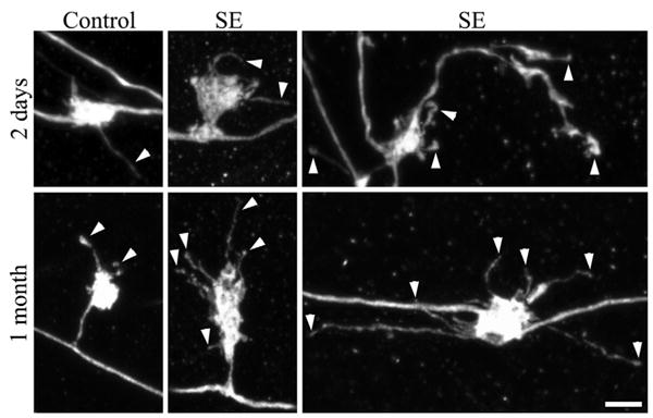
FIGURE 2.

Confocal reconstructions of granule cell giant mossy fiber boutons. Giant boutons are from control animals and animals that underwent status epilepticus (SE) either 2 days or 1 month earlier. The 1 month animals shown exhibited minimal loss of hippocampal principal neurons after status epilepticus (Cumulative damage score = 2). Two days after status epilepticus, the number of filopodia per giant mossy fiber bouton was significantly increased (arrowheads). One month after status epilepticus, giant mossy fiber bouton area, the number of filopodia per giant mossy fiber bouton (arrowheads), and filopodia length were significantly increased relative to age-matched saline controls. Scale bar = 3 μm. [Color figure can be viewed in the online issue, which is available at www.interscience.wiley.com.]
