Abstract
WEB SITE FEATURE
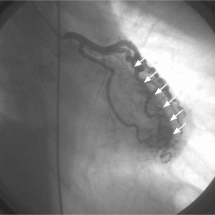
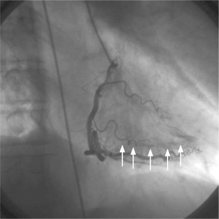
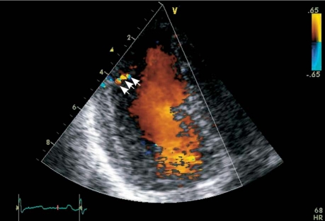
A 55-year-old man came to our hospital with exertional chest pain, dyspnea, and fatigue that had lasted 3 months. On physical examination, his blood pressure was 130/70 mmHg, and his heart rate was 85 beats/min. Electrocardiography showed sinus rhythm and no specific changes in the ST segment or the T wave. Echocardiography revealed ventricles of normal size and function. Coronary angiography revealed multiple fistulae that arose from all 3 major coronary arteries and drained exclusively into the left ventricle (Figs. 1 and 2). The coronary arteries were free of atherosclerotic disease. We performed repeat echocardiography, focusing on the fistulae, which indeed drained into the left ventricle (Fig. 3). The patient was discharged from the hospital with instructions to take 100 mg of metoprolol daily. He has experienced no anginal symptoms for 1 year.
Fig. 1. Coronary angiography shows fistulae from the left anterior descending coronary artery to the left ventricle (arrows).
Real-time motion image is available at www.texasheart.org/journal.
Fig. 2. Coronary angiographyshows fistulae from the right coronary artery to the left ventricle (arrows).
Real-time motion image is available at www.texasheart.org/journal.
Fig. 3. Transthoracic color-flow Doppler echocardiography (apical 4-chamber view) shows a fistula draining into the left ventricle (arrows).
Comment
Coronary–cameral fistulae are rare and are predominantly congenital communications between the coronary arterial circulation and the chambers or great vessels of the heart. It is rare that a coronary artery fistula causes myocardial ischemia. Therapeutic approaches are designed to reduce the myocardial oxygen demand and thereby ameliorate the demand–supply mismatch. Symptomatic relief has been achieved with β-blockers or with calcium-channel blockers.1,2
Sufficiently enlarged coronary arteries can be detected by use of 2-dimensional echocardiography. In children, the diagnosis of coronary artery fistula can often be made with transthoracic 2-dimensional and color-flow Doppler echocardiography. However, in adults, transesophageal echocardiography may be more sensitive.3,4 Coronary arteriography is the best method by which to determine the origin of such fistulae; indeed, in our patient, this method showed multiple coronary artery fistulae.
Patients who have small fistulae have an excellent long-term prognosis.5 Larger fistulae, when they are hemodynamically significant, should be closed by means of surgery or transcatheter embolization.6–8 In our patient, the fistulae were small and not suitable for percutaneous closure, and, because metoprolol therapy relieved the patient's symptoms, we did not consider closing the fistulae.
Supplementary Material
Footnotes
Address for reprints: Sakir Arslan, MD, Ataturk Universitesi Tip Fakultesi Kardiyoloji Anabilim Dali, 25070 Erzurum, Turkey
E-mail: drsakirarslan@gmail.com
References
- 1.Heper G, Kose S. Increased myocardial ischemia during nitrate therapy caused by multiple coronary artery-left ventricle fistulae? Tex Heart Inst J 2005;32(1):50–2. [PMC free article] [PubMed]
- 2.Wolf A, Rockson SG. Myocardial ischemia and infarction due to multiple coronary-cameral fistulae: two case reports and review of the literature. Cathet Cardiovasc Diagn 1998;43(2): 179–83. [DOI] [PubMed]
- 3.Yang Y, Li Z, Wang X. Assessment of coronary artery fistula by color Doppler echocardiography. Echocardiography 1998; 15(1):67–72. [DOI] [PubMed]
- 4.Krishnamoorthy KM, Rao S. Transesophageal echocardiography for the diagnosis of coronary arteriovenous fistula. Int J Cardiol 2004;96(2):281–3. [DOI] [PubMed]
- 5.Sherwood MC, Rockenmacher S, Colan SD, Geva T. Prognostic significance of clinically silent coronary artery fistulas. Am J Cardiol 1999;83(3):407–11. [DOI] [PubMed]
- 6.Kassaian SE, Alidoosti M, Sadeghian H, Dehkordi MR. Transcatheter closure of a coronary fistula with an Amplatzer vascular plug: should a retrograde approach be standard? Tex Heart Inst J 2008;35(1):58–61. [PMC free article] [PubMed]
- 7.McMahon CJ, Nihill MR, Kovalchin JP, Mullins CE, Grifka RG. Coronary artery fistula. Management and intermediate-term outcome after transcatheter coil occlusion. Tex Heart Inst J 2001;28(1):21–5. [PMC free article] [PubMed]
- 8.Wang S, Wu Q, Hu S, Xu J, Sun L, Song Y, Lu F. Surgical treatment of 52 patients with congenital coronary artery fistulas. Chin Med J (Engl) 2001;114(7):752–5. [PubMed]
Associated Data
This section collects any data citations, data availability statements, or supplementary materials included in this article.


