Abstract
We studied the effect of environmental parameters on the survival of Burkholderia pseudomallei. There was a small increase in bacterial count for up to 28 days in sterilized distilled water or rain water, in water at 20°C or 40°C, and in buffered solutions of pH 4 or higher. Counts of culturable B. pseudomallei declined at pH 3, in the presence of seawater or water with concentrations of 4% salt or higher, and under refrigeration. The morphological appearances of B. pseudomallei changed under conditions that maintained culturable numbers from bacilli to coccoid cells and spiral forms under pH or salt stress. These observations indicate that B. pseudomallei can endure nutrient-depleted environments as well as a wide range of pH, salt concentrations, and temperatures for periods of up to 28 days. The relative stability of B. pseudomallei under these conditions underlines the tenacity of this species and its potential for natural dispersal in water: in surface water collections, in managed water distribution systems, and through rainfall. These survival properties help explain the recent expansion of the known melioidosis endemic zone in Australia and may have played a part in recent melioidosis outbreaks.
Introduction
The Gram-negative bacterium Burkholderia pseudomallei is known to survive austere environmental conditions for prolonged periods. Contaminated water supplies were a feature of two Australian melioidosis outbreaks,1,2 but as yet, there is only limited in vitro data on B. pseudomallei survival in liquids.3–6 B. pseudomallei has been shown to maintain culturable bacterial numbers over prolonged periods in the absence of a carbon source and can survive in distilled water for several years, after which culturable bacteria have been recovered.7 Other B. pseudomallei environmental survival studies have concentrated on survival on environmental surfaces8 and in the presence of a limited range of changing environmental conditions, although experimental design limits how far these findings can be extrapolated.6 These studies also provide some insight into the behavior of B. pseudomallei in dry conditions and may explain its ability to persist in soil between rainy seasons in the melioidosis-endemic zone. However, few studies address survival in water on the land surface, between soil particles in the rhizosphere, in water droplets, or in seawater. The continued survival of B. pseudomallei at 20°C and 40°C has been previously reported.6 B. pseudomallei isolates have been recovered from water sources ranging from pH 2–9.9 Tong and others6 showed survival of eight strains of B. pseudomallei isolated from soil and water in China in pH 3 saline for a maximum of 7 days.6 Other Gram-negative bacteria including Pseudomonas fluorescens have been shown to survive for extended periods of time in distilled water up to 16 years,10 but extended survival has not been reported previously in salt water. Our previous studies on B. pseudomallei survival in chlorine-treated water focused on potable water treatment because of concerns about the adequacy of water treatment during an investigation into an outbreak of melioidosis in Western Australia (WA).3,4,11 None of the studies cited above dealt with bacterial survival in the range of waters potentially relevant to known water-related outbreaks1,12,13 nor the other environmental variables (temperature range, pH, and NaCl concentration) highlighted as potential environmental contributors during the investigation of one specific outbreak.2,11,14 The aim of this study was to investigate the viability and persistence of B. pseudomallei in a liquid environment with particular reference to fresh potable water, seawater, rainwater, temperature, and pH variation to understand the role these factors might play in raising or lowering the threat of environmental exposure to B. pseudomallei and therefore, to subsequent development of disease.
Materials and Methods
Bacteria.
The following strains of B. pseudomallei were studied: NCTC 10276 (clinical reference strain from a British citizen believed to have encountered B. pseudomallei in rural India obtained from the WA Culture Collection), NCTC 13177 (clinical isolate from the third septicaemic patient in the WA outbreak of 1997, lodged in the WA Culture Collection by T.J.J.I. in December of 1997, and submitted to the National Collection of Type Cultures, Mill Hill, London, U.K. in 1998), and a persistently mucoid isolate BCC 11 (persistently mucoid phenotype, clinical isolate from septicaemic strain, provided by Dr. G. Lum, Royal Darwin Hospital, Northern Territory). B. pseudomallei NCTC 13177 and DM98 have both been fully sequenced (GenBank. NZ_ABBQ00000000. Read, T et al. Genomics, Naval Medical Research Center/Biological Defense Research Directorate, 12300 Washington Ave., 2nd Floor, Rockville, MD 20852, USA). All strains were stored at −70°C in brain–heart infusion broth supplemented with 15% glycerol. After incubation on 5% horse blood agar (BA) for 24 hours at 37°C, a single colony was transferred to 15 mL tripticase soy broth (TSB) and incubated for 24 hours at 37°C. The pellicle that formed on the broth surface was removed carefully before washing the culture in sterile distilled water (SDW). Cells were suspended again in each of the liquids indicated in Results or in broth medium to obtain a starting concentration of approximately 106 CFU/mL.
Solutions studied.
SDW (PathWest Media preparation service, Perth, Western Australia, Australia) was used as a reference liquid. Kimberley rain water (KRW) was obtained during a summer downpour by collection into a wide neck container placed 1 m above the ground near Argyle Village, Western Australia. This rainwater was sterilized by repeated filtration through a 0.8-µm/0.2-µm Acrodisc Supor Membrane syringe filter (Pall Corporation, Newquay, United Kingdom). Artificial sea water (ASW) was obtained by reconstituting Sea Salts, an artificial seawater base (Sigma-Aldrich, St. Louis, MO), with demineralized hypo-osmolar water and then autoclaving the resulting solution. Fresh water has a salt content of < 0.05% w/v, and brine has a salt content of > 5%. Brackish water is 0.05–3% salt, and saline water is 3-5% salt. Washed bacterial cells were re-suspended in 5 mL of each of the water preparations (SDW, KRW, or ASW) and maintained in sealed containers at room temperature for the duration of the experiment. Solutions for pH studies were prepared in citrate–phosphate buffer (citric acid; BDH Chemicals, Kilsyth, Australia; dibasic sodium phosphate; Ajax Chemicals, Sydney, Australia) and adjusted to a pH of 3, 4, 5, 6, or 7 in accordance with standard methods.15 Solutions with concentrations of 40%, 4%, 0.4%, 0.04%, and 0.004% artificial sea salts (S9883-500g; Sigma-Aldrich) were prepared with hypo-osmolar water and autoclaved.
Survival studies.
Survival of B. pseudomallei strains was monitored over a 28-day period in three types of water. Survival was tested in SDW and filter-sterilized KRW. Further survival studies were conducted for a similar period of time under conditions of varying pH, temperature, or salt concentration as suggested by the original outbreak investigations.2,11 To inoculate the solution under investigation, 106 CFU/mL of washed B. pseudomallei NCTC 10276, NCTC 13177, or DM98 were used. At 0, 1, 2, 5, and 8 hours and 1, 2, 5, 7, 14, 21, and 28 days after inoculation, a 100-μL aliquot was aseptically removed from each of the 5-mL experimental cultures. The number of viable cells was determined by plating serial dilutions (from 10−2 to 10−4) of each aliquot onto Plate Count Agar medium (PCA; PathWest Media) with a spiral-plating device (Don Whitley Scientific Ltd., Shipley, United Kingdom.). All data points in each experiment were obtained in triplicate, and statistical calculations were made with GraphPad Prism version 4.00 for Windows (GraphPad Software, San Diego, CA). P values were calculated using the unpaired t test, and results were considered significant when P < 0.05.
Fluorescence microscopy.
The BacLight Bacterial Viability Kit (Invitrogen Molecular Probes, Eugene, WA) was used to stain B. pseudomallei for epifluorescent microscopy. Briefly, the green fluorescent dye SYTO 9 caused apple-green fluorescence of bacterial cells when live and was quenched by red fluorescence when propidium iodide penetrated the cytoplasm. Red cells were classified as dead, whereas green cells were classed as live. A 5-μL aliquot was taken from each bacteria/liquid series after 10 days of incubation and stained according to the manufacturers' directions. The mixture was transferred to a clean microscope slide, covered with an acid-washed glass cover slip, sealed with nail polish along the cover slip edge, and left in the dark for 15 minutes. These preparations were examined under bright field and fluorescent illumination at ×1000 total magnification on an Olympus BX60FS (Olympus Optical Company Ltd, Tokyo, Japan) microscope with digital image capture. The approximate red-to-green cell ratio (live-to-dead, respectively) as well as cell shape and size were recorded by direct visual examination of a minimum of 20 high-powered fields. Images were captured by simultaneous fluorescent and differential interference contrast (DIC) microscopy and superimposed to obtain a viability assessment of each bacterial cell.
Transmission electron microscopy.
A 20-mL aliquot of each experimental culture was removed after 10 days of incubation and washed by centrifugation at 3000 rcf for 10 minutes. The pellet was re-suspended in 2.5% glutaraldehyde in phosphate buffer (0.05 M; pH 7) and shaken overnight at 4°C. The cells were washed three times in phosphate buffer to remove the fixative. A 100-μL aliquot was removed, spread onto 5% horse BA, and incubated to ensure non-viability. The cell suspension was mixed with 1.1-μm polystyrene beads (Sigma-Aldrich) and deposited on formvar-coated electron microscope (EM) grids (ProSciTech, Townsville, Australia); the excess liquid was removed. Grids were negatively stained with 1% phosphotungstic acid. Excess stain was removed by rinsing in SDW, and the grids were blotted dry. Samples were examined on a transmission electron microscope (Philips CM10, Eindhoven, The Netherlands) fitted with MegaView III Soft Imaging System (Olympus Soft Imaging Solutions GmbH, Münster, Germany).
Scanning electron microscopy.
At 10 days, a 20-mL aliquot of each experimental culture was centrifuged at 3000 rcf for 10 minutes. The resulting pellets were re-suspended in 1 mL of SDW; 5 μL aliquots were placed on Alcian blue-coated cover slips. The cover slips were coated with Alcian blue by first placing a drop of 1% aqueous Alcian blue on laboratory film (Parafilm, Pechiney Plastic Packaging Co., Chicago, IL) and then inverting the cover glass onto the Alcian blue droplet for 20 minutes on each side. This was followed by three washes in SDW and drying. After 1 hour, cover slips with deposited bacterial suspension were flooded with 2.5% glutaraldehyde for 30 minutes. Samples were rinsed in phosphate buffer (0.05 M; pH 7.4), then post-fixed for 30 minutes in 1% aqueous osmium tetroxide (ProSciTech), and rinsed another time in phosphate buffer. Dehydration was performed in an ethanol gradient (25%, 50%, 70%, 95%, and 100%). Cover slips in super-dry ethanol were critical-point dried, mounted on aluminum pin-type SEM mounts (12.6-mm diameter; ProSciTech), and coated with 2-nm platinum vapor (Polaron ES100, Watford, United Kingdom). Cover slips were examined on a scanning electron microscope (Zeiss SUPRA 55 Variable Pressure SEM, Oberkochen, Germany) at an accelerating voltage of 1.50 kV.
Results
Natural versus distilled water.
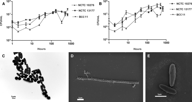
The effect of SDW and KRW was investigated over a period of 28 days. The comparative survival of all three strains of B. pseudomallei is shown inFigure 1. Over a period of 28 days, bacterial counts increased by at least two-fold in SDW and up to 30-fold for NCTC 13177 and BCC 11 in KRW (P = 0.0004 for NCTC 13177; t = 36.50, df = 2, and P < 0.0001 for BCC 11). All three strains were compared in SDW (t = 11.35, df = 5, P < 0.0001 for NCTC 10276; t = 3.311, df = 3, P < 0.0001 NCTC 13177; t = 18.63, df = 4, P < 0.0001 for BCC 11). The relatively high starting concentration of NCTC 10276 in KRW dropped after 2 hours and thereafter continued increasing similarly to the other strains during the reminder of the experiments. The majority (> 90%) of bacteria incubated in SDW or KRW were bacilli 1–2 μm in length (Figure 1C) with smooth cell walls and uniform size. After prolonged survival in water, occasional spiral forms were observed (Figure 1D), although the majority of cells were still intact bacilli (Figure 1E).
Figure 1.

(A) Survival of three reference strains of B. pseudomallei in SDW over a 28-day period. (B) Survival of three reference strains of B. pseudomallei in filter-sterilized KRW over a 28-day period. (C) Washed B. pseudomallei NCTC 13177 in SDW at time = 0. The dark circular objects are 1-μm diameter standard-sizing beads. (D) Spiral bacilli seen as occasional morphologic variants in SDW solutions after incubation for 28 days. (E) Conventional bacillary morphology of B. pseudomallei at time = 0 by scanning electron microscopy.
Survival at varying temperatures.
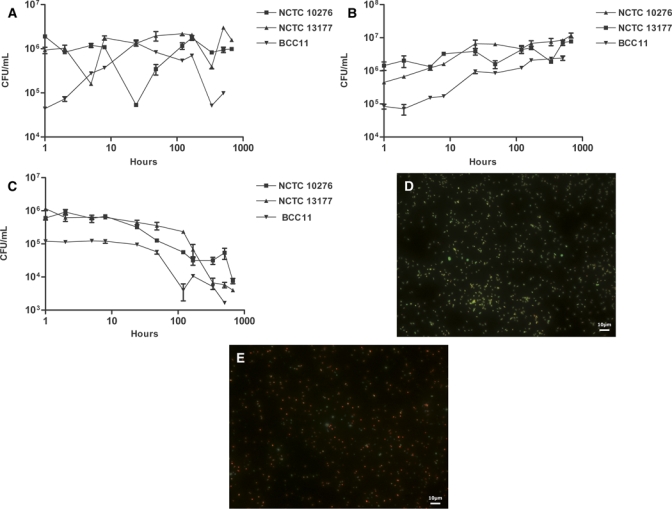
The survival of the same three strains of B. pseudomallei was observed at 40°C, 20°C, and 2°C over a 28-day period (Figure 2). All three strains survived the entire period at all three temperatures. However, from time 0–28 days at 2°C, there was a significant (100- to 9000-fold) decrease in the recovered viable count for all three strains (t = 4.508, df = 3, P < 0.0001 for NCTC 10276; t = 5.303, df = 3, P < 0.0001 for NCTC 13177; t = 13.45, df = 2, P < 0.0001 for BCC 11). The bacterial count at 20°C increased 7- to 30-fold for all three strains (t = 2.983 df = 6, P = 0.0245 for NCTC 10276; t = 10.21, df = 4, P = 0.0005 for NCTC 13177; t = 3.792, df = 3, P = 0.0322 for BCC 11). At 40°C, NCTC 13177 (t = 16.27, df = 4, P < 0.0001) and BCC 11 (t = 19.23, df = 2, P = 0.0027) showed increases of approximately 1.8-fold in viable count over the 28-day time period, whereas NCTC 10276 did not. The majority of cells (circa 80%) in Figure 2E were not fluorescent green, although only a small minority (10–20%) were red, indicating a loss of membrane integrity and thus, cell death.
Figure 2.

(A) Survival of three reference strains of B. pseudomallei in SDW over a 28-day period at a constant 40°C. (B) Survival of three reference strains of B. pseudomallei in SDW over a 28-day period at a constant 20°C. (C) Survival of three reference strains of B. pseudomallei in SDW over a 28-day period at a constant 2°C. (D) Suspension of B. pseudomallei NCTC 13177 cells at 0 days in SDW showing predominant viable cells (green). (E) Suspension of B. pseudomallei NCTC 13177 cells after 28 days in SDW showing a similar ratio of live to dead cells (i.e., predominantly non-viable cells [red] against scanty viable cells [green]).
Survival at varying pH.
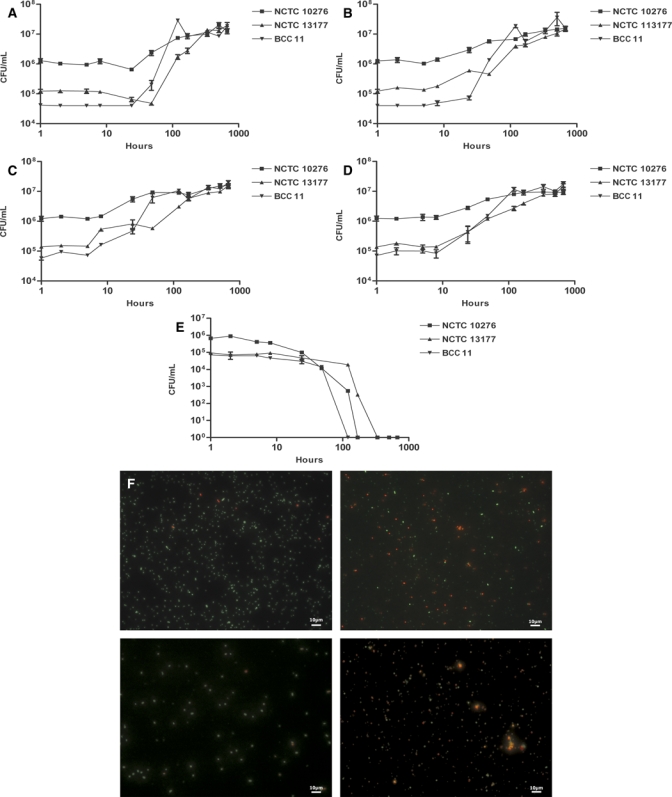
The survival of three strains of B. pseudomallei was studied at pH ranging from 3 to 7 over a period of 28 days (Figure 3). All three strains did not survive until the end of the experiment (28 days) at pH 3 (Figure 3E). NCTC 10276, NCTC 13177, and BCC 11 were not recovered at pH 3 after 7 days, 14 days, or 5 days incubation, respectively. The three strains survived for the entire experimental period at the other pH values tested. A significant increase of between 9- and 400-fold in the bacterial count was observed at all other pH values tested: NCTC 10276 at pH 4 (t = 5.230, df = 4, P = 0.0064), pH 5 (t = 4.632, df = 4, P = 0.0098), pH 6 (t = 5.478, df = 4, P = 0.0054), and pH 7 (t = 3.656, df = 4, P = 0.0216); NCTC 13177 at pH 4 (t = 4.435, df = 3, P = 0.0213), pH 5 (t = 4.954, df = 3, P = 0.0158), pH 6 (t = 4.848, df = 5, P = 0.0047), and pH 7 (t = 6.452, df = 6, P = 0.0007); BCC 11 at pH 4 (t = 3.716, df = 5, P = 0.0138), pH 5 (t = 4.286, df = 4, P = 0.0128), pH 6 (t = 5.936, df = 5, P = 0.0019), and pH 7 (t = 7.978, df = 4, P = 0.0013). Coccoid cells predominated at acidic pH. Cells with atypical shapes, including spiral forms, were seen under the stress provided at pH 3 (Figure 3F, bottom right). Spiral forms remained only a very small fraction of cells present. Fluorescent dye treatment at pH 3 showed that a small minority of cells were viable at that pH after 28 days (Figure 3F, bottom right), whereas the majority were either non-viable or compromised. After extended incubation at pH 3, the approximate cell number (Figure 3E and F, bottom right) vastly exceeded the viable count as determined by CFU/mL.
Figure 3.

(A) Survival of three reference strains of B. pseudomallei in SDW buffered to pH 7 over a 28-day period. (B) Survival of three reference strains of B. pseudomallei in SDW buffered to pH 6 over a 28-day period. (C) Survival of three reference strains of B. pseudomallei in SDW buffered to pH 5 over a 28-day period. (D) Survival of three reference strains of B. pseudomallei in SDW buffered to pH 4 over a 28-day period. (E) Survival of three reference strains of B. pseudomallei in SDW buffered to pH 3 over a 28-day period. (F) Live/dead bacterial cell viability results showing B. pseudomallei at time = 0 and pH 7 (top left), time = 28 days and pH 7 (top right), time = 0 and pH 3 (bottom left), and time = 28 days and pH 3 (bottom right). At pH 7, predominantly live cells are lost and replaced by a mixture of live and dead cells, whereas at pH 3, live cells and a lot of indeterminate cells are replaced by mainly non-viable cells. However, after 28 days at pH 3, apparently viable bacterial cells are still present.
Effect of salt concentration.
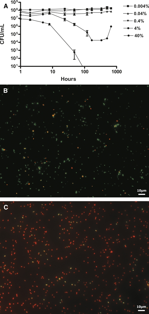
B. pseudomallei NCTC 13177 was not culturable in 40% salt after 48 hours (Figure 4A). The number of CFU/mL recovered after a 28-day period of incubation in 4% salt was significantly lower than at the beginning of the experiment (t = 4.181, df = 3, P = 0.0249). Survival of B. pseudomallei was shown at a range of salt concentrations (0.4%, 0.04%, and 0.004%) over a 28-day period. At these salt concentrations, the number of culturable cells did not differ significantly (P > 0.05) from each other or from the no-salt control from time 0 to the end of the 28-day period. The effects of stress produced by high salt concentration on cell viability and morphology can be seen in Figure 4C.
Figure 4.

(A) Survival of B. pseudomallei NCTC 13177 at increasing ranges of sea-salt concentrations over a 28-day period. (B) Live/dead bacterial cell viability results showing predominantly viable B. pseudomallei cells at time = 0 in 40% sea salts. (C) Bacterial cells after exposure to sea salt for a 28-day incubation period showing mainly non-viable bacteria. Note the presence of apparently viable bacterial cells despite an absence of culturable bacteria.
Discussion
Previous survival studies of B. pseudomallei have concentrated on either live bacteria survival in potable water or dried bacteria survival under varying environmental conditions on solid surfaces.3,4,6–8 It was previously indicated that local drinking water or seawater could have contributed to an outbreak of melioidosis.2,11,14 In that outbreak, the implicated water supply was drinking water of extreme acidity (pH = 3.5–4.0). The affected community was close to the sea, so the surface water was contaminated by seawater. Mid-day temperatures were around 42°C in the shade, and nighttime temperatures rarely fell below 20°C at that time of year.2,11,14
In the present study, we examined B. pseudomallei survival by varying the temperature, pH, and salt concentration of the suspending liquid to simulate the physical environment encountered by B. pseudomallei at the time of the melioidosis outbreak.14 Although the potable water supply implicated in the outbreak was of an unusually acidic pH,14 results obtained here indicate that a pH of 4.0 is unable to completely suppress the strain of B. pseudomallei that caused the outbreak (NCTC 13177).
An increase in culturable B. pseudomallei count was recorded for all strains in KRW and SDW, which is in agreement with previous observations of an increase in viable bacterial by CFU/mL during the first 3 weeks of B. pseudomallei survival in SDW.7 No exogenous nutrients were supplied to the bacteria in our water–survival experiments, and preparatory nutrient medium was removed by washing the bacteria in SDW. It was postulated that survival and increase in number of B. pseudomallei in the absence of nutrients may be caused by dying cells providing enough substrates for continued replication of the surviving organisms.7 An alternative reason for the modest increase in the observed count of colony-forming units is reductive division. Reductive division is a rise in cell numbers without a rise in biomass, and it is commonly associated with a change in bacterial morphology from rods to cocci.16
It is known that B. pseudomallei cells are of optimal size for inhalation and subsequent retention in the lungs.17 The size of B. pseudomallei in water may, thus, determine persistence and respirability of aerosolized bacterial cells, although cells are likely to change in size when airborne. The reduced survivability of B. pseudomallei in ASW raises an additional possibility that seashore communities such as the one affected in the WA melioidosis outbreak11 may not benefit from a suppression of B. pseudomallei by sea water after the salt content has been diluted by fresh water onshore. Moreover, the in vitro suppressive effect took hours to cause a reduction in bacterial count, ruling out the probability of any immediate antibacterial effect. The salt–survival curve showed reduced survival at a concentration of 4%. Bacterial numbers decreased significantly (P = 0.0249), but culturable cells were still present at the end of the experimental period, which is in accordance with the ASW survival study and previously reported data.5 Tolerance of relatively high salt conditions may allow B. pseudomallei to survive for prolonged periods at high salt concentrations, such as those encountered in coastal locations.14 Survival at the opposite end of the NaCl concentration spectrum has effectively been observed in SDW for several years without demonstrable loss of bacterial count.7 Other Gram-negative bacteria have been shown to survive for extended periods of time in SDW, including Pseudomonas fluorescens for 16 years,10 but extended survival has not been reported previously in salt water.
Results from this study expand on previous observations on the ability of B. pseudomallei to survive in a range of environmental conditions.6,9 All strains of B. pseudomallei survived for at least 5 days in relatively acidic conditions, although survival at pH 3 varied with the strains tested. B. pseudomallei isolates have been recovered from water sources ranging from pH 2 to 9.6 Tong and others6 showed survival of eight strains of B. pseudomallei isolated from soil and water in China in pH 3 saline for a maximum of 7 days. The strains used in the present study survived for a shorter period. Possible explanations include strain-to-strain variation, differences in inoculum preparation (this study took great care to wash bacteria before inoculation of liquids), and variations in trace micronutrients present in water used for survival experiments. Moreover, an acid pH may be encountered in a wide range of environments such as in the acidic vacuoles of the Acanthamoeba species,18 in soil,19 and in water.6,14
The continued survival of B. pseudomallei at 20°C and 40°C is consistent with previous observations.6 This temperature range reflects the ground temperatures prevailing in the region between 20°N and 20°S of the equator where melioidosis is endemic.20 At 2°C, all three strains of B. pseudomallei survived for the 28-day duration of the experiment. In contrast, Tong and others6 found B. pseudomallei strains that survived at 0°C for a mean of 18 days, although one strain survived for 42 days. It is, therefore, likely that there is strain-to-strain variation in low temperature survival of B. pseudomallei. B. pseudomallei has been isolated from a non-endemic temperate location over a period of 25 years,21 showing long-term survival in subtropical conditions where ground temperature can fall as low as 0°C in winter.
Bacterial cells subjected to stressful conditions were thought to become sublethally damaged and unsuited to culture.7,22,23 In the current study, fluorescence microscopy using a red/green viability stain revealed bacterial cells that stained yellow or orange, suggesting an intermediate or indeterminate state. Berney and others24 found that the bacterial outer membrane can act as a barrier to the epifluorescent SYTO 9 stain and that outer membrane damage is characteristic of intermediate cellular states for stationary phase Escherichia coli and Salmonella enterica serovar Typhimurium. Viable cells damaged by stressful environmental conditions or in stationary phase may, therefore, have been underestimated by the current study's culture-based method. Thus, bacterial survival may be greater than shown by the culture-based methods used in the present study.
The morphological variations seen in vitro under extreme conditions appear to be an exaggerated version of normal bacterial morphology, with the exception of the occasional spiral forms that we observed. These bizarre shapes did not appear under conditions closer to the normal range of temperature, pH, and salinity encountered in environmental samples. The significance of spiral forms, blebs, and other unusual morphological variations has yet to be determined. At present, we can only surmise that these represent alterations in the cell wall, but it is not clear whether or not these are an adaptive response or the consequence of environmentally mediated damage. The effect of environmental conditions on bacterial-cell integrity, metabolism, gene expression, and stress response needs more detailed characterization with additional bacterial strains and quantitative cell biology methods.
The present study concentrated on key aspects of the physical environment considered to have contributed to the genesis of a melioidosis outbreak that occurred in Western Australia. Temperature, water type, and pH are all likely to have affected the persistence of B. pseudomallei in the environment and thus, will have made their contribution to the sequence of events that resulted in a total of five cases of septicaemia, three of which were fatal. The likely distribution of the infective agent through the community's water supply argues in favor of the relevance of bacterial survival in liquid media. However, the behavior of B. pseudomallei in surface water or raindrops at the microenvironment level is difficult to model. This study provides some insight into bacterial survival, persistence, and dissemination in liquid and semi-solid environments and thus, lays the foundation for an understanding of variation in B. pseudomallei viability and morphology before dissemination and dispersal in rain, wet soil, and surface-accumulated water. The notable ability of B. pseudomallei to persist and even multiply across a wide range of liquid environmental conditions is a likely determinant of the environmental threat of B. pseudomallei infection.
Footnotes
Authors' addresses: Jeannie Robertson, School of Health Sciences, Curtin University, Bentley, Western Australia, E-mail: jean.robertson@postgrad.curtin.edu.au. Avram Levy and Timothy J. J. Inglis, Division of Microbiology and Infectious Diseases, PathWest Laboratory Medicine Western Australia, Nedlands, Western Australia, Australia, E-mails: a-levy@cyllene.uwa.edu.au and tim.inglis@health.wa.gov.au. Jose-Luis Sagripanti, Edgewood Chemical and Biological Center, Aberdeen Proving Ground, Edgewood, MD, E-mail: joseluis.sagripanti@us.army.mil.
References
- 1.Currie BJ, Mayo M, Anstey NM, Donohoe P, Haase A, Kemp DJ. A cluster of melioidosis cases from an endemic region is clonal and is linked to the water supply using molecular typing of Burkholderia pseudomallei isolates. Am J Trop Med Hyg. 2001;65:177–179. doi: 10.4269/ajtmh.2001.65.177. [DOI] [PubMed] [Google Scholar]
- 2.Inglis TJ, Garrow SC, Henderson M, Clair A, Sampson J, O'Reilly L, Cameron B. Burkholderia pseudomallei traced to water treatment plant in Australia. Emerg Infect Dis. 2000;6:56–59. doi: 10.3201/eid0601.000110. [DOI] [PMC free article] [PubMed] [Google Scholar]
- 3.Howard K, Inglis TJ. The effect of free chlorine on Burkholderia pseudomallei in potable water. Water Res. 2003;37:4425–4432. doi: 10.1016/S0043-1354(03)00440-8. [DOI] [PubMed] [Google Scholar]
- 4.Howard K, Inglis TJJ. Disinfection of Burkholderia pseudomallei in potable water. Water Res. 2005;39:1085–1092. doi: 10.1016/j.watres.2004.12.028. [DOI] [PubMed] [Google Scholar]
- 5.Inglis TJ, Sagripanti JL. Environmental factors that affect survival and persistence of Burkholderia pseudomallei. Appl Environ Microbiol. 2006;72:6865–6875. doi: 10.1128/AEM.01036-06. [DOI] [PMC free article] [PubMed] [Google Scholar]
- 6.Tong S, Yang S, Lu Z, He W. Laboratory investigation of ecological factors influencing the environmental presence of Burkholderia pseudomallei. Microbiol Immunol. 1996;40:451–453. doi: 10.1111/j.1348-0421.1996.tb01092.x. [DOI] [PubMed] [Google Scholar]
- 7.Wuthiekanun V, Smith MD, White NJ. Survival of Burkholderia pseudomallei in the absence of nutrients. Trans R Soc Trop Med Hyg. 1995;89:491. doi: 10.1016/0035-9203(95)90080-2. [DOI] [PubMed] [Google Scholar]
- 8.Shams AM, Rose LJ, Hodges L, Arduino MJ. Survival of Burkholderia pseudomallei on environmental surfaces. Appl Environ Microbiol. 2007;73:8001–8004. doi: 10.1128/AEM.00936-07. [DOI] [PMC free article] [PubMed] [Google Scholar]
- 9.Finkelstein RA, Atthasampunna P, Chulasamaya M. Pseudomonas (Burkholderia) pseudomallei in Thailand, 1964–1967: geographic distribution of the organism, attempts to identify cases of active infection, and presence of antibody in representative sera. Am J Trop Med Hyg. 2000;62:232–239. doi: 10.4269/ajtmh.2000.62.232. [DOI] [PubMed] [Google Scholar]
- 10.Liao CH, Shollenberger LM. Survivability and long-term preservation of bacteria in water and in phosphate-buffered saline. Lett Appl Microbiol. 2003;37:45–50. doi: 10.1046/j.1472-765x.2003.01345.x. [DOI] [PubMed] [Google Scholar]
- 11.Inglis TJ, Garrow SC, Adams C, Henderson M, Mayo M. Dry-season outbreak of melioidosis in Western Australia. Lancet. 1998;352:1600. doi: 10.1016/S0140-6736(05)61047-1. [DOI] [PubMed] [Google Scholar]
- 12.Inglis T, Mee B, Chang B. The environmental microbiology of melioidosis. Rev Med Microbiol. 2001;12::13–20. [Google Scholar]
- 13.Rolim DB. Melioidosis, northeastern Brazil. Emerg Infect Dis. 2005;11:1458–1460. doi: 10.3201/eid1109.050493. [DOI] [PMC free article] [PubMed] [Google Scholar]
- 14.Inglis TJ, Garrow SC, Adams C, Henderson M, Mayo M, Currie BJ. Acute melioidosis outbreak in western Australia. Epidemiol Infect. 1999;123:437–443. doi: 10.1017/s0950268899002964. [DOI] [PMC free article] [PubMed] [Google Scholar]
- 15.Collee JG, Duguid JP, Fraser AG, Marmion BP. Practical Medical Microbiology. 13th Edition. Edinburgh: Churchill Livingstone; 1989. pp. 90–99. (pH measurements and buffers, oxidation-reduction potentials, suspension fluids and preparation of glassware). [Google Scholar]
- 16.Young KD. The selective value of bacterial shape. Microbiol Mol Biol Rev. 2006;70:660–703. doi: 10.1128/MMBR.00001-06. [DOI] [PMC free article] [PubMed] [Google Scholar]
- 17.Sagripanti JL, Carrera M, Robertson JN, Levy A, Inglis TJJ. Size distribution of Burkholderia pseudomallei. Proceedings of the 5th World Melioidosis Congress, November 2007. Khon Kaen; Thailand: 2007. [Google Scholar]
- 18.Lamothe J, Thyssen S, Valvano MA. Burkholderia cepacia complex isolates survive intracellularly without replication within acidic vacuoles of Acanthamoeba polyphaga. Cell Microbiol. 2004;6:1127–1138. doi: 10.1111/j.1462-5822.2004.00424.x. [DOI] [PubMed] [Google Scholar]
- 19.Kanai K, Kondo E, Dejsirilert S, Naigowit P. In: Melioidosis—Prevailing Problems and Future Directions. Puthucheary SD, Malik MA, editors. Kuala Lumpur, Malaysia: Malaysian Society of Infectious Diseases and Chemotherapy; 1994. pp. 26–38. (Growth and survival of Pseudomonas pseudomallei in acidic environment with possible relation to the ecology and epidemiology of melioidosis). [Google Scholar]
- 20.Redfearn MS, Palleroni NJ, Stanier RY. A comparative study of Pseudomonas pseudomallei and Bacillus mallei. J Gen Appl Microbiol. 1966;43:293–313. doi: 10.1099/00221287-43-2-293. [DOI] [PubMed] [Google Scholar]
- 21.Currie B, Smith-Vaughan H, Golledge C, Buller N, Sriprakash KS, Kemp DJ. Pseudomonas pseudomallei isolates collected over 25 years from a non-tropical endemic focus show clonality on the basis of ribotyping. Epidemiol Infect. 1994;113:307–312. doi: 10.1017/s0950268800051736. [DOI] [PMC free article] [PubMed] [Google Scholar]
- 22.Roszak DB, Colwell RR. Survival strategies of bacteria in the natural environment. Microbiol Mol Biol Rev. 1987;51:365–379. doi: 10.1128/mr.51.3.365-379.1987. [DOI] [PMC free article] [PubMed] [Google Scholar]
- 23.Xu H-S, Roberts N, Singleton FL, Attwell RW, Grimes DJ, Colwell RR. Survival and viability of nonculturable Escherichia coli and Vibrio cholerae in the estuarine and marine environment. Microb Ecol. 1983;8:313–324. doi: 10.1007/BF02010671. [DOI] [PubMed] [Google Scholar]
- 24.Berney M, Hammes F, Bosshard F, Weilenmann H-U, Egli T. Assessment and interpretation of bacterial viability by using the LIVE/DEAD BacLight Kit in combination with flow cytometry. Appl Environ Microbiol. 2007;73:3283–3290. doi: 10.1128/AEM.02750-06. [DOI] [PMC free article] [PubMed] [Google Scholar]


