Abstract
OBJECTIVE:
To review the etiology, clinical features and management of acute infectious conjunctivitis in children after the newborn period.
DATA SOURCES:
Articles obtained from MEDLINE published before March 2000.
DATA SELECTION AND EXTRACTION:
Representative articles on the etiology and clinical features were selected. Twenty-four clinical trials were also selected. From these articles, the main findings from three placebo controlled trials and two comparative clinical trials involving children are summarized in detail. The main findings from 19 comparative clinical trials in adults are briefly summarized.
DATA SYNTHESIS AND CONCLUSIONS:
Acute infectious conjunctivitis caused by bacteria or viruses is a very common problem in children after the neonatal period. The most common bacterial pathogens are nontypable Haemophilus influenzae and Streptococcus pneumoniae. Diagnostic microbiology tests are not indicated for uncomplicated cases but may be useful for very young or very ill children if there is no response to initial therapy; for nosocomial cases; for cases suspected to be caused by sexually transmitted pathogens; and for outbreaks. Conjunctivitis is usually a mild, self-limited disease, but topical antibiotics are superior to placebo in reducing the duration and severity of symptoms. Most topical agents have equivalent efficacy; therefore, the selection of first-line agents should include inexpensive drugs with few adverse effects. Good choices include polymyxin/gramicidin, polymyxin/trimethoprim or sulfacetamide. Referral to an ophthalmologist should be considered in situations in which the diagnosis of uncomplicated conjunctivitis is in doubt or if there is no prompt response to therapy.
Keywords: Conjunctivitis, Drug therapy, Etiology
Abstract
OBJECTIF :
Examiner l’étiologie, les caractéristiques cliniques et la prise en charge de la conjonctivite infectieuse aiguë chez les enfants après la période néonatale.
SOURCES DES DONNÉES :
Articles obtenus dans MEDLINE publiés avant mars 2000.
SÉLECTION ET EXTRACTION DES DONNÉES :
Des articles représentatifs de l’étiologie et des caractéristiques cliniques ont été sélectionnés. Vingt-quatre essais cliniques ont également été retenus. Tirés de ces articles, un résumé détaillé des principales observations de trois essais contrôlés contre placebo et de deux essais cliniques comparatifs portant sur des enfants ainsi qu’un bref résumé des principales observations de 19 essais cliniques comparatifs portant sur des adultes sont présentés.
SYNTHÈSE DES DONNÉES ET CONCLUSION :
La conjonctivite infectieuse aiguë causée par des bactéries ou des virus représente une pathologie très courante chez les enfants après la période néonatale. Les pathogènes bactériens les plus courants sont l’Haemophilus influenzae et le Streptococcus pneumoniae ne pouvant être typés. Les examens de microbiologie diagnostique ne sont pas indiqués lorsque les cas sont simples, mais peuvent être utiles dans les situations suivantes : chez les enfants très jeunes ou très malades qui ne réagissent pas au traitement initial, qui souffrent d’une maladie nosocomiale, dont le cas est présumé comme secondaire à des pathogènes transmis sexuellement et en cas de flambées. Règle générale, la conjonctivite est une maladie bénigne et résolutive, mais les antibiotiques topiques donnent de meilleurs résultats qu’un placebo pour réduire la durée et la gravité des symptômes. La plupart des agents topiques sont d’une efficacité équivalente. Par conséquent, les agents de première ligne devraient être sélectionnés parmi les médicaments peu coûteux aux effets secondaires rares. Parmi les choix judicieux, soulignons la polymyxine et la gramicidine, la polymyxine et le triméthoprime, ou la sulfacétamide. Il faut envisager une consultation avec un ophtalmologiste lorsque le diagnostic de conjonctivite simple est mis en doute ou que le traitement ne procure pas une amélioration rapide de l’état du patient.
Acute conjunctivitis may be defined as conjunctival redness with or without increased tearing or discharge that is less than 14 days in duration (1). Acute infectious conjunctivitis is a common problem in childhood, and is the most common acute eye infection treated by primary care physicians and paediatricians. The present paper reviews the etiology, clinical features and diagnosis of acute infectious conjunctivitis. Data from clinical trials of conjunctivitis treatment are summarized and presented to aid readers in making evidence-based treatment decisions. Neonatal conjunctivitis is not discussed.
ETIOLOGY
Acute conjunctivitis is almost always caused by bacteria or viruses. One study showed that bacteria caused acute conjunctivitis in 80% of cases, viruses in 13% of cases and allergy in 2% of cases; no etiology was identified in 5% of cases (1).
Several bacterial pathogens may cause conjunctivitis. In acute, uncomplicated purulent conjunctivitis, approximately 50% of cases are caused by nontypable Haemophilus influenzae, approximately 25% of cases are caused by Streptococcus pneumoniae and less than 5% of cases are caused by Moraxella catarrhalis (1,2). Other less common organisms include Staphylococcus species, Gram-negative bacilli, other Gram-positive bacteria and Chlamydia species. All of these organisms have been found in conjunctival and lid cultures from children with conjunctivitis but not in matched controls without conjunctivitis (1,2). The combination of conjunctivitis and acute otitis media is most commonly caused by nontypable H influenzae, and occasionally by S pneumoniae (3–9).
Other bacteria can cause conjunctivitis in specific settings. Niesseria gonorrhea causes conjunctivitis in sexually active adolescents and in individuals who are victims of sexual abuse. It is rarely, if ever, acquired in a nonsexual manner, except when it is acquired at birth (9,10). Niesseria meningiditis is a rare cause of bacterial conjunctivitis (2,10,11). Enteric Gram-negative bacteria are a rare cause of conjunctivitis that may be acquired nosocomially due to poor hygiene and after antimicrobial therapy (2,10). Staphylococcus aureus and coagulase-negative staphylococci are often recovered from lid cultures but not from the conjunctiva (1,2). The organisms have been associated with blepharitis and are perhaps more common in adults. Viridans streptococci and other Gram-positive bacteria have been cultured from eye swabs, but their role as a cause of conjunctivitis is unproven (1,5,11). The clinical conjunctivitis syndromes of trachoma and adult inclusion conjunctivitis are caused by Chlamydia trachomatis (11).
Several viruses cause conjunctivitis. Adenoviruses (types 3, 4, 5 and 7) cause pharyngoconjunctival fever (2,7,11). Other adenoviruses (types 8, 19 and 37) cause keratoconjunctivitis (2,11). Herpes simplex virus (HSV) also causes keratoconjunctivitis (2,7,11). Enteroviruses (coxsackie A24 or enterovirus 70) cause an acute hemorrhagic conjunctivitis (11). Other viruses that may cause conjunctivitis include measles, influenza, molluscum contagiosum and rubella (11).
CLINICAL FEATURES AND DIFFERENTIAL DIAGNOSIS
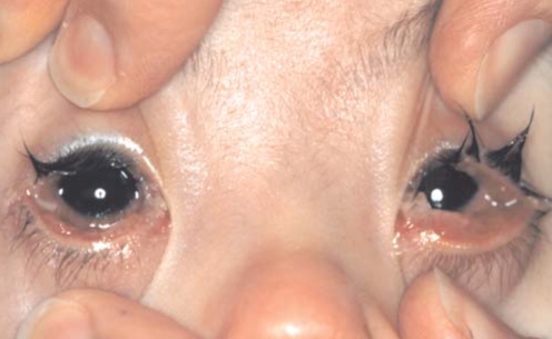
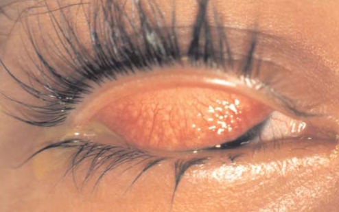
There are certain clinical features that help distinguish the etiology of conjunctivitis. Infectious conjunctivitis (bacterial or viral) is associated with conjunctival hyperemia and ocular discharge (7). The palpebral involvement is typically much greater than the bulbar involvement. Clinical features can sometimes be used to distinguish bacterial from viral conjunctivitis, as outlined in Table 1 (1,2,7). The typical appearance of bacterial and viral conjunctivitis is shown in Figures 1 and 2. As noted above, there is a connection between bacterial conjunctivitis and acute otitis media, and therefore, physicians should examine the ears of all children who have conjunctivitis.
TABLE 1:
Clinical features of bacterial and viral conjunctivitis
| Clinical feature | Bacterial conjunctivitis | Viral conjunctivitis |
|---|---|---|
| Bilateral at onset | In 75% of cases | In 35% of cases |
| Conjunctival discharge | Mucopurulent | Watery |
| Conjunctival membrane apparent | Late onset | Early onset |
| Preauricular nodes and/or pharyngitis | No | Yes |
| Concurrent otitis media | In 20% to 75% of cases | In 0% of cases |
| Slit lamp conjunctival findings | Mixed and/or nonspecific | Follicular |
Figure 1).
Typical appearance of bacterial conjunctivitis
Figure 2).
Typical appearance of viral conjunctivitis (upper eyelid everted)
There are unique clinical features associated with some bacteria and viruses. N gonorrhea causes ‘hyperacute’ conjunctivitis with abrupt onset, rapid progression, prominent lid edema, excessive purulent discharge and a tender globe (2,7,10,11). N meningiditis conjunctivitis is generally unilateral with purulent discharge (2,10,11). In approximately 20% of patients with N meningiditis conjunctivitis, systemic disease develops later. Chlamydia conjunctivitis is associated with urethritis and cervicitis. It causes a subacute infection that is follicular in nature. Inclusion conjunctivitis is a sexually transmitted disease with ocular signs of a chronic conjunctival reaction that is most prominent in the lower lid, with a scant mucopurulent discharge (11). It is usually found in teenagers and young adults, but it can also be seen in neonates infected at delivery. A significant percentage of patients with chlamydia conjunctivitis also have concomitant asymptomatic gonococcal infection (11). Trachoma affects about 400 million individuals worldwide and is the leading cause of preventable blindness (11). It mostly occurs in underprivileged countries with inadequate sanitation (11). The hallmark finding is a follicular conjunctivitis, and blindness is caused by a process of progressive scarring and cicatrisation of the eyelids and adnexa.
Adenoviruses (types 3, 4, 5 and 7) cause conjunctivitis in patients younger than 10 years of age, and outbreaks of conjunctivitis do occur (2,7,11). The viral infection causes the conjunctivitis, and also leads to pharyngitis and fever. Other adenoviruses (types 8, 19 and 37) more commonly cause epidemics in older children and adults (2,11). Infections caused by these types of adenoviruses are not associated with upper respiratory symptoms or fever. The predominant finding is corneal involvement with the sensation that a foreign body is present and photophobia. The illness evolves over seven to 10 days, but local discomfort can continue for weeks to months. One may see marked eyelid swelling with this type of infection.
HSV can cause serious illnesses with vision-threatening complications with or without systemic manifestations. Herpes simplex infection is usually seen in the one- to five-year-old patient and is usually unilateral (2,7,11). Infection with HSV should be considered in any child with an acute follicular conjunctivitis and watery discharge. This type of infection can also be seen in neonatal herpes infection, with the disease involving the skin, eyes and mucous membranes. There may be vesicular lesions on the eyelids and a keratitis (dendritic). Enteroviral conjunctivitis is of abrupt onset, bilateral, with lid edema and profuse watery discharge (11). There often is pinpoint or confluent subconjunctival hemorrhage. Associated neurological involvement in the form of neuropathies may be present.
The differential diagnosis of acute conjunctivitis includes systemic diseases that have associated conjunctivitis and allergic conjunctivitis. Systemic disorders that are associated with conjunctivitis include Kawasaki syndrome, Stevens-Johnson syndrome, ataxia-telangiectasia, Lyme disease and juvenile rheumatoid arthritis (7). In patients with the above disorders, the bulbar involvement is much greater than the palpebral involvement, and there is no eye discharge. In allergic conjunctivitis, patients have prominent itching as a symptom and may have a stringy discharge (7). In allergic conjunctivitis, acute triggers, seasonal variation or evidence of atopy may be present. Allergic conjunctivitis and vernal keratoconjunctivitis share many of the signs and symptoms of infectious conjunctivitis, but the presence of itchiness as a symptom and papillary changes on the palpebral conductive should help differentiate this from infectious causes. Episcleritis, scleritis and anterior uveitis, all of which may have associated rheumatological findings, can also mimic infectious conjunctivitis. Scleritis and anterior uveitis are more ominous disorders that may have ramifications for sight if not diagnosed early. The clinical features of infectious conjunctivitis compared with systemic diseases with associated conjunctivitis and allergic conjunctivitis are summarized in Table 2.
TABLE 2:
Clinical features of infectious conjunctivitis, allergic conjunctivitis and systemic disorders with conjunctivitis
| Clinical feature | Infectious conjunctivitis | Allergic conjunctivitis | Systemic disorders with conjunctivitis |
|---|---|---|---|
| Conjunctival involvement | Palpebral greater than bulbar | Papillary changes on palpebra | Bulbar greater than palpebral |
| Eye discharge | Watery to mucopurulent | May be stringy | Often absent |
| Eye itchiness | No | Yes | No |
| Acute triggers, seasonal variation and history of atopy | No | Yes | No |
In addition, more serious ocular conditions can mimic infectious conjunctivitis. When these more ominous disorders are not diagnosed appropriately, they can lead to permanent vision loss. Acute angle closure glaucoma and congenital glaucoma can mimic infectious conjunctivitis. Both disorders can lead to permanent vision loss if they are not appropriately diagnosed. Orbital cellulitis can be misdiagnosed early in its evolution, and retinoblastoma can mimic conjunctivitis and orbital cellulitis. Whenever a patient does not respond rapidly to therapy for infectious conjunctivitis; has evidence of HSV, gonococcal or chlamydia infection; or has evidence of a noninfectious systemic disorder, a referral to an ophthalmologist should be made.
DIAGNOSTIC TESTS
Diagnostic microbiological testing for acute infectious conjunctivitis is not routinely indicated. Most cases are readily diagnosed on the basis of clinical features, and effective empirical therapy can be provided without a microbiological diagnosis. In the majority of cases, cultures do not provide clinically important information. Indications for performing stains and a bacterial culture include the following: age younger than two months; a very ill patient; a lack of response to initial therapy; nosocomial infections; sexual abuse; and outbreaks of conjunctivitis (2,11). If bacterial cultures are taken, culture swabs and two smears for Gram and acridine orange stains should be collected, and the swabs should be cultured on blood and chocolate (12,13). The most appropriate sample is obtained with a conjunctival scraping using an appropriate swab (12,13). Testing for chlamydia conjunctivitis is performed by identifying the antigen from a monoclonal antibody test (12,13). The conjunctivae are scraped for cells using a spatula, and the sample is then placed on a special slide that is provided with the testing kit.
Viral cultures are not useful unless HSV is considered or during an outbreak investigation because no effective treatment is available, except for HSV (12,13). If HSV is thought to be the causative agent, an urgent eye examination by an ophthalmologist is necessary. A swab stick or spatula is rubbed firmly against the conjunctiva, and to maximize the yield of cultures, the culture media should be inoculated directly or the sample should be immediately taken to the laboratory in viral transport media (13). The swabs are then either inoculated directly into tissue culture, used in immunoflourescence tests for a particular viral antigen or used for a nucleic acid, amplification-based (eg, polymerase chain reaction) diagnosis. A combination of these techniques can be used to provide a rapid and sensitive result (12,13).
MANAGEMENT
An algorithm for the diagnosis and treatment of acute conjunctivitis is presented in Figure 3. The initial step in the management of both bacterial and viral conjunctivitis is the use of symptomatic measures to both control symptoms and prevent the spread of the conjunctivitis. Important symptomatic treatments include the use of an eye toilette, warm compresses, artificial tears, strict hand washing and the avoidance of bright lights (if photophobia is present).
Figure 3).
Algorithm for the management of acute conjunctivitis in children. HSV Herpes simplex virus
Acute bacterial conjunctivitis is mostly a self-limited illness that lasts seven to 14 days, but antimicrobial treatment will reduce symptoms and shorten the duration of the clinical disease (2,14). Topical antimicrobial agents are the usual method of treating bacterial conjunctivitis. Numerous placebo controlled trials have shown both statistical and clinical benefit from topical antimicrobial agents (Table 3) (14–16). The topical antimicrobial selection for the treatment of bacterial conjunctivitis includes aminoglycosides (gentamycin, tobramycin, neomycin and framycetin), fluoroquinolones (ciprofloxacin, ofloxacin and norfloxacin), sulfacetamide, chloramphenicol, erythromycin and combination agents, including neomycin and polymyxin with bacitracin or gramicidin, and polymyxin and trimethoprim or bacitracin or gramicidin.
TABLE 3:
Placebo controlled clinical trials for drug treatment of acute bacterial conjunctivitis
| Study | Number enrolled | Age range (years) | Study drug | Outcome(s) compared | Outcome measure day* | Result (drug versus placebo) (%)† | P |
|---|---|---|---|---|---|---|---|
| Gigliotti et al, 1984 (14) | 66 | Birth to 18 | Polysporin (Warner-Lambert Consumer Healthcare, Canada) | Clinical cure | 3 to 5 | 62 versus 28 | <0.02 |
| Clinical improvement | 8 to 10 | 91 versus 72 | NS | ||||
| Bacteriological cure | 3 to 5 | 71 versus 19 | <0.001 | ||||
| 8 to 10 | 79 versus 31 | <0.001 | |||||
| Leibowitz, 1991 (15) | 288 | Not stated | Ciprofloxacin | Bacteriological cure | 3 | 94 versus 60 | <0.001 |
| Miller et al, 1992 (16) | 284 | At least 18 | Norfloxacin | Clinical cure or improvement | 5 | 88 versus 72 | <0.01 |
| Bacteriological cure | 2 to 3 | 65 versus 26 | <0.01 |
Day after onset of study that outcomes were measured;
Percentage of patients in each study group (study drug versus placebo) that achieved a beneficial outcome; NS Not significant
In choosing a topical antimicrobial agent, certain factors need to be considered, including the efficacy, the route of administration (ointment versus drops), dose interval and duration of therapy, side effects, and cost (2,7,11,17). Any antimicrobial agent combined with a steroid should be avoided. The corticosteroid in these combination topical agents may hinder the eradication of the organism. In addition, steroids can cause the dendritic ulcer of herpes keratitis to worsen (this condition may be mistaken for acute bacterial conjunctivitis), and prolonged use leads to increased intraocular pressure (17). Limited data exist about the susceptibility of the most common organisms that cause acute conjunctivitis to the antimicrobial agents that are routinely used (Table 4).
TABLE 4:
In vitro antimicrobial susceptibility (%) of topical antimicrobial agents to common pathogens causing bacterial conjunctivitis
| Drug | Estimated percentage of antimicrobial susceptibility | |||
|---|---|---|---|---|
| Haemophilus influenzae | Streptococcus pneumoniae | Moraxella catarrhalis | Streptococcus aureus | |
| Neomycin | 85 | <5 | Most | 95 |
| Polymyxin | 83 | 0 | Not reported | 11 |
| Bacitracin | 0 | 100 | Not reported | 99 |
| Gramicidin | 0 | Most | Not reported | 50 |
| Sulfacetamide | 70 | 75 | Most | 96 |
| Trimethoprim | 95 | 64 | 0 | 91 |
| Gentamicin and other aminoglycosides | 85 | <5 | Most | 95 |
| Ciprofloxacin | 100 | 90 | 100 | 90 |
| Ofloxacin | 100 | 90 | 99 | 90 |
| Chloramphenicol | 95 | 95 | 100 | Most |
| Neosporin (Glaxo Wellcome Inc, Canada) | ||||
| Neomycin and Bacitracin or Gramicidin |
85 0 0 |
<5 100 Most |
Most Not reported Not reported |
95 99 50 |
| Polysporin (Warner-Lambert Consumer Healthcare, Canada) | ||||
| Polymyxin and Bacitracin or Gramicidin |
83 0 0 |
0 100 Most |
Not reported Not reported Not reported |
11 99 50 |
| Polytrim (Allergan Inc, Canada)) | ||||
| Polymyxin and Trimethoprim |
83 95 |
0 64 |
Not reported 0 |
11 91 |
Data from multiple sources
Few clinical trials have been performed to compare two or more antimicrobial agents. Lohr et al (18) studied 158 patients from two months to 21 years of age (88% of patients were 12 years of age or younger). All patients had culture-proven bacterial conjunctivitis (S pneumoniae or H influenzae), and all were given polymyxin/trimethoprim (Polytrim, Allergan Inc, Canada), sulfacetamide or gentamicin drops, one drop to each eye every 3 h while awake. After 10 days, the clinical cure rates of polymyxin/trimethoprim, sulfacetamide and gentamicin were 93%, 93% and 97%, respectively (P was not significant), and the bacteriological cure rates were 83%, 72% and 68%, respectively (P was not significant) (18). Gross et al (19) studied 141 children aged 12 years or younger with culture-proven bacterial conjunctivitis (45% with H influenzae, 30% with S pneumoniae and 25% with another infection) and compared ciprofloxacin with tobramycin drops, one to two drops every 2 h while awake for two days and then every 4 h for another five days. After seven days, the clinical cure rates of ciprofloxacin and tobramycin were 99% and 97%, respectively, and the bacteriological cure rates were 90% and 84%, respectively.
Many other comparison studies involving adults have compared aminoglycosides, quinolones, chloramphenicol, combination antibiotics and other agents (16,20–38). In almost all of the studies, no differences were found in the clinical or bacteriological cure rates. One study found that trimethoprim/polymyxin was superior to chloramphenicol (39), another study found that ciprofloxacin was superior to rifamycin (36) and a different study found that fusidic acid was superior to chloramphenicol or framycetin (40).
Side effects of topical ophthalmic antimicrobial agents are common, but, fortunately, they are mostly minor. Local irritation is the most commonly reported adverse effect (sulfacetamide, especially burns with instillation) (17). Moderate to severe drug side effects are rare and drug specific. Neomycin may cause allergic reactions, especially five to six days after use (2,41). All aminoglycosides can cause corneal toxicity, and it is not clear whether this is due to prolonged use or other factors (2,11,17). There have been reported cases of idiosyncratic aplastic anemia with topical chloramphenicol use (11,42). Cases of Stevens-Johnson syndrome have been reported with the use of sulfacetamide (43,44). Arthropathy has not been reported with the topical use of fluoroquinolones.
Because clinical efficacy is more or less equivalent among many topical antimicrobial agents, the important factors in choosing a first-line topical antimicrobial agent are convenience, side effects and cost. Brand name antimicrobial agents are more expensive than generic formulations but are not routinely more effective. The least expensive antimicrobial agents include polymyxin/gramicidin (Polysporin, Warner-Lambert Consumer Healthcare, Canada), sulfacetamide and gentamicin. Thus, appropriate first-line agents include polymyxin/gramicidin (Polysporin), polymyxin/trimethoprim (Polytrim) or sulfacetamide. In Canada, the first two formulations are available over the counter, but sulfacetamide requires a prescription. Alternative agents include aminoglycosides and neomycin/gramicidin (Neosporin, Glaxo Wellcome Inc, Canada) or fluoroquinolones. Although effective, there is no role for chloramphenicol in the treatment of conjunctivitis because of the small risk of serious hematological toxicity.
Systemic antimicrobial agents have also been found to be effective in the treatment of conjunctivitis and the prevention of otitis media, but the routine use of systemic antimicrobial agents in the treatment of bacterial conjunctivitis is not recommended (2,9,11,41). There may be a role for systemic antibiotics in the treatment of conjunctivitis in the otitisprone child. Systemic antimicrobial agents are also indicated for the therapy of neonatal conjunctivitis due to N gonorrhea or C trachomatis (45). The incidental benefit of treating a focal or systemic infection with systemic antimicrobial agents is that, if bacterial conjunctivitis is present, it also will be treated (2). Therapy with intravenous antimicrobial agents provides levels of antibiotic in the conjunctiva that are more than sufficient to eradicate bacterial pathogens. With oral therapy, the levels of antibiotic in the conjunctiva are more likely to be considerably lower but still adequate (2).
For the treatment of viral conjunctivitis, the mainstay of management is symptomatic control. There is no evidence to support the use of topical antimicrobial agents. For herpes keratoconjunctivitis, the topical medications that are used are trifluridine (Viroptic, Glaxo Wellcome Inc) and idoxuridine (Herplex, Allergan Inc) (11,13). Currently, there are no other topical or systemic antimicrobial agents available for the management of viral conjunctivitis.
Finally, any patient treated for suspected bacterial or viral conjunctivitis that does not improve significantly with initial therapy should be referred to an ophthalmologist for further evaluation. As an example, HSV, varicella, epidemic keratoconjunctivitis and chlamydia infections should be followed by an ophthalmologist because these diseases can have significant vision-threatening corneal complications associated with them. Severe bacterial conjunctivitis can also be associated with bacterial keratitis and corneal ulcers, which may not respond to standard antibiotic therapy.
A review of the management of ‘the red eye’ was recently published (46). The review presented several helpful practical pointers; however, it focused on the management of adults and did not critically review clinical trials of drug therapy. In addition, the author recommended the use of antibacterial drops for the management of presumed viral conjunctivitis because “in many cases … the patient will not accept a recommendation that no therapy be administered” (46). In the opinion of the authors of the present paper, this belief conflicts with the rapidly changing beliefs and behaviours of physicians, and the public in Canada toward the acceptance of more appropriate use of antibiotics in children.
SUMMARY
Acute conjunctivitis is a common childhood condition and is most frequently infectious. A diagnosis can usually be made on the basis of clinical features, but laboratory testing may be necessary in complicated or severe cases. Most cases of viral and bacterial conjunctivitis are self-limited. Symptomatic measures may reduce symptoms and prevent the spread of the infection. Topical antimicrobial therapy is effective to reduce symptoms and eradicate bacterial pathogens. Inexpensive topical antimicrobial agents, such as sulfacetamide, are appropriate first-line agents with few side effects. Referral to an ophthalmologist is indicated in severe cases, in cases with unusual clinical features or in cases that do not respond to appropriate therapy.
REFERENCES
- 1.Weiss A, Brinser JH, Nazar-Stewart V. Acute conjunctivitis in childhood. J Pediatr. 1993;122:10–4. doi: 10.1016/s0022-3476(05)83479-1. [DOI] [PubMed] [Google Scholar]
- 2.Weiss A. Acute conjunctivitis in childhood. Curr Probl Pediatr. 1994;24:4–11. doi: 10.1016/0045-9380(94)90022-1. [DOI] [PubMed] [Google Scholar]
- 3.Coffey JD., Jr Otitis media in the practice of pediatrics. Bacteriological and clinical observations. Pediatrics. 1966;38:25–32. [PubMed] [Google Scholar]
- 4.Bodor FF. Conjunctivitis-otitis syndrome. Pediatrics. 1982;69:695–8. [PubMed] [Google Scholar]
- 5.Gigliotti F, Williams WT, Hayden FG, et al. Etiology of acute conjunctivitis in children. J Pediatr. 1981;98:531–6. doi: 10.1016/s0022-3476(81)80754-8. [DOI] [PubMed] [Google Scholar]
- 6.Harrison CJ, Hedrick JA, Block SL, Gilchrist MJ. Relation of the outcome of conjunctivitis and the conjunctivitis-otitis syndrome to identifiable risk factors and oral antimicrobial therapy. Pediatr Infect Dis J. 1987;6:536–40. doi: 10.1097/00006454-198706000-00010. [DOI] [PubMed] [Google Scholar]
- 7.Wagner RS. The differential diagnosis of the red eye. Contemp Pediatr. 1991:1–10. [Google Scholar]
- 8.Wald ER, Serdy C, Guerra N, Blatt S. Short course oral antibiotic treatment of bacterial conjunctivitis. Pediatr Res. 1993;33(Part 2):124A. [Google Scholar]
- 9.Wald ER. Conjunctivitis in infants and children. Pediatr Infect Dis J. 1997;16(Suppl 2):S17–20. doi: 10.1097/00006454-199702001-00005. [DOI] [PubMed] [Google Scholar]
- 10.Limberg MB. A review of bacterial keratitis and bacterial conjunctivitis. Am J Ophthalmol. 1991;112(Suppl 4):2S–9S. [PubMed] [Google Scholar]
- 11.Donahue SP, Khoury JM, Kowalski RP. Common ocular infections. A prescriber’s guide. Drugs. 1996;52:526–40. doi: 10.2165/00003495-199652040-00006. [DOI] [PubMed] [Google Scholar]
- 12.Byrne KA, Burd EM, Tabbara KF, Hyndiuk RA. Diagnostic Microbiology and Cytology of the Eye. 1st edn. Newton: Butterworth-Heinemann; 1995. [Google Scholar]
- 13.Seal DV, Bron JB, Hay J. Ocular Infection Investigation and Treatment in Practice. 1st edn. London: Martin Dunitz Ltd; 1998. [Google Scholar]
- 14.Gigliotti F, Hendley O, Morgan J, et al. Efficacy of topical antibiotic therapy in acute conjunctivitis in children. J Pediatr. 1984;104:623–6. doi: 10.1016/s0022-3476(84)80566-1. [DOI] [PubMed] [Google Scholar]
- 15.Leibowitz HM. Antibacterial effectiveness of ciprofloxacin 0.3% ophthalmic solution in the treatment of bacterial conjunctivitis. Am J Ophthalmol. 1991;112(Suppl 4):29S–33S. [PubMed] [Google Scholar]
- 16.Miller IM, Wittreich J, Vogel R, Cook TJ. The safety and efficacy of topical norfloxacin compared with placebo in the treatment of acute, bacterial conjunctivitis. The Norfloxacin-Placebo Ocular Study Group. Eur J Ophthalmol. 1992;2:58–66. doi: 10.1177/112067219200200202. [DOI] [PubMed] [Google Scholar]
- 17.Wallace DK, Steinkuller PG. Ocular medications in children. Clin Pediatr. 1998;37:645–52. doi: 10.1177/000992289803701101. [DOI] [PubMed] [Google Scholar]
- 18.Lohr JA, Austin RD, Grossman M, Hayden GF, Knowlton GM, Dudley SM. Comparison of three topical antimicrobials for acute bacterial conjunctivitis. Pediatr Infect Dis J. 1988;7:626–9. doi: 10.1097/00006454-198809000-00005. [DOI] [PubMed] [Google Scholar]
- 19.Gross RD, Hoffman RO, Lindsay RN. A comparison of ciprofloxacin and tobramycin in bacterial conjunctivitis in children. Clin Pediatr. 1997:435–44. doi: 10.1177/000992289703600801. [DOI] [PubMed] [Google Scholar]
- 20.Bloom PA, Leeming JP, Power W, Laidlaw DA, Collum LM, Easty DL. Topical ciprofloxacin in the treatment of blepharitis and blepharaconjunctivitis. Eur J Ophthalmol. 1994;4:6–12. doi: 10.1177/112067219400400102. [DOI] [PubMed] [Google Scholar]
- 21.Adenis JP, Colin J, Verin P, Riss I, Saint-Blancat P. Ciprofloxacin ophthalmic solution in the treatment of conjunctivitis and blepharitis: A comparison with fusidic acid. Eur J Ophthalmol. 1996;6:368–74. doi: 10.1177/112067219600600404. [DOI] [PubMed] [Google Scholar]
- 22.Adenis JP, Brasseur G, Demailly P, et al. Comparative evaluation of efficacy and safety of ciprofloxacin and norfloxacin ophthalmic solutions. Eur J Ophthalmol. 1996;6:287–92. doi: 10.1177/112067219600600312. [DOI] [PubMed] [Google Scholar]
- 23.Power WJ, Collum LM, Easty DL, et al. Evaluation of efficacy and safety of ciprofloxacin ophthalmic solution versus chloramphenicol. Eur J Ophthalmol. 1993;3:77–82. doi: 10.1177/112067219300300205. [DOI] [PubMed] [Google Scholar]
- 24.Leibowitz HM, Hyndiuk RA, Smolin GR, Nozik RA, Hunter GJ. Tobramycin in external eye disease: A double-masked study vs gentamicin. Curr Eye Res. 1981;1:259–66. doi: 10.3109/02713688108999445. [DOI] [PubMed] [Google Scholar]
- 25.Miller IM, Vogel R, Cook TJ, Wittreich J. Topically administered norfloxacin compared with topically administered gentamicin for the treatment of external ocular bacterial infections. The Worldwide Norfloxacin Ophthalmic Study Group. Am J Ophthalmol. 1992;15:638–44. doi: 10.1016/s0002-9394(14)74787-7. [DOI] [PubMed] [Google Scholar]
- 26.Jacobson JA, Call NB, Kasworm EM, Dirks MS, Turner RB. Safety and efficacy of topical norfloxacin versus tobramycin in the treatment of external ocular infections. Antimicrob Agents Chemother. 1988;32:1820–4. doi: 10.1128/aac.32.12.1820. [DOI] [PMC free article] [PubMed] [Google Scholar]
- 27.Gwon A. Topical ofloxacin compared with gentamicin in the treatment of external ocular infection. Ofloxacin Study Group. Br J Ophthalmol. 1992;76:714–8. doi: 10.1136/bjo.76.12.714. [DOI] [PMC free article] [PubMed] [Google Scholar]
- 28.Gwon A. Ofloxacin vs tobramycin for the treatment of external ocular infection. Ofloxacin Study Group II. Arch Ophthalmol. 1992;110:1234–7. doi: 10.1001/archopht.1992.01080210052022. [DOI] [PubMed] [Google Scholar]
- 29.Bron AJ, Leber G, Rizk SN, et al. Ofloxacin compared with chloramphenicol in the management of external ocular infection. Br J Ophthalmol. 1991;75:675–9. doi: 10.1136/bjo.75.11.675. [DOI] [PMC free article] [PubMed] [Google Scholar]
- 30.Foulks GN, Austin R, Knowlton G. Clinical comparison of topical solutions containing trimethoprim in treating ocular surface bacterial infections. J Ocul Pharmocol. 1988;4:111–5. doi: 10.1089/jop.1988.4.111. [DOI] [PubMed] [Google Scholar]
- 31.Lamberts DW, Buka T, Knowlton GM. Clinical evaluation of trimethoprim-containing ophthalmic solutions in humans. Am J Ophthalmol. 1984;98:11–6. doi: 10.1016/0002-9394(84)90181-8. [DOI] [PubMed] [Google Scholar]
- 32.Genee E, Schlechtweg C, Bauerreiss P, Gibson JR. Trimethoprim-polymyxin eye drops versus neomycin-oolymyxin-gramicidin eye drops in the treatment of presumptive bacterial conjunctivitis – a double-blind study. Ophthamologica. 1982;184:92–6. doi: 10.1159/000309190. [DOI] [PubMed] [Google Scholar]
- 33.van Rensburg SF, Gibson JR, Harvey SG, Burke CA. Trimethoprim-polymyxin ophthalmic solution versus chloramphenicol ophthalmic solution in the treatment of bacterial conjunctivitis. Pharmatherapeutica. 1982;3:274–7. [PubMed] [Google Scholar]
- 34.Behrens-Baumann W, Quentin CD, Gibson JR, Calthrop JG, Harvey SG, Booth K. Trimethoprim-polymyxin B sulphate ophthalmic ointment in the treatment of bacterial conjunctivitis: A double-blind study versus chloramphenicol ophthalmic ointment. Curr Med Res Opin. 1988;11:227–31. doi: 10.1185/03007998809114240. [DOI] [PubMed] [Google Scholar]
- 35.Nozik RA, Smolin G, Knowlton G, Austin R. Trimethoprim-polymyxin B ophthalmic solution in treatment of surface ocular bacterial infections. Ann Ophthalmol. 1985;17:746–8. [PubMed] [Google Scholar]
- 36.Adenis JP, Colin J, Verin P, Saint-Blancat P, Malet F. Ciprofloxacin ophthalmic solution versus rifamycin ophthalmic solution for the treatment of conjunctivitis and blepharitis. Eur J Ophthalmol. 1995;5:82–7. doi: 10.1177/112067219500500203. [DOI] [PubMed] [Google Scholar]
- 37.Behrens-Baumann W, Begall T. Antiseptics versus antibiotics in the treatment of the experimental conjunctivitis caused by Staphylococcus aureus. Ger J Ophthalmol. 1993;2:409–11. [PubMed] [Google Scholar]
- 38.Jarudi N, Golden B, Hoyme J, Tyson MD, Harter JG. Comparison of antibiotic therapy in presumptive bacterial conjunctivitis. Am J Ophthalmol. 1975;79:790–4. doi: 10.1016/0002-9394(75)90738-2. [DOI] [PubMed] [Google Scholar]
- 39.Gibson JR. Trimethoprim-polymyxin B ophthalmic solution in the treatment of presumptive bacterial conjunctivitis – a multicentre trial of its efficacy versus neomycin-polymyxin B-gramicidin and chloramphenicol ophthalmic solutions. J Antimicrob Chemother. 1983;11:217–21. doi: 10.1093/jac/11.3.217. [DOI] [PubMed] [Google Scholar]
- 40.Dirdal M. Fucithalmic in acute conjunctivitis. Open, randomized comparison of fusidic acid, chloramphenicol and framycetin eye drops. Acta Ophthalmologica. 1987;65:129–33. doi: 10.1111/j.1755-3768.1987.tb06989.x. [DOI] [PubMed] [Google Scholar]
- 41.Steinert RF. Current therapy for bacterial keratitis and bacterial conjunctivitis. Am J Ophthamol. 1991;112(Suppl 4):10S–4S. [PubMed] [Google Scholar]
- 42.Rayner SA, Buckley RJ. Ocular chloramphenicol and aplastic anemia. Is there a link? Drug Saf. 1996;14:273–6. doi: 10.2165/00002018-199614050-00001. [DOI] [PubMed] [Google Scholar]
- 43.Genvert GI, Cohen EJ, Donnenfeld ED, Bleecher MH. Erythema multiforme after use of topical sulfacetamide. Am J Ophthalmol. 1985;99:465–8. doi: 10.1016/0002-9394(85)90014-5. [DOI] [PubMed] [Google Scholar]
- 44.Gottschalk HR, Stone OJ. Stevens-Johnson syndrome from ophthalmic sulfonamide. Arch Dermatol. 1976;112:513–4. [PubMed] [Google Scholar]
- 45.Laboratory Centre for Disease Control (LCDC) Expert Working Groups on Canadian Guidelines for Sexually Transmitted Disease . Canadian STD Guidelines. Ottawa: Minister of Public Works and Government Services, Canada; 1998. Ophthalmia neonatorum; pp. 123–6. [Google Scholar]
- 46.Leibowitz HM. The red eye. N Engl J Med. 2000;343:345–51. doi: 10.1056/NEJM200008033430507. [DOI] [PubMed] [Google Scholar]


