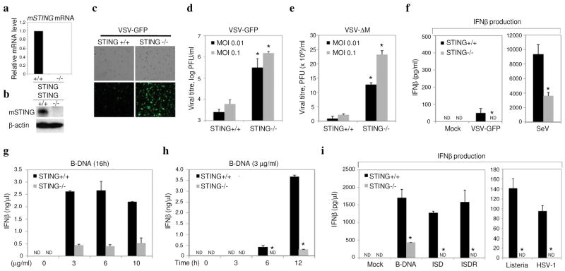
Fig 3. Loss of STING affects host defense.
a. qRT-PCR analysis of mSTING mRNA in STING −/− or control MEFs. b. Immunoblot of mSTING in −/− cells or controls. c. Fluorescence microscopy (GFP) of mSTING −/− or controls infected with VSV-GFP (MOI 0.1). d. Viral titers from (c). e. Viral titers following VSVΔM infection. f. Endogenous IFNβ levels from STING −/− or controls after infection with VSV-GFP (MOI 1) or Sendai Virus (SeV MOI 1) g. STING −/− MEF’s or controls were treated with transfected B-DNA and IFNβ measured by ELISA. h. Time course analysis of (g). i. STING −/− or controls were exposed to transfected B-form DNA, interferon stimulatory DNA (ISD), or ISD reversed nucleotide sequene (ISDR), Listeria monocytogenes (MOI 50), HSV (MOI 5) for 12 hours, and IFNβ measured by ELISA. Asterisks indicate significant difference (P < 0.05) as determined by Student’s t-test. Error bars; +/− s.d. ND; not detectable.

