Otosclerosis and Paget’s diseases are among the bony disorders that can affect the otic capsule. Otosclerosis is a disorder of the bony labyrinth and usually the stapes [1], while Paget’s disease may develop in long bones, in the axial skeleton and in the skull and may involve the otic capsule as well [2]. The association of both entities in the same patient is a rare finding. Kelemen, in 1977, reported a pair of temporal bones that revealed widespread Paget's disease with otosclerosis on both sides. Kelemen also reported that stapedial fixation was produced on the right by otosclerosis, and on the left by Paget's disease.
The incidence of clinical otosclerosis in the Caucasian population is about 0.1%. The ratio of incidence of clinical: histological otosclerosis in the same population is evaluated to be 1:10. The greatest age period at risk for otosclerosis is between 20 and 40 years. Paget's disease occurs more commonly in the elderly and has been reported to involve the temporal bone in 30% of those afflicted. Paget's disease can involve the middle ear structures creating a conductive hearing loss, and extend into the otic capsule creating a sensorineural hearing loss and a vestibular dysfunction.
Defects in osteoprotegerin (OPG) have been implicated in the molecular pathogenesis in both diseases. Osteoprotegerin is expressed at unusually high levels in the inner ear, and has been demonstrated to be a potent inhibitor of bone resorption in vivo, therefore preventing remodeling of the inner ear capsule.
Case Report
A 59 year old female patient with a seven-year history of progressive bilateral hearing impairment, thought to be due to otosclerosis, underwent initially left stapedectomy surgery, followed by right stapedectomy two years later. She maintained bilateral air bone gaps closure with a speech discrimination of 100% until before her death from metastatic breast adenocarcinoma. The patient did not have vestibular symptoms. On radiographic studies, she was found to have Pagetoid changes of the left ilium, and demineralization with poor cortical detail of the petrous apices bilaterally suggestive of Paget’s disease in her temporal bones.
Histopathologic Findings
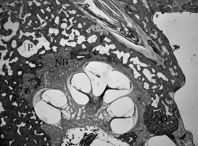
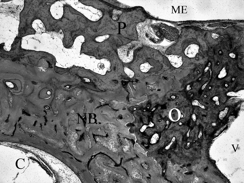
The histopathological findings are reported for the right temporal bone. The right temporal bone shows Pagetoid changes anteromedially to the cochlea. Pagetoid changes involved also the bone adjacent to the utricle and the bony walls of the semicircular canals. An otosclerotic focus is seen in the area just anterior to the oval window. Mostly the cochlear endosteum is not involved by Paget’s disease or by otosclerosis in this bone, except in the proximal basal turn of the cochlea, where there is some encroachment of the otosclerosis on the endosteum adjacent to the spiral ligament. This case shows a rare association of both pathological entities, in the same temporal bone. There is no clear or definite relationship between the 2 entities except that they coexisted in this temporal bone.
Fig. 1.
Light microscopic horizontal section (20× Hematoxylin and Eosin) showing the presence of Otosclerosis (O) and Paget’s disease (P) in the same temporal bone. Normal Bone (NB) is also shown.
Fig. 2.
High power (100x Hematoxylin and Eosin) horizontal section showing Otosclerosis (O), Paget’s bone (P), and normal bone (NB). C=Cochlea; V=Vestibule, ME=Middle Ear.
REFERENCES
- 1.Kelemen G. Temporal bone showing otosclerosis, Paget’s disease and adenocarcinoma. Ann Otol Rhinol Laryngol. 1977;86:381–385. doi: 10.1177/000348947708600316. [DOI] [PubMed] [Google Scholar]
- 2.Schuknecht FH. Disorders of Bone. In: Lea, Febiger, editors. Pathology Of The Ear. 1993. pp. 365–414. [Google Scholar]
- 3.Sorensen MS, Frisch T, Bretlau P. Dynamic bone studies of the labyrinthine capsule in relation to otosclerosis. Adv Otorhinolaryngol. 2007;65:53–58. doi: 10.1159/000098670. [DOI] [PubMed] [Google Scholar]
- 4.Zehnder AF, Kristiansen AG, Adams JC, et al. Osteoprotegerin in the inner ear may inhibit bone remodeling in the otic capsule. Laryngoscope. 2005;115:172–177. doi: 10.1097/01.mlg.0000150702.28451.35. [DOI] [PubMed] [Google Scholar]


