

Abstract
A complete occlusion of the internal carotid artery (ICA) is an important cause of cerebrovascular disease. A never‐symptomatic ICA occlusion has a relatively benign course, whereas symptomatic occlusion increases future risk of strokes. Ultrasonography, magnetic resonance imaging and contrast angiography are useful diagnostic tests, and functional imaging of the brain (eg, with positron emission tomography) helps to understand haemodynamic factors involved in the pathophysiology of brain ischaemia. Recently, there has been a resurgence of interest in the role of extracranial–intracranial bypass surgery for the treatment of completely occluded ICA. With advances in the measurement of cerebral haemodynamics, it may be possible to identify high‐risk patients who could benefit from the bypass surgery.
The accurate prevalence and incidence rates of internal carotid artery (ICA) occlusion are difficult to ascertain as ICA occlusion can remain asymptomatic. In a retrospective, population‐based study on patients with symptomatic ICA occlusion, an incidence rate of 6/100 000 was reported.1 The authors of the study consider it to be an underestimation as many patients with transient ischaemic attack (TIA) do not seek medical attention and some patients with stroke or TIA do not undergo carotid imaging. The same study suggests that about 15% of large‐vessel infarctions may be caused by ICA occlusion. In a study in Manchester, UK, Mead et al2 reported ICA occlusion in 50 of 380 patients with ischaemic stroke consecutively over a 1‐year period. In a study on the correlation of arteriographic findings and symptoms in patients with cerebrovascular disease, 25% of patients with ischaemic stroke had ICA occlusion.3 The prevalence of asymptomatic ICA occlusion is unknown.
Clinical features of ICA occlusion
The clinical spectrum of ICA occlusion ranges from being a completely asymptomatic occlusion (often noted on the “wrong” side during investigation of a TIA or stroke) to a devastating stroke or death. It is possible that in patients with adequate collateral flow, ICA occlusion may remain asymptomatic. TIA or stroke caused by ICA occlusion can present with clinical features similar to those due to any other aetiology. An episode of transient monocular blindness (amaurosis fugax) is highly suggestive of ICA disease proximal to the ophthalmic branch. ICA occlusion is an independent predictor of neurological worsening in stroke.4
A thorough history may disclose a haemodynamic basis of symptoms in some patients. Any condition that further reduces cerebral perfusion—for example, rising from a sitting or lying position (orthostatic TIAs), postprandial hypotension, fluid or blood loss, exercise (cerebral claudication) or cardiac failure—may precipitate ischaemic symptoms in these patients. A rare manifestation of haemodynamic compromise in patients with ICA occlusion is limb shaking.5 The patients complain of repetitive involuntary movements of one or both limbs on one side. This is often misdiagnosed as partial seizures. An electroencephalogram during such episodes does not show any epileptiform activity and cerebral blood flow (CBF) is reduced.6,7 Even rarer are the symptoms of retinal ischaemia (spells of monocular reduced vision) that occur on looking into bright light (retinal claudication),8 presumably resulting from an increase in metabolic demand in the retina on the background of an already reduced perfusion. Importantly, limb shaking and retinal claudication occur in only a minority of patients,9 and have a high positive predictive value but a poor negative value for diagnosing ICA occlusion.
Some patients develop unaccustomed headaches in chronic ICA occlusion as a result of the development of collateral circulation. In ICA occlusion, collateral flow from the branches of external carotid artery (ECA) may cause ipsilateral brisk pulsations at the angle of the jaw, brow and cheek, the so‐called ABC pulsations of Fisher.10 However, collateralisation may predominantly occur through the circle of Willis and, therefore, ABC pulsations may not be brisk.
Although amaurosis fugax is a symptom of acute retinal ischaemia most often caused by embolism from the ICA, in 4–18% of patients with occlusion or severe stenosis of the ICA, a syndrome of chronic ocular ischaemia may develop.11 Patients complain of progressive loss of visual acuity. Venous stasis retinopathy is the term used to describe ophthalmoscopic appearance in the syndrome of chronic ocular ischaemia and is characterised by mid‐peripheral microaneurysms and small dot‐and‐blot intraretinal haemorrhages, arterial narrowing and venous dilatation, and in advanced stages, cotton‐wool exudates and neovascularisation of the optic disc, retina and iris. Nearly one third of patients with symptomatic ICA occlusion show evidence of venous stasis retinopathy, although clinical manifestations are rare.11 Venous stasis retinopathy can be distinguished from diabetic retinopathy by its unilaterality, mid‐peripheral location and low retinal artery pressure. It is important to recognise this condition in order to start secondary prevention strategies.
ICA occlusion can also manifest as syncopal episodes.12
It is suggested that chronic cerebral ischaemia caused by ICA occlusion could cause dementia.13 Improvement in cognitive function after external carotid–internal carotid bypass surgery has been reported by some13 but not by others.14
Pathogenesis of cerebral and retinal ischaemia in ICA occlusion
ICA occlusion can produce brain or retinal ischaemia by the following mechanisms:
Embolism from the distal or proximal stump or from atherosclerotic plaques in the common carotid artery or ECA, which traverse through the collateral pathways involving the ECA. Arguments in favour of this hypothesis are the cessation of symptoms after excision or clipping of the proximal stump, and with antithrombotic treatment.15 In addition, some pathological evidence is consistent with embolism from the distal tail of the occluded ICA.16 Embolism is probably the most common mechanism, accounting for nearly two thirds of strokes in ICA occlusion.17
A compromised CBF, the so‐called perfusion failure with distal insufficiency or misery perfusion syndrome, may have a role in the pathogenesis of ischaemia in patients with ICA occlusion. In chronic ICA occlusion, collateral circulation may maintain cerebral perfusion. Failure of collateralisation may contribute to the haemodynamic compromise. Recent studies using functional brain imaging with positron emission tomography or single‐photon emission computed tomography showed haemodynamic compromise as an independent risk factor for future strokes or TIAs.18 ICA occlusion is frequently associated with borderzone infarcts,19 implicating haemodynamic failure.
Both embolic and haemodynamic factors may act synergistically in the same patient.17 Animal studies have shown that for a given embolic event, the size of the infarct is markedly increased in presence of a pre‐existing hemodynamic impairment.20
Compensatory mechanisms can prevent ischaemia in ICA occlusion. Development of collateral circulation is one of them. The most important source of collateral flow is provided by the contralateral ICA via the circle of Willis. The blood flows in an anterograde manner up the contralateral ICA and then across the circle of Willis to the anterior communicating artery. From here, it goes in an anterograde manner along the cortical branches of the anterior cerebral artery and in a retrograde manner along the anterior cerebral artery to the middle cerebral artery (MCA), and then distally into the MCA territory in the usual anterograde manner. The other important source of collateral flow is from the orbital branches of the ipsilateral ECA. Anterograde flow up in the ECA to the orbit (mainly via its maxillary branches, but also via facial, frontal branches or leptomeningeal branches) allows links with the ophthalmic branch (OA) of the ICA. Blood flows in a retrograde manner in the ophthalmic branch to join the supraophthalmic part of the ICA, where it flows in the usual anterograde fashion. Less commonly, collateral circulation may come from the vertebrobasilar systems or the cortical surface branches.
If collateralisation is inadequate, CBF can be maintained despite reduced cerebral perfusion pressure by dilatation of the resistance vessels (cerebral autoregulation). This autoregulatory vasodilatation can be diagnosed by reduced or absent CBF response to vasodilative stimuli such as hypercapnia or acetazolamide—cerebrovascular reactivity. In patients with ICA occlusion, a vasodilative stimulus does not increase CBF, as autoregulation has already caused maximal vasodilatation in response to a reduced cerebral perfusion pressure. When autoregulatory vasodilatation fails to maintain normal CBF, oxygen extraction fraction (OEF) of the affected brain tissue increases to maintain normal cerebral metabolism, the so‐called stage II haemodynamic failure or misery perfusion.21 An increased OEF has shown to be a powerful independent risk factor for subsequent ischaemic strokes in patients with symptomatic ICA occlusion.18 The combined annual stroke risk in the subgroups with impaired haemodynamic measurements of any severity was 12.5% for all strokes and 9.5% for ipsilateral strokes, suggesting a definitely worse prognosis in patients with impaired cerebral perfusion than in those without.16
Diagnosis of ICA occlusion
It is technically challenging to accurately differentiate total occlusion from a near‐total occlusion (also termed preocclusive stenosis). This is important from the points of view of treatment and prognosis. The patients with symptomatic near‐occlusion are considered to be at a high risk of future embolisation and can derive benefit from carotid endarterectomy. However, carotid endarterectomy is not an option in complete ICA occlusion. Therefore, missing a residual lumen may deprive a patient of a potentially beneficial surgery, and misdiagnosing a complete occlusion as near‐occlusion has the risk of subjecting the patient to unnecessary angiography or surgery. However, a recent article reported lesser benefits of surgery in near‐occlusion.22
Ultrasonography is usually the initial imaging modality for the evaluation of symptomatic ICA disease. B‐mode (brightness modulation) ultrasonography is quite accurate for assessing luminal narrowing and for differentiating normal artery or artery with non‐significant plaque disease from a severely stenotic lesion (stenosis >70%). However, it may be difficult to differentiate near‐occlusion from a complete occlusion. Addition of continuous and pulsed Doppler techniques to B‐mode in duplex systems provides qualitative and quantitative information about the haemodynamic variables (eg, velocity changes, post‐stenotic turbulence). ICA occlusion is characterised by the absence of any signal along the extracranial course of the ICA (fig 1). Sometimes, a low‐velocity signal with a marked reversed signal and absent diastolic flow can be recorded at the origin of the ICA—the stump flow. The accuracy of carotid duplex ultrasonography in diagnosing total carotid occlusion is reported as 97%, with a positive predictive value of 96%, a negative predictive value of 98%, a sensitivity of 91% and a specificity of 99%.23 Newer techniques including power and colour Doppler ultrasonography improve detection of near‐occlusions.24 The use of a contrast agent with duplex ultrasound imaging techniques has improved reliability in distinguishing total occlusions from near‐total occlusions.25 Ultrasonography may not reliably depict high‐up occlusions (beyond the available sonographic window). Low‐velocity flow with a high resistance pattern when measured in a patent cervical ICA may suggest such a lesion, but similar waveforms could be found in a severe high‐grade distal stenosis. Transoral ultrasonography may help in detecting distal ICA occlusion.26

Figure 1 Occlusion of the right internal carotid artery with no evidence of flow using pulsed Doppler imaging.
Magnetic resonance angiography (MRA), with or without gadolinium enhancement, is increasingly used in the evaluation of stroke. Unlike conventional angiography, MRA does not show the vessel anatomy but creates an image of flow in the vessels. Time‐of‐flight MRA depends on the movement of blood. The magnetisation of protons in the moving tissue appears bright. Total occlusion is defined as a flow signal termination on all sequences at any point along the intracranial or extracranial ICA with no flow signal intensity distally (fig 2). In case of near‐occlusion, a focal flow gap is noted. Gadolinium‐enhanced MRA is at least as sensitive as ultrasonography in diagnosing total occlusions in the neck, and probably better than ultrasonography at diagnosing near‐occlusions.27 However, supraclinoid occlusions may not be reliably diagnosed.
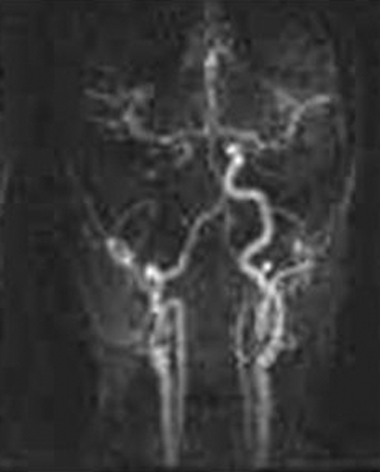
Figure 2 Magnetic resonance angiography showing occluded right proximal internal carotid artery (arrow). The collateral flow to the right hemisphere from the circle of Willis can be seen.
Recently, computed tomography–angiography (CTA) has been used to diagnose ICA disease. Single‐row detector CTA can distinguish total ICA occlusion from hairline residual lumen with a high degree of accuracy.28 Multislice CTA showed an excellent correlation with catheter angiography in diagnosing total versus near‐ICA occlusion.29 When used in combination with duplex ultrasonography, CTA may obviate the need for catheter angiography.30
Digital subtraction angiography is the “gold standard” imaging modality for the evaluation of carotid occlusive disease. However, it is an invasive procedure and therefore should be used only when ultrasonography or MRA fail to provide a definitive distinction between a near‐occlusion and a complete occlusion. Angiography may be the best imaging technique for making appropriate therapeutic decisions in patients with diffuse narrowing (string lesions) on MRA and those with low‐velocity high‐resistance flow in the ICA on ultrasonography.27
Treatment of symptomatic ICA occlusion
Acute ICA occlusion is a therapeutic challenge as a result of poor neurological outcome and paucity of effective therapeutic options available. Patients presenting with stroke should be admitted to a stroke unit. Hypertension should not be corrected in the acute phase unless in a malignant range. Hypotension should be avoided in view of its potential to severely compromise cerebral perfusion in ICA occlusion. Some clinicians use short‐term (about 6 weeks) anticoagulation with heparin and warfarin to reduce embolisation from the fresh clot followed by antiplatelet drugs, although evidence for this strategy is unavailable. In patients presenting soon (<3 h) after the onset of stroke with no evidence of cerebral haemorrhage or a large infarction on brain imaging, intravenous thrombolysis using recombinant tissue plasminogen activator may be considered. However, a large clot usually fails to lyse. In a retrospective study, most patients did not recanalise their ICA occlusion after intravenous recombinant tissue plasminogen activator treatment.31 The recanalisation rate of an associated proximal MCA clot, found in 45% of patients, was good and accounted for a good outcome.
Thrombolytic treatment using a combination of intravenous and intra‐arterial routes or using the intra‐arterial only route has been reported to be effective in distal ICA occlusion, particularly when given soon after the onset of stroke.32
In a small study on 21 patients with strokes due to major arterial occlusions, an intravenous bolus of tirofiban and heparin followed by intra‐arterial administration of urokinase coupled with mechanical thrombolysis was shown to be successful in re‐establishing vessel patency with a good functional outcome.33
A high revascularisation rate has been reported with angioplasty and stenting (endovascular treatment) for acute carotid occlusions. Sugg et al34 reported a 64% immediate recanalisation rate with endovascular treatment in patients with ICA occlusion treated within 3 h of onset of stroke.
However, as yet, there are no large controlled trials showing efficacy of any of these approaches, and an acute ICA occlusion is mostly managed conservatively.
Established ICA occlusion
As it is technically difficult to open a chronically occluded ICA, the management of a chronic ICA occlusion mainly includes strategies to reduce the risk of future strokes and other cardiovascular events. This usually means treatments to reduce embolic risk, but in some cases also to improve cerebral perfusion.
Modification of risk factors
As with other cardiovascular diseases, modifications of risk factors—for example, hypertension, diabetes, hyperlipidaemia and smoking—are vital secondary prevention measures. However, aggressively correcting hypertension should be avoided when clinical features (eg, limb shaking, orthostatic TIAs, syndrome of chronic ocular ischaemia) and investigations suggest a haemodynamic origin for the symptoms. This is in view of the fact that a relatively slight decrease in blood pressure can precipitate cerebral ischaemia. A recent study showed adverse outcome of correcting hypertension in bilateral but not in unilateral ICA occlusion.35
Antithrombotic and anticoagulant agents
In patients with previous stroke or TIA, antiplatelet treatment leads to a 22% reduction in the risk of future non‐fatal stroke, non‐fatal myocardial infarction or a vascular death per 1000 patients treated.36 Aspirin has shown to reduce risk of future cardiovascular events by about 13% (95% confidence interval 4% to 21%) in patients with TIAs or non‐disabling ischaemic stroke from any cause.37 On this basis, aspirin is also used in patients with symptomatic ICA occlusion. Addition of dipyridamole may further reduce the risk of strokes.38 In genuinely aspirin‐intolerant people, clopidogrel may be considered.39 Addition of clopidogrel to aspirin probably increases the risk of bleeding, with no major additional benefit.40 No studies have compared aspirin with oral anticoagulants in ICA occlusion. Although anticoagulants are often used in ICA occlusion caused by dissection, the evidence base for this is poor. Similarly, it is unclear whether antithrombotic agents reduce the risk of future strokes in the subset of patients with a haemodynamic basis for the cerebral ischaemia.
Revascularisation procedures in ICA occlusion
Direct procedure for revascularisation
External carotid–internal carotid bypass surgery: the superficial temporal artery to middle cerebral artery bypass can improve CBF in patients with symptomatic unilateral carotid occlusion.41 However, a large, randomised clinical trial failed to show any benefit of this bypass over contemporary medical treatment in preventing stroke in patients with symptomatic ICA occlusion.42 One of the criticisms of this study is that it did not identify patients with compromised CBF who could have derived greater benefit from the operation.43 Therefore, an ongoing Carotid Occlusion Surgery Study is aiming to re‐examine its effectiveness in reducing subsequent ipsilateral ischaemic stroke in patients with recent cerebral ischaemic symptoms and increased OEF as measured by positron emission tomography.44 An increased OEF distal to a symptomatic ICA occlusion is a requirement for randomisation to surgery or medical treatment. A newer technique using a venous transplant for a bypass between the proximal superficial temporal artery and the most distal, intracranial part of the ICA or the proximal MCA results in a larger increase in blood flow (“high‐flow” external carotid–internal carotid bypass) and may be more effective at restoring CBF.45
Indirect procedures
Endarterectomy or angioplasty stenting of a haemodynamically relevant stenosis of the contralateral ICA: the contralateral ICA is often the most important source of collateral flow in ICA occlusion. In patients with ICA occlusion and a severe stenosis of the contralateral ICA, carotid endarterectomy of the contralateral ICA resulted in a long‐term cerebral haemodynamic improvement not only on the side of surgery but also on the side of the ICA occlusion.46 Gonzalez et al47 have recently reported endovascular treatment (angioplasty and stenting) of contralateral ICA to be safe and effective in patients with a symptomatic ICA occlusion and a severe stenosis of the contralateral ICA.47 However, no large, controlled studies have evaluated the efficacy and safety of these therapeutic approaches in ICA occlusion.
Endarterectomy of the ipsilateral ECA: in some patients with ICA occlusion, collateral flow may predominantly be provided by the branches of ECA. A stenotic lesion in ECA may lead to ischaemic symptoms in these patients.15 In a collective review of case series, Gertler et al48 reported safety and efficacy of endarterectomy of ipsilateral ECA in a subgroup of patients with symptomatic ICA occlusion. Resolution of symptoms was seen in 83% of patients, with another 7% showing marked improvement. The overall neurological complication rate was 5%, with more recent reports being associated with improved mortality and morbidity, implying a learning curve.
Outcome of ICA occlusion
The subject of future risk of stroke or TIA in patients with ICA occlusion is complex. A never‐symptomatic ICA occlusion is considered to have a benign course.49 However, symptomatic ICA occlusion increases the risk of future cerebrovascular events. In a study on angiographically proved ICA occlusion, the rate of recurrent stroke was 4.8% at 1 year, 12.2% at 3 years and 17.1% at 5 years.50 In a meta‐analysis of 20 follow‐up studies on patients with symptomatic ICA occlusion, the annual risk of stroke was 5.5% and that of ipsilateral stroke was 2.1%.16 Those with evidence of haemodynamic compromise on functional imaging are at an even higher risk (all strokes, 12.5%; and ipsilateral stroke, 9.5%). Patients with bilateral carotid occlusion have a high risk of stroke.
Conclusion
ICA occlusion is an important cause of cerebral or retinal ischaemia. The clinical course of ICA occlusion is variable, from being a completely asymptomatic to one leading to devastating strokes. Our understanding of the role of embolism and haemodynamic factors in the pathogenesis has greatly improved in recent years, largely due to advances in morphological and functional imaging techniques. This will hopefully help in developing treatments targeted to the underlying mechanism.
Abbreviations
CBF - cerebral blood flow
CTA - computed tomography–angiography
ECA - external carotid artery
ICA - internal carotid artery
MCA - middle cerebral artery
MRA - magnetic resonance angiography
OEF - oxygen extraction fraction
TIA - transient ischaemic attack
Footnotes
Competing interests: None declared.
References
- 1.Flaherty M L, Flemming K D, McClelland R.et al Population‐based study of symptomatic internal carotid artery occlusion. Incidence and long‐term follow‐up. Stroke 200435e349. [DOI] [PubMed] [Google Scholar]
- 2.Mead G E, Shingler H, Farrell A.et al Carotid disease in acute stroke. Age Ageing 199827677–682. [DOI] [PubMed] [Google Scholar]
- 3.Thiele B L, Young J V, Chikos P M.et al Correlation of arteriographic findings and symptoms in cerebrovascular disease. Neurology 1980301041–1046. [DOI] [PubMed] [Google Scholar]
- 4.Weimar C, Mieck T, Buchthal J.et al Neurologic worsening during the acute phase of ischemic stroke. Arch Neurol 200562393–397. [DOI] [PubMed] [Google Scholar]
- 5.Yanagihara T, Piepgras D G, Klass D W. Repetitive involuntary movement associated with episodic cerebral ischemia. Ann Neurol 198518244–250. [DOI] [PubMed] [Google Scholar]
- 6.Yanagihara T, Klass D W. Rhythmic involuntary movement as a manifestation of transient ischemic attacks. Trans Am Neurol Assoc 198110646–48. [PubMed] [Google Scholar]
- 7.Tatemichi T K, Young W L, Prohovnik I.et al Perfusion insufficiency in limb‐shaking transient ischemic attacks. Stroke 199021341–347. [DOI] [PubMed] [Google Scholar]
- 8.Furlan A J, Whisnant J P, Kearns T P. Unilateral visual loss in bright light: an unusual symptom of carotid artery occlusive disease. Arch Neurol 197936675–676. [DOI] [PubMed] [Google Scholar]
- 9.Klijn C J, Kappelle L J, van Huffelen A C.et al Recurrent ischemia in symptomatic carotid occlusion: prognostic value of hemodynamic factors. Neurology 2000551806–1812. [DOI] [PubMed] [Google Scholar]
- 10.Fisher C M. Facial pulses in internal carotid artery occlusion. Neurology 197020476–478. [DOI] [PubMed] [Google Scholar]
- 11.Klijn C J, Kappelle L J, van Schooneveld M J.et al Venous stasis retinopathy in symptomatic carotid artery occlusion: prevalence, cause, and outcome. Stroke 200233695–701. [DOI] [PubMed] [Google Scholar]
- 12.Kashiwazaki D, Kuroda S, Terasaka S.et al Carotid occlusive disease presenting with loss of consciousness. No Shinkei Geka 20053329–34. [PubMed] [Google Scholar]
- 13.Tatemichi T K, Desmond D W, Prohovnik I.et al Dementia associated with bilateral carotid occlusions: neuropsychological and haemodynamic course after extracranial to intracranial bypass surgery. J Neurol Neurosurg Psychiatry 199558633–636. [DOI] [PMC free article] [PubMed] [Google Scholar]
- 14.Younkin D, Hungerbuhler J P, O'Connor M.et al Superficial temporal‐middle cerebral artery anastomosis: effects on vascular, neurologic, and neuropsychological functions. Neurology 198535462–469. [DOI] [PubMed] [Google Scholar]
- 15.Barnett H J M, Peerless S J, Kaufmann J C E. ‘Stump' of internal carotid artery: a source for further cerebral embolic ischemia. Stroke 19789448–456. [DOI] [PubMed] [Google Scholar]
- 16.Klijn C J, Kappelle L J, Tulleken C A.et al Symptomatic carotid artery occlusion. A reappraisal of hemodynamic factors. Stroke 1997282084–2093. [DOI] [PubMed] [Google Scholar]
- 17.Pessin M S, Hinton R C, Davis K R.et al Mechanisms of acute carotid Stroke. Ann Neurol 19796245. [DOI] [PubMed] [Google Scholar]
- 18.Yamauchi H, Fukuyama H, Nagahama Y.et al Evidence for misery perfusion and risk for recurrent stroke in major cerebral arterial occlusive diseases from PET. J Neurol Neurosurg Psychiatry 19966118–25. [DOI] [PMC free article] [PubMed] [Google Scholar]
- 19.Hupperts R M M, Lodder J, Heuts‐van Raak E P M.et al Borderzone brain infarcts on CT taking into account the variability in vascular supply areas. Cerebrovasc Dis 19966294–300. [Google Scholar]
- 20.Omae T, Mayzel‐Oreg O, Li F.et al Inapparent hemodynamic insufficiency exacerbates ischemic damage in a rat microembolic stroke model. Stroke 2000312494–2499. [DOI] [PubMed] [Google Scholar]
- 21.Baron J C, Bousser M G, Rey A.et al Reversal of focal “misery perfusion syndrome” by extra‐intracranial artery bypass in hemodynamic cerebral ischemia: a case study with O‐15 positron emission tomography. Stroke 198112454–459. [DOI] [PubMed] [Google Scholar]
- 22.Fox A J, Eliasziw M, Rothwell P M.et al Identification, prognosis, and management of patients with carotid artery near occlusion. AJNR Am J Neuroradiol 2005262086–2094. [PMC free article] [PubMed] [Google Scholar]
- 23.AbuRahma A F, Pollack J A, Robinson P A.et al The reliability of color duplex ultrasound in diagnosing total carotid occlusion. Am J Surg 1997174185–187. [DOI] [PubMed] [Google Scholar]
- 24.Furst G, Saleh A, Wenserski F.et al Reliability and validity of noninvasive imaging of internal carotid artery occlusion. Stroke 1999301444–1449. [DOI] [PubMed] [Google Scholar]
- 25.Ohm C, Bendick P J, Monash J.et al Diagnosis of total internal carotid occlusions with duplex ultrasound and ultrasound contrast. Vasc Endovascular Surg 200539237–243. [DOI] [PubMed] [Google Scholar]
- 26.Isa K, Yasaka M, Kimura K.et al Transoral carotid ultrasonography for evaluating internal carotid artery occlusion. Intern Med 200544567–571. [DOI] [PubMed] [Google Scholar]
- 27.El‐Saden S M, Grant E G, Hathout G M.et al Imaging of the internal carotid artery: the dilemma of total versus near total occlusion. Radiology 2001221301–308. [DOI] [PubMed] [Google Scholar]
- 28.Lev M H, Romero J M, Goodman D N.et al Total occlusion versus hairline residual lumen of the internal carotid arteries: accuracy of single section helical CT angiography. AJNR Am J Neuroradiol 2003241123–1129. [PMC free article] [PubMed] [Google Scholar]
- 29.Chen C J, Lee T H, Hsu H L.et al Multi‐Slice CT angiography in diagnosing total versus near occlusions of the internal carotid artery: comparison with catheter angiography. Stroke 20043583–85. [DOI] [PubMed] [Google Scholar]
- 30.Herzig R, Burval S, Krupka B.et al Comparison of ultrasonography, CT angiography, and digital subtraction angiography in severe carotid stenoses. Eur J Neurol 200411774–781. [DOI] [PubMed] [Google Scholar]
- 31.Christou I, Felberg R A, Demchuk A M.et al Intravenous tissue plasminogen activator and flow improvement in acute ischemic stroke patients with internal carotid artery occlusion. J Neuroimaging 200212119–123. [DOI] [PubMed] [Google Scholar]
- 32.Zaidat O O, Suarez J I, Santillan C.et al Response to intra‐arterial and combined intravenous and intra‐arterial thrombolytic therapy in patients with distal internal carotid artery occlusion. Stroke 2002331821–1826. [DOI] [PubMed] [Google Scholar]
- 33.Mangiafico S, Cellerini M, Nencini P.et al Intravenous glycoprotein IIb/IIIa inhibitor (tirofiban) followed by intra‐arterial urokinase and mechanical thrombolysis in stroke. AJNR Am J Neuroradiol 2005262595–2601. [PMC free article] [PubMed] [Google Scholar]
- 34.Sugg R M, Malkoff M D, Noser E A.et al Endovascular recanalization of internal carotid artery occlusion in acute ischemic stroke. AJNR Am J Neuroradiol 2005262591–2594. [PMC free article] [PubMed] [Google Scholar]
- 35.Rothwell P M, Howard S C, Spence J D. Relationship between blood pressure and stroke risk in patients with symptomatic carotid occlusive disease. Stroke 2003342583–2590. [DOI] [PubMed] [Google Scholar]
- 36.Antithrombotic Trialists' Collaboration Collaborative meta‐analysis of randomised trials of antiplatelet therapy for prevention of death, myocardial infarction, and stroke in high risk patients. BMJ 200232471–86. [DOI] [PMC free article] [PubMed] [Google Scholar]
- 37.Diener H C. Stroke prevention: anti‐platelet and anti‐thrombolytic therapy. Neurol Clin 200018343–355. [DOI] [PubMed] [Google Scholar]
- 38.Sivenius J, Cunha L, Diener H C.et al Antiplatelet therapy is effective regardless of age. ESPS2 Working Group. Acta Neurol Scand 19999954–60. [DOI] [PubMed] [Google Scholar]
- 39.Cannon C P. CAPRIE Investigators. Effectiveness of clopidogrel versus aspirin in preventing acute myocardial infarction in patients with symptomatic atherothrombosis (CAPRIE trial). Am J Cardiol 200290760–762. [DOI] [PubMed] [Google Scholar]
- 40.Diener H C, Bogousslavsky J, Brass L M.et al Aspirin and clopidogrel compared with clopidogrel alone after recent ischaemic stroke or transient ischaemic attack in high‐risk patients (MATCH): randomised, double‐blind, placebo‐controlled trial. Lancet 2004364331–337. [DOI] [PubMed] [Google Scholar]
- 41.Schmiedek P, Piepgras A, Leinsinger G.et al Improvement of cerebrovascular reserve capacity by EC‐IC arterial bypass surgery in patients with ICA occlusion and hemodynamic cerebral ischemia. J Neurosurg 199481236–244. [DOI] [PubMed] [Google Scholar]
- 42.The EC/IC Bypass Study Group Failure of extracranial‐intracranial arterial bypass to reduce the risk of ischemic stroke: results of an international randomized trial. N Engl J Med 19853131191–1200. [DOI] [PubMed] [Google Scholar]
- 43.Sundt T M. Was the international randomized trial of extracranial‐intracranial arterial bypass representative of the population at risk? N Engl J Med 1987316814–816. [DOI] [PubMed] [Google Scholar]
- 44.Grubb R L, Jr, Powers W J, Derdeyn C P.et al The carotid occlusion surgery study. Neurosurg Focus 200314e9. [DOI] [PubMed] [Google Scholar]
- 45.Klijn C J, Kappelle L J, van der Zwan A.et al Excimer laser‐assisted high‐flow extracranial/intracranial bypass in patients with symptomatic carotid artery occlusion at high risk of recurrent cerebral ischemia: safety and long‐term outcome. Stroke 2002332451–2458. [DOI] [PubMed] [Google Scholar]
- 46.Baracchini C, Meneghetti G, Manara R.et al Cerebral hemodynamics after contralateral carotid endarterectomy in patients with symptomatic and asymptomatic carotid occlusion: a 10‐year follow‐up. J Cereb Blood Flow Metab 20067899–905. [DOI] [PubMed] [Google Scholar]
- 47.Gonzalez A, Gonzalez‐Marcos J R, Martinez E.et al Safety and security of carotid artery stenting for severe stenosis with contralateral occlusion. Cerebrovasc Dis 200520(Suppl 2)123–128. [DOI] [PubMed] [Google Scholar]
- 48.Gertler J P, Cambria R P. The role of external carotid endarterectomy in the treatment of ipsilateral internal carotid occlusion: collective review. J Vasc Surg 19876158–167. [DOI] [PubMed] [Google Scholar]
- 49.Powers W J, Derdeyn C P, Fritsch S M.et al Benign prognosis of never‐symptomatic carotid occlusion. Neurology 200054878–882. [DOI] [PubMed] [Google Scholar]
- 50.Sacquegna T, De Carolis P, Pazzaglia P.et al The clinical course and prognosis of carotid artery occlusion. J Neurol Neurosurg Psychiatry 1982451037–1039. [DOI] [PMC free article] [PubMed] [Google Scholar]