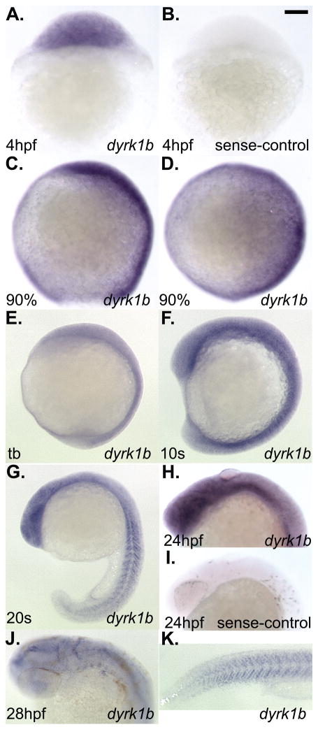
Figure 2. Whole-mount in situ hybridization analysis of dyrk1b expression in wildtype zebrafish embryos.
A) ubiquitous expression of dyrk1b at 4hpf. B) sense-strand negative control for staining also at 4hpf. C) lateral view of 90% epiboly stage, D) dorsal view of 90% epiboly stage. E) lateral view of tailbud (tb) stage. F) lateral views of 10 somites (10s) stage. G) lateral view of 20 somites (20s) stage. H) lateral view of 24hpf stage. I) sense-strand negative control for staining also at 24hpf. J) dorsolateral view of 28hpf stage. K) lateral tail view of the same animal shown in panel J. Scale bar in panel B = 100 micrometers length.

