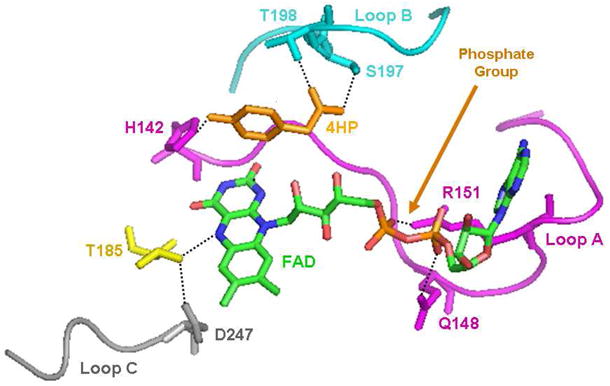
Figure 8.

Illustration of three large loops in the structure of the oxygenase from T. thermophilus (adapted from the structure 2YYJ in the PDB) that are not found in the structure of the oxygenase from A. baumannii (7). FAD and HPA bound to the protein are shown in CPK colors, loop A (between strands β5 and β6 and interacting with part of FAD) is shown in scarlet, loop B (between strands β8 and β9 and interacting with HPA) is shown in cyan, and loop C (between strands β10 and β11 and possibly interacting with the flavin hydroperoxide) is shown in grey. Some potential hydrogen bonds are shown as dotted line.
