Abstract
Memory formation is a temporally graded process during which transcription and translation steps are required in the first hours after acquisition. Although persistence is a key characteristic of memory storage, its mechanisms are scarcely characterized. Here, we show that long-lasting but not short-lived inhibitory avoidance long-term memory is associated with a delayed expression of c-Fos in the hippocampus. Importantly, this late wave of c-Fos is necessary for maintenance of inhibitory avoidance long-term storage. Moreover, inhibition of transcription in the dorsal hippocampus 24 h after training hinders persistence but not formation of long-term storage. These findings indicate that a delayed phase of transcription is essential for maintenance of a hippocampus-dependent memory trace. Our results support the hypothesis that recurrent rounds of consolidation-like events take place late after learning in the dorsal hippocampus to maintain memories.
Keywords: gene expression, noradrenaline, Inhibitory Avoidance, BDNF, alpha-amanitin
Information storage in the brain is a temporally graded process involving several phases. Long-term (LTM), but not short-term memory (STM), requires de novo RNA synthesis around training (1–3). Additionally, transcription of specific plasticity-related genes is dynamically regulated by neural activity during memory formation (4, 5).
Immediate early genes (IEGs), including the inducible transcription factors (ITF) c-fos and zif268, are the first group of genes expressed after synaptic activation (6). c-Fos forms heterodimeric complexes with Jun proteins to build up the transcription factor AP-1, which regulates the expression of a variety of effector genes (7, 8). Rapid and transient expression of c-Fos is associated with the initial steps of LTM in different learning tasks (6, 8–10), and deletion of the c-fos gene or inhibition of its expression at the time of training impairs memory (10–12).
Despite the growing amount of data accumulated during the last four decades regarding LTM formation, there is no satisfactory explanation for the fact that memories outlast the lifetime of the changes in specific patterns of synaptic weights that originally encoded them. We have shown that after inhibitory avoidance (IA) training, an aversively motivated hippocampus-dependent learning task, there is a BDNF- and protein synthesis-dependent late phase in dorsal hippocampus critical for persistence but not formation of LTM (13, 14). We also found an increase in c-fos protein levels at that late posttraining time (13), suggesting that transcriptional events might be triggered during the persistence phase of memory storage. In the present study, we determined whether or not there was a late transcription phase involved in LTM persistence and whether or not c-Fos expression in the dorsal hippocampus was functionally associated with that phase.
Results
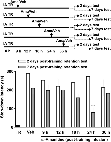
To investigate whether or not there is more than one wave of c-Fos expression in a persistent memory and to determine the role of transcription, in general, and c-Fos expression, in particular, on persistence of LTM storage we used a one-trial IA. This task has been extensively used for studying posttraining memory processing because of its rapid hippocampus-dependent acquisition and its hippocampus-dependent recall (9). At different posttraining times, we infused the specific inhibitor of RNA polymerase II α-amanitin into the dorsal CA1 region at a dose known to block memory formation (1, 2). There were no differences in training performance in any group studied (P = 0.95; Newman–Keuls after ANOVA; n = 10–12). Infusion of α-amanitin 24 h posttraining impaired memory retention 7 d thereafter (P < 0.005 with respect to vehicle-injected animals; n = 12) (Fig. 1), leaving intact retention performance at 2 d. No change in memory retention was observed when α-amanitin was given 9, 12, 18, or 36 h after training with respect to the corresponding vehicle group at each time point. These findings indicate that a late wave of mRNA synthesis in the dorsal hippocampus is necessary to maintain but not to form a persistent IA memory.
Fig. 1.
Hippocampal mRNA synthesis is required 24 h after training for LTM persistence but not for memory formation. (Upper) Schematic procedure. (Lower) Animals were infused into dorsal hippocampus with vehicle (Veh) or α-amanitin (0.5 μM; 1 μl/side) 9, 12, 18, 24, or 36 h after training. Data are expressed as mean ± SEM of training (TR, black bar) or test-session step-down latency, 2 d (white bars) or 7 d (gray bars) after training. **P < 0.01 vs. Veh; two-tailed Student’s t test, n = 10–12.
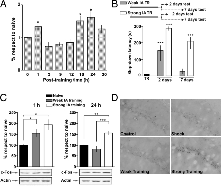
We then asked if the delayed c-Fos expression seen after IA training could be associated to a transcription-dependent mechanism involved in LTM persistence. To determine the temporal course of the changes in c-Fos expression associated with IA training, we measured c-Fos protein levels at different posttraining times. Persistent memories are associated with two waves of c-Fos expression: an early and short wave around 1 h posttraining, and a second wave that peaks 18–24 h after training (P < 0.05) (Fig. 2A). The second wave is also transient, because no changes were found at the 30-h time point. No significant differences were obtained between the naïve group and the shocked nontrained group (1 h: naïve = 100 ± 6.7%, shocked = 87 ± 5.7%, P = 0.18; 18 h: naïve = 100 ± 15%, shocked = 103 ± 7.9%, P = 0.87; 24 h: naïve = 100 ± 6.3%, shocked = 88 ± 12.3%, P = 0.4; Student’s t test; n = 5–6).
Fig. 2.
Memory processing is associated with two waves of c-Fos expression. (A) Time course of changes in hippocampal c-Fos levels after strong IA training. Bars indicate the percentage of change with respect to the naïve group for rats trained and killed immediately, 1, 3, 9, 12, 18, 24, or 30 h after training. Data are expressed as mean ± SEM. *P < 0.05; two-tailed Student’s t test; n = 5–8. (B) Strong (0.7 mA), but not weak (0.3 mA), IA training generates a persistent LTM. (Upper) Schematic procedure. (Lower) Data are expressed as mean ± SEM of TR (black bars) or test-session step-down latency at 2 or 7 d after weak (gray bars) or strong (white bars) IA training. ***P < 0.0001 vs. TR; two-tailed Student’s t test, n = 10. (C) Strong and weak training induced c-Fos expression 1 h after training (Left). In contrast, strong, but not weak, IA training results in increased c-Fos 24 h posttraining (Right). (Upper) Bars show normalized mean percentage levels of c-Fos with respect to the naïve group. Data are expressed as mean ± SEM of naïve (black bar), weak (gray bar), and strong (white bar) IA training. *P < 0.05; **P < 0.01; ***P < 0.001 vs. naïve in Newman–Keuls test after ANOVA; n = 6. (Lower) Representative Western blots showing c-Fos and actin levels. (D) Strong, but not weak, IA training is associated with increased c-Fos immunoreactivity in dorsal CA1 24 h after training.
We next asked whether or not the second wave of c-Fos expression was involved in memory persistence. If the delayed wave of c-Fos expression is required specifically for maintenance of a persistent LTM, then it should not occur after a training session that is unable to induce a long-lasting LTM trace. Differences in LTM persistence can be induced by modifying the amount or strength of training. IA training using a strong foot shock (0.7 mA; 3 s; strong training) (Fig. 2B), which generates a persistent LTM as tested 7 d after training (14), increased c-Fos expression in the dorsal hippocampus 24 h posttraining (P < 0.01 compared with naïve) (Fig. 2C). However, training with a mild foot shock (0.3 mA, 3 s; weak training), which produces a rapidly decaying LTM (Fig. 2B), did not change c-Fos levels at 24 h posttraining (Fig. 2C). In addition, immunohistochemical assays of c-Fos expression revealed higher c-Fos immunoreactivity in strong IA-trained rats compared with weak-trained shock and naive control groups (Fig. 2D). Strong and weak training induced the first wave of c-Fos expression around 1 h after training (P < 0.05) (Fig. 2C). This indicates that formation of lasting and nonlasting LTM is associated with an early phase of c-Fos expression, whereas the late posttraining c-Fos expression phase is linked specifically to persistence of LTM storage. To further substantiate this claim, two additional control experiments were done. First, a context, no foot–shock experiment was performed in which the animals were placed on the platform but did not receive a foot shock when they stepped down to the grid. Second, a delayed foot–shock experiment was performed in which the animals were placed on the platform, no foot shock was given, the subjects were returned to their home cage, and then, 1 h later, they received the foot shock. Immunoblot analysis revealed significant differences in c-Fos levels 24 h after strong IA training with respect to all control groups (P < 0.05) (Table 1), confirming that late c-Fos expression is associated with a training experience that generates a persistent memory.
Table 1.
Strong IA training induces a late increase in c-Fos expression
| Group | Percentage change |
| Naïve | 100.0 ± 7.7 |
| Shock | 88.0 ± 12.3 |
| Context with no foot shock | 93.4 ± 7.5 |
| Delayed foot shock | 91.2 ± 8.6 |
| Strong IA training | 156.1 ± 11.1* |
Rats were trained and killed 24 h later. Hippocampi were dissected out and homogenized for Western blot analysis. Values (mean ± SEM) indicate the percentage of change with respect to naïve groups. (*P < 0.05; Newman–Keuls test after ANOVA; n = 5.)
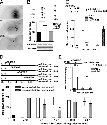
To address the question of whether or not the expression of c-Fos is required for memory persistence, we used an antisense oligonucleotide (ASO) (15) that specifically blocks de novo expression of c-Fos by binding to c-fos mRNA. We determined the extent of diffusion and stability of c-fos ASO. Rats received 2 nmol (1 μl/side) of biotinylated c-fos ASO into dorsal CA1 and were killed 2, 5, or 24 h later. c-fos ASO was consistently observed in the dorsal hippocampus at 2 h and remained detectable for at least 5 h. Twenty-four hours after infusion, the biotinylated ASO was undetectable (Fig. 3A).
Fig. 3.
Delayed expression of c-Fos is required for persistence but not memory formation. (A) Biotinylated ASO anatomical distribution and relative concentrations at different times after infusion. By 2 h, the ASO diffused throughout the dorsal hippocampus and remained there for at least 3 more h. After 24 h, the ASO was cleared out from the hippocampus. (B–E) Schematic of the procedure used. (B) Animals infused into dorsal hippocampus with MSO (white bars) or ASO (gray bars) 12 h after training (Upper). Twenty-four hours later, the dorsal hippocampus was dissected out and used for Western blot analysis (Lower). Bars show the normalized percentage levels with respect to naïve animals. *P < 0.05 in Newman–Keuls test after ANOVA; n = 6. (C) Animals were infused in the dorsal hippocampus with MSO (white bars) or ASO (gray bars) 4 h before training. Data are expressed as mean ± SEM of TR (black bars) or test session step-down latency 2 or 7 d after IA training. The dotted bar represents mean test-session step-down latency of ASO-infused animals that were retrained and tested 24 h later. *P < 0.05; **P < 0.01 vs. MSO group; Student’s t test; n = 10–12 per group. (D) Animals were infused in the dorsal hippocampus with MSO (white bars) or ASO (gray bars; 2 nmol/side) 8, 12, 18, or 24 h after training. Data are expressed as mean ± SEM of TR (black bars) or test session step-down latency 2 or 7 d after IA training. **P < 0.01 vs. MSO group; Student’s t test; n = 10–12 per group. (E) Intrahippocampal infusion of c-fos ASO 12 h posttraining impairs LTM persistence in a permanent way. Data are expressed as mean ± SEM of TR (black bars) or test-session step-down latency 7 and 14 d after IA training. *P < 0.05 vs. MSO group; Student’s t test; n = 10–12 per group.
Infusion of c-fos ASO into dorsal hippocampus 12 h posttraining prevented the IA training-induced increase in c-Fos expression 24 h after training (P < 0.05) (Fig. 3B). No effect was seen on c-Fos expression when infusing c-fos missense oligonucleotide (MSO), a control sequence containing the same nucleotides but in scrambled order. These findings indicated that a single intrahippocampal infusion of c-fos ASO blocks de novo expression of c-Fos induced by training. We next determined whether or not inhibition of the delayed expression of c-Fos was sufficient to block memory persistence. As a control, we confirmed and extended previous findings (11, 12), showing that intra-CA1 infusion of c-fos ASO, but not of c-fos MSO, 4 h before training prevented the first wave of training-induced increase in c-Fos and also impeded LTM formation as revealed in two test sessions carried out at 2 and 7 d after training (each animal was tested only one time) (Fig. 3C). One day after the last test session (eighth day), rats previously infused with ASO were submitted again to a strong training protocol and were tested 24 h thereafter. They exhibited retention test latencies similar to those found 2 d after the original training (Fig. 3C), suggesting that c-fos ASO totally prevented IA memory formation and that ASO infusion did not affect the functionality of hippocampus.
Infusion of c-fos ASO into the dorsal hippocampus 12 h after training did not alter IA memory retention 2 d posttraining but significantly impaired LTM retention at 7 d (gray bars; t = 3.3; P < 0.003; n = 12 per group) (Fig. 3D). In addition, animals injected with c-fos ASO 12 h after training that were amnesic when tested 7 d posttraining and also showed a memory deficit when retested at 14 d (Fig. 3E). Because no spontaneous recovery was found, these results indicate that c-fos ASO given 12 h after training resulted in a long-lasting memory deficit. Therefore, memory persistence but not memory formation is affected by the inhibition of the second wave of c-Fos expression. Importantly, no effect was seen on retention when c-fos ASO was infused into CA1 8, 18, or 24 h after training (Fig. 3D).
The expression of Zif268 can be reduced by c-fos ASO (16). We found no changes in learning-induced increase of Zif268 levels in the hippocampus after c-fos ASO infusion (MSO = 152 ± 10% and ASO = 161 ± 17% [percentages with respect to naïve]; P > 0.05 when MSO vs. ASO and P < 0.05 when MSO or ASO vs. naïve in a Newman–Keuls test after ANOVA; n = 5–6). Unlike c-fos ASO, infusion of zif268 ASO 12 h posttraining did not affect LTM 7 d after IA training (MSO 178 ± 48 s; ASO 195 ± 32 s; P = 0.76 in Student’s t test; n = 9).
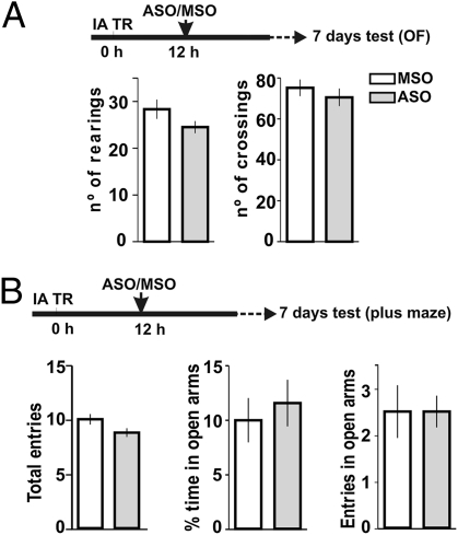
Performance during IA test sessions can be altered by changes in locomotor activity and anxiety, which can be potentially affected by c-fos ASO. For that reason, animals that received c-fos ASO 12 h after IA training were subjected to the open field and elevated, and plus, they had maze tests 7 d later. c-fos ASO did not affect anxiety state or exploratory behavior in a novel environment and did not modify basal locomotor activity (Fig. 4 A and B), suggesting that the observed memory deficit is directly caused by inhibition of c-Fos expression that is required for persistence of the memory trace. Moreover, it is not likely that the lower retention scores observed at 7 d posttraining were caused by modifications in behavioral performance, because administration of c-fos ASO at posttraining time points surrounding the critical period did not cause any deficit in memory retention (Fig. 3D).
Fig. 4.
Infusion of c-fos ASO 12 h after IA training does not affect locomotor activity, anxiety state, or exploratory behavior. (A) Number of rearings (Left) and crossings (Right) during a 5-min open-field (OF) session for animals that had received c-fos MSO (open bars) or c-fos ASO (gray bars; 2 nmol/μl; 1 μl/side) in dorsal CA1 12 h posttraining 7 d before the OF session. Data are expressed as mean ± SEM number of crossings or rearings (n = 8). (B) Total number of entries (Left), time spent in open arms (Center), and number of entries into the open arms (Right) during a 5-min plus maze session for rats that had received bilateral intra-CA1 infusion of c-fos MSO (white bars) or c-fos ASO (gray bars) 7 d before the OF session (n = 8).
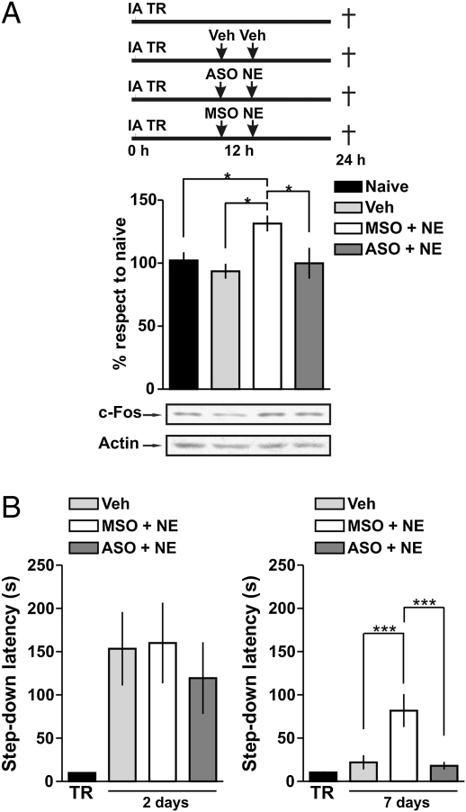
We next asked whether or not induction of the second wave of c-Fos expression can promote memory persistence. Stimulation of adrenergic receptors increases c-Fos levels in the hippocampus (17). To analyze the role of c-Fos in the promotion of LTM persistence, we used a weak training protocol that leaves a rapidly decaying LTM (Fig. 2B). Infusion of norepinephrine (NE; 0.3 μg/side) into dorsal CA1 12 h after training induced c-Fos expression (Fig. 5A, white bar) and promoted LTM persistence (P < 0.01) (Fig. 5B). The intra-CA1 infusion of c-fos ASO, but not c-fos MSO, 1 h before the administration of NE abolished the NE-dependent increase in c-Fos expression and the induction of a persistent memory (Fig. 5 A and B). None of the effects observed at 7 d were found when rats were tested 2 d after training (Fig. 5B). These findings indicated that NE-induced persistence of memory storage is mediated by de novo synthesis of c-Fos during a late posttraining time window. These results also indicate that the increased expression of c-Fos in the dorsal hippocampus promotes memory persistence, transforming a nonlasting LTM trace into a persistent one.
Fig. 5.
Delayed posttraining infusion of NE promotes LTM persistence. (A) Schematic procedure. (Top) Animals infused into dorsal hippocampus with vehicle (Veh; light gray bar), MSO (white bar), or ASO (dark gray bar; 2 nmol/μl; 1 μl/side) 11 h after training. One hour later, they were also injected with Veh (light gray bar) or NE (white and dark gray bars; 0.6 μg/μl; 0.5 μl/side). Twenty-four hours after training, the dorsal hippocampus was dissected out and used for Western blot analysis (Bottom). Bars show the normalized percentage levels with respect to naïve (black bar) animals. *P < 0.05 in Newman–Keuls test after ANOVA (n = 6). (B) Animals infused in the dorsal hippocampus with vehicle (Veh; light gray bar), MSO (white bar), or ASO (dark gray bar) 11 h after training. One hour later, they were also injected with Veh (light gray bar) or NE (white and dark gray bars). Data are expressed as mean ± SEM of TR (black bars) or test-session step-down latency 2 (Left) or 7 d (Right) after training. ***P < 0.001 in Newman–Keuls test after ANOVA (n = 8–10).
Discussion
The main finding of the present work is that de novo mRNA synthesis that involves a delayed wave of c-Fos expression in the rat hippocampus is required during a restricted time window around 24 h after training for the persistence of LTM storage but not for memory formation. These results show that the hippocampus is still engaged in memory processing after LTM is already formed and that a previously unknown phase of c-Fos expression plays an important role in maintenance of a memory trace over time. Together with recent reports in rats (13, 14) and Aplysia (18), our findings endorse the hypothesis that LTM storage is achieved by recurrent rounds of consolidation-like mRNA and protein synthesis-dependent processes (19, 20).
A classical view in modern biology is that a rapid expression of IEGs is tightly coupled to cellular activity. In the brain, expression of IEGs plays a role in regulating synaptic plasticity, and some IEGs, including c-Fos, have been associated with the first transient transcriptional steps involved in memory formation (4, 5, 12). We found that there are two posttraining waves of c-Fos expression in the hippocampus: an early one (6, 10) around 1 h after training, which is involved in IA LTM formation, and a late wave, peaking around 24 h after the strong training protocol, which is specifically involved in memory persistence.
The early wave of c-Fos expression is part of a rapid pulse of increased gene transcription involving a broad functional repertoire of molecules, including transcription factors, growth factors, signal transduction molecules, and cytoskeletal components (6, 21). IEG induction has been attributed to the experience of novelty or saliency of the context in different behavioral studies (6, 22). The explicit assumption of this hypothesis is that the functional role of a given IEG is to participate in memory formation of the experience that induced its expression. However, there are alternative interpretations. One that has attracted much attention is the possible role of IEGs as coincident detectors and/or metaplasticity signals (6, 22).
The delayed wave of c-Fos in strongly trained animals is, in part, unexpected. There are reports claiming that ITFs such as c-fos increase over 6 h after noxious stimulation (23), that a delayed expression of c-Fos occurred after a single seizure (24), or that a wave of c-Fos expression appeared in 4–7 d in the hippocampus and was association with excitotoxic cell death (25). However, the present findings represent evidence of a late and transient wave of c-Fos expression associated with a neurobiologic-relevant event such as memory processing. Our data suggest that this wave is at the core of the molecular mechanisms underlying memory persistence. Strekalova et al. (26), however, found no changes in c-Fos levels 48 h after contextual fear in mice. The apparent discrepancy may be explained by the fact that we measured c-Fos at 24 h posttraining instead 48 h. Importantly, the late increase in c-Fos is only detectable in persistent LTMs. In works such as that of Strekalova et al. (26), memory is only evaluated at a single posttraining time point, making it impossible to know the persistence of the memory trace.
Based on previous findings (13, 14), we propose that BDNF induction and ERK activation 12 h after training are crucial for the initiation of a signaling cascade, which includes the induction of c-Fos and a transcription-dependent phase around 24 h after training. Recent studies provide evidence that memory storage is associated with synapses remodeling and growth of new synaptic connections (27, 28). Thus, late c-Fos-dependent transcription could be necessary for the expression of effector genes involved in the synaptic remodeling related to memory persistence. In this context, it has been shown that there is an increase in the number of CA1 dendritic spines 48 h after a contextual fear conditioning (29).
Guzowski and coworkers (20) suggested that distinct phases of memory processing are associated with different patterns of gene expression. They found that a core–neural gene–expression network is engaged in information processing. Among the commonly up-regulated genes are the IEGs c-fos, zif268, arc, homer 1a, and jun B. Consistent with this, ongoing plasticity by means of the late expression of “plasticity genes” like c-Fos is able to increase the capacity to process and store information (30). Studies with artificial neural networks have shown that changes in synaptic weights within a dynamic neuronal ensemble can increase persistence of a memory trace (31). Our findings suggest that to maintain modifications in synaptic weights within a network, activity at the time of the initial encoding steps must be repeated later on. It has been reported that persistence of contextual fear conditioning depends on the integrity of ERK oscillations for at least 1 week posttraining (32).
The requirement of mRNA synthesis and c-Fos for LTM persistence several hours after the protein synthesis-dependent phase indicates that there is a complex dynamic regulation of transcription and translation processes involved in memory storage. Several works have described changes in transcription–factor activation in memory consolidation. cAMP-response element binding protein (CREB) activation, which has been consistently implicated in memory processing (33) and is considered a molecular marker for memory (34), exhibited a protracted increase after learning (35–37). CCAAT enhancer-binding protein (C/EBP) was also increased between 9 and 28 h after training (36). Thus, it seems likely that CREB and C/EBP activation is required at some point either to initiate the process of late consolidation or as a readout of this process.
The role of noradrenergic modulation on memory formation has been described (4). However, its role in memory persistence is unknown. We found that intra-CA1 infusion of NE 12 h after weak IA training transforms a nonlasting LTM trace into a persistent one through the induction of c-Fos expression. In addition, dopamine D1 receptor activation increases hippocampal BDNF and c-Fos expression and controls LTM persistence (38, 39). We suggest that the increased expression of c-Fos 18–24 h after training is part of a common final pathway activated by catecholamines to regulate the maintenance of LTM storage.
Our findings open new avenues of research on late memory consolidation in the hippocampus. A prevailing view is that there are two types of memory consolidation: (i) a fast one, called synaptic or cellular consolidation, involves events occurring during and early after training and lasts from several hours to a couple of days (19), and (ii) a slow, system consolidation that entails the participation of several neocortical regions and their interactions with the hippocampus (19, 40, 41). Systems-level consolidation lasts many days, weeks, or months in most learning tasks. Stabilization of LTM is achieved by gradually binding together the multiple cortical regions that store memory as a whole. If systems-level consolidation is the main mechanism of LTM storage, an obvious requisite is that the memory trace must last long enough in the hippocampus as to permit the initial steps of memory consolidation in the neocortex (42). Therefore, an important question arises: are the late protein synthesis-, BDNF- and transcription-dependent phases that occur between 12–24 h after training a necessary link between cellular and systems-level consolidation? Further experiments using different approaches are needed to properly address this and related questions regarding the molecular events that underlie the persistence of a long-lasting memory.
Materials and Methods
Subjects.
Male Wistar rats (2.5 months/220–250 g) from our colony were used. Animals were housed five to a cage at 23°C, with water and food ad libitum, under a 12-h light/dark cycle (lights on at 7:00 AM). The procedures followed the guidelines of the National Institutes of Health Guide for the Care and Use of Laboratory Animals and were approved by the Animal Care and Use Committees of the University of Buenos Aires and the Pontifical Catholic University of Rio Grande do Sul.
Surgery.
Rats were implanted under deep thionembutal anesthesia with 22-gauge guide cannulae in the dorsal CA1 region of the hippocampus at coordinates A −4.3, L ± 3.0, and V 1.4 of the atlas of Paxinos and Watson (43). The cannulae were fixed to the skull with dental acrylic.
Inhibitory Avoidance.
After recovery from surgery, the animals were handled one time per day for 2 d and then trained as described (35). The apparatus was a 50 × 25 × 25-cm acrylic box with a 5-cm high, 7-cm wide, and 25-cm long platform on the left end of a series of bronze bars that made up the floor of the box. For training, animals were placed on the platform; as they stepped down to the grid, they received a 3-s, 0.7-mA scrambled foot shock (strong training) or a 3-s, 0.3-mA scrambled foot shock (weak training). Rats were tested 2 or 7 d after training. All animals were tested only one time. In the test sessions, the foot shock was omitted. Animals were trained between 7:00 AM and 9:00 AM
Histological Analysis.
For analysis of oligonucleotide (ODN) spread after injection, rats were injected with 2 nmol/μl (1 μl/side) of biotinylated c-fos antisense ODN (c-fos ASO), and 2, 5, or 24 h later, they were anesthetized and perfused transcardially with 0.9% saline followed by 4% paraformaldehyde. The brains were isolated and sliced, and the ASO was detected by avidin–biotin staining (13). For c-Fos immunohistochemistry, rats were anesthetized 24 h after training and perfused transcardially with 0.9% saline followed by 4% paraformaldehyde. Brain sections were incubated with an anti-c-Fos antibody (1:2,000; Santa Cruz Biotechnology Inc.) as described elsewhere (11).
Open-Field and Elevated Plus Maze Tests.
The open field was a 50 × 50 × 39-cm arena with black plywood walls and a brown floor divided into nine squares by black lines. The number of line crossings and rearings were measured during a 5-min test session. The total number of entries into the four arms, the number of entries, and the time spent in the open arms were recorded over a 5-min session in an elevated plus maze test.
Drugs and Infusion Procedures.
Rats received bilateral intra-CA1 infusions of saline or α-amanitin (46 ng per side; Sigma) 9, 12, 18, 24, or 36 h after training (44). ODN (Genbiotech) were HPLC-purified phosphorothioated end-capped 15-mer sequences that were resuspended in sterile saline to a concentration of 2 nmol/μl. Both ODNs were subjected to a BLAST search on the National Center for Biotechnology Information BLAST server using the GenBank database (c-fos ASO 5′-GAA CAT CAT GGT CGT-3′, c-fos MSO 5′-GTA CCA ATC GGG ATT-3′). ASO is specific for rat c-fos mRNA. The control MSO sequence did not generate any full matches to identify gene sequences in the database. NE HCl was prepared in saline (45) (0.3 μg per side; Sigma). Examination of cannula placement was performed as described (35). Only the behavioral data from animals with cannulae located in the intended site were analyzed.
Immunoblot Assays.
Tissue was homogenized, and samples of homogenates were subjected to SDS/PAGE as described before (14). Membranes were incubated first with anti-c-Fos antibody (1:3,000; Santa Cruz Biotechnology Inc.) and then stripped and incubated with anti-Actin antibody (1:5,000; Santa Cruz Biotechnology Inc.). Film densitometry analysis was performed by using an MCID Image Analysis System (version 5.02, Imaging Research Inc.).
Data Analysis.
Statistical analysis was performed by unpaired Student’s t test or one-way ANOVA comparing mean step-down latencies of the drug-treated groups and vehicles at each time point studied. Immunoblot data were analyzed by one-way ANOVA followed by a Newman–Keuls multiple comparison test. All data are presented as mean ± SEM.
Acknowledgments
We thank Janine Rossato and Guido Dorman. This work was supported by grants from the National Agency of Scientific and Technological Promotion of Argentina (to J.H.M. and M.C.) and the National Research Council of Brazil (to I.I. and M.C.).
Footnotes
The authors declare no conflict of interest.
References
- 1.Duvarci S, Nader K, LeDoux JE. De novo mRNA synthesis is required for both consolidation and reconsolidation of fear memories in the amygdala. Learn Mem. 2008;15:747–755. doi: 10.1101/lm.1027208. [DOI] [PMC free article] [PubMed] [Google Scholar]
- 2.Igaz LM, Vianna MR, Medina JH, Izquierdo I. Two time periods of hippocampal mRNA synthesis are required for memory consolidation of fear-motivated learning. J Neurosci. 2002;22:6781–6789. doi: 10.1523/JNEUROSCI.22-15-06781.2002. [DOI] [PMC free article] [PubMed] [Google Scholar]
- 3.Squire LR, Barondes SH. Actinomycin-D: Effects on memory at different times after training. Nature. 1970;225:649–650. doi: 10.1038/225649a0. [DOI] [PubMed] [Google Scholar]
- 4.McGaugh JL. Memory—a century of consolidation. Science. 2000;287:248–251. doi: 10.1126/science.287.5451.248. [DOI] [PubMed] [Google Scholar]
- 5.Kandel ER. The molecular biology of memory storage: A dialogue between genes and synapses. Science. 2001;294:1030–1038. doi: 10.1126/science.1067020. [DOI] [PubMed] [Google Scholar]
- 6.Guzowski JF. Insights into immediate-early gene function in hippocampal memory consolidation using antisense oligonucleotide and fluorescent imaging approaches. Hippocampus. 2002;12:86–104. doi: 10.1002/hipo.10010. [DOI] [PubMed] [Google Scholar]
- 7.Greenberg ME, Ziff EB. Stimulation of 3T3 cells induces transcription of the c-fos proto-oncogene. Nature. 1984;311:433–438. doi: 10.1038/311433a0. [DOI] [PubMed] [Google Scholar]
- 8.Kubik S, Miyashita T, Guzowski JF. Using immediate-early genes to map hippocampal subregional functions. Learn Mem. 2007;14:758–770. doi: 10.1101/lm.698107. [DOI] [PubMed] [Google Scholar]
- 9.Izquierdo I, Medina JH. Memory formation: The sequence of biochemical events in the hippocampus and its connection to activity in other brain structures. Neurobiol Learn Mem. 1997;68:285–316. doi: 10.1006/nlme.1997.3799. [DOI] [PubMed] [Google Scholar]
- 10.Tischmeyer W, Grimm R. Activation of immediate early genes and memory formation. Cell Mol Life Sci. 1999;55:564–574. doi: 10.1007/s000180050315. [DOI] [PMC free article] [PubMed] [Google Scholar]
- 11.He J, Yamada K, Nabeshima T. A role of Fos expression in the CA3 region of the hippocampus in spatial memory formation in rats. Neuropsychopharmacology. 2002;26:259–268. doi: 10.1016/S0893-133X(01)00332-3. [DOI] [PubMed] [Google Scholar]
- 12.Lamprecht R, Dudai Y. Transient expression of c-Fos in rat amygdala during training is required for encoding conditioned taste aversion memory. Learn Mem. 1996;3:31–41. doi: 10.1101/lm.3.1.31. [DOI] [PubMed] [Google Scholar]
- 13.Bekinschtein P, et al. Persistence of long-term memory storage requires a late protein synthesis- and BDNF- dependent phase in the hippocampus. Neuron. 2007;53:261–277. doi: 10.1016/j.neuron.2006.11.025. [DOI] [PubMed] [Google Scholar]
- 14.Bekinschtein P, et al. BDNF is essential to promote persistence of long-term memory storage. Proc Natl Acad Sci USA. 2008;105:2711–2716. doi: 10.1073/pnas.0711863105. [DOI] [PMC free article] [PubMed] [Google Scholar]
- 15.Dragunow M, Lawlor P, Chiasson B, Robertson H. c-fos antisense generates apomorphine and amphetamine-induced rotation. Neuroreport. 1993;5:305–306. doi: 10.1097/00001756-199312000-00031. [DOI] [PubMed] [Google Scholar]
- 16.Dragunow M, Tse C, Glass M, Lawlor P. c-fos antisense reduces expression of Krox 24 in rat caudate and neocortex. Cell Mol Neurobiol. 1994;14:395–405. doi: 10.1007/BF02088826. [DOI] [PMC free article] [PubMed] [Google Scholar]
- 17.Bing G, Stone EA, Zhang Y, Filer D. Immunohistochemical studies of noradrenergic-induced expression of c-fos in the rat CNS. Brain Res. 1992;592:57–62. doi: 10.1016/0006-8993(92)91658-2. [DOI] [PubMed] [Google Scholar]
- 18.Miniaci MC, et al. Sustained CPEB-dependent local protein synthesis is required to stabilize synaptic growth for persistence of long-term facilitation in Aplysia. Neuron. 2008;59:1024–1036. doi: 10.1016/j.neuron.2008.07.036. [DOI] [PMC free article] [PubMed] [Google Scholar]
- 19.Dudai Y, Eisenberg M. Rites of passage of the engram: Reconsolidation and the lingering consolidation hypothesis. Neuron. 2004;44:93–100. doi: 10.1016/j.neuron.2004.09.003. [DOI] [PubMed] [Google Scholar]
- 20.Miyashita T, Kubik S, Lewandowski G, Guzowski JF. Networks of neurons, networks of genes: An integrated view of memory consolidation. Neurobiol Learn Mem. 2008;89:269–284. doi: 10.1016/j.nlm.2007.08.012. [DOI] [PMC free article] [PubMed] [Google Scholar]
- 21.Lanahan A, Worley P. Immediate-early genes and synaptic function. Neurobiol Learn Mem. 1998;70:37–43. doi: 10.1006/nlme.1998.3836. [DOI] [PubMed] [Google Scholar]
- 22.Clayton DF. The genomic action potential. Neurobiol Learn Mem. 2000;74:185–216. doi: 10.1006/nlme.2000.3967. [DOI] [PubMed] [Google Scholar]
- 23.Redburn JL, Leah JD. Accelerated breakdown and enhanced expression of c-Fos in the rat brain after noxious stimulation. Neurosci Lett. 1997;237:97–100. doi: 10.1016/s0304-3940(97)00820-3. [DOI] [PubMed] [Google Scholar]
- 24.Bing G, et al. Long-term expression of Fos-related antigen and transient expression of delta FosB associated with seizures in the rat hippocampus and striatum. J Neurochem. 1997;68:272–279. doi: 10.1046/j.1471-4159.1997.68010272.x. [DOI] [PubMed] [Google Scholar]
- 25.Smeyne RJ, et al. Continuous c-fos expression precedes programmed cell death in vivo. Nature. 1993;363:166–169. doi: 10.1038/363166a0. [DOI] [PubMed] [Google Scholar]
- 26.Strekalova T, et al. Memory retrieval after contextual fear conditioning induces c-Fos and JunB expression in CA1 hippocampus. Genes Brain Behav. 2003;2:3–10. doi: 10.1034/j.1601-183x.2003.00001.x. [DOI] [PubMed] [Google Scholar]
- 27.Lamprecht R, LeDoux J. Structural plasticity and memory. Nat Rev Neurosci. 2004;5:45–54. doi: 10.1038/nrn1301. [DOI] [PubMed] [Google Scholar]
- 28.Alonso M, Medina JH, Pozzo-Miller L. ERK1/2 activation is necessary for BDNF to increase dendritic spine density in hippocampal CA1 pyramidal neurons. Learn Mem. 2004;11:172–178. doi: 10.1101/lm.67804. [DOI] [PMC free article] [PubMed] [Google Scholar]
- 29.Restivo L, Vetere G, Bontempi B, Ammassari-Teule M. The formation of recent and remote memory is associated with time-dependent formation of dendritic spines in the hippocampus and anterior cingulate cortex. J Neurosci. 2009;29:8206–8214. doi: 10.1523/JNEUROSCI.0966-09.2009. [DOI] [PMC free article] [PubMed] [Google Scholar]
- 30.Fusi S, Abbott LF. Limits on the memory storage capacity of bounded synapses. Nat Neurosci. 2007;10:485–493. doi: 10.1038/nn1859. [DOI] [PubMed] [Google Scholar]
- 31.Abraham WC, Robins A. Memory retention—the synaptic stability versus plasticity dilemma. Trends Neurosci. 2005;28:73–78. doi: 10.1016/j.tins.2004.12.003. [DOI] [PubMed] [Google Scholar]
- 32.Eckel-Mahan KL, et al. Circadian oscillation of hippocampal MAPK activity and cAMP: Implications for memory persistence. Nat Neurosci. 2008;11:993–994. doi: 10.1038/nn.2174. [DOI] [PMC free article] [PubMed] [Google Scholar]
- 33.Silva AJ, Kogan JH, Frankland PW, Kida S. CREB and memory. Annu Rev Neurosci. 1998;21:127–148. doi: 10.1146/annurev.neuro.21.1.127. [DOI] [PubMed] [Google Scholar]
- 34.Viola H, et al. Phosphorylated cAMP response element-binding protein as a molecular marker of memory processing in rat hippocampus: Effect of novelty. J Neurosci. 2000;20:RC112. doi: 10.1523/JNEUROSCI.20-23-j0002.2000. [DOI] [PMC free article] [PubMed] [Google Scholar]
- 35.Bernabeu R, et al. Involvement of hippocampal cAMP/cAMP-dependent protein kinase signaling pathways in a late memory consolidation phase of aversively motivated learning in rats. Proc Natl Acad Sci USA. 1997;94:7041–7046. doi: 10.1073/pnas.94.13.7041. [DOI] [PMC free article] [PubMed] [Google Scholar]
- 36.Taubenfeld SM, et al. Fornix-dependent induction of hippocampal CCAAT enhancer-binding protein [beta] and [delta] Co-localizes with phosphorylated cAMP response element-binding protein and accompanies long-term memory consolidation. J Neurosci. 2001;21:84–91. doi: 10.1523/JNEUROSCI.21-01-00084.2001. [DOI] [PMC free article] [PubMed] [Google Scholar]
- 37.Guzowski JF, McGaugh JL. Antisense oligodeoxynucleotide-mediated disruption of hippocampal cAMP response element binding protein levels impairs consolidation of memory for water maze training. Proc Natl Acad Sci USA. 1997;94:2693–2698. doi: 10.1073/pnas.94.6.2693. [DOI] [PMC free article] [PubMed] [Google Scholar]
- 38.Rossato JI, Bevilaqua LR, Izquierdo I, Medina JH, Cammarota M. Dopamine controls persistence of long-term memory storage. Science. 2009;325:1017–1020. doi: 10.1126/science.1172545. [DOI] [PubMed] [Google Scholar]
- 39.Kang DK, Kim KO, Lee SH, Lee YS, Son H. c-Fos expression by dopaminergic receptor activation in rat hippocampal neurons. Mol Cells. 2000;10:546–551. doi: 10.1007/s10059-000-0546-y. [DOI] [PubMed] [Google Scholar]
- 40.Morris RG. Elements of a neurobiological theory of hippocampal function: The role of synaptic plasticity, synaptic tagging and schemas. Eur J Neurosci. 2006;23:2829–2846. doi: 10.1111/j.1460-9568.2006.04888.x. [DOI] [PubMed] [Google Scholar]
- 41.Tse D, et al. Schemas and memory consolidation. Science. 2007;316:76–82. doi: 10.1126/science.1135935. [DOI] [PubMed] [Google Scholar]
- 42.Medina JH, Bekinschtein P, Cammarota M, Izquierdo I. Do memories consolidate to persist or do they persist to consolidate? Behav Brain Res. 2008;192:61–69. doi: 10.1016/j.bbr.2008.02.006. [DOI] [PubMed] [Google Scholar]
- 43.Paxinos G, Watson C. The Rat Brain in Stereotaxic Coordinates. Compact 3rd Ed. Vol 1. San Diego: Academic Press; 1997. pp. 33–37. [Google Scholar]
- 44.Strocchi P, Montanaro N, Dall’olio R. Effect of alpha-amanitin on brain RNA and protein synthesis and on retention of avoidance conditioning. Pharmacol Biochem Behav. 1977;6:433–437. doi: 10.1016/0091-3057(77)90181-2. [DOI] [PubMed] [Google Scholar]
- 45.Bevilaqua L, et al. Drugs acting upon the cyclic adenosine monophosphate/protein kinase A signaling pathway modulate memory consolidation when given late after training into rat hippocampus but not amygdala. Behav Pharmacol. 1997;8:331–338. doi: 10.1097/00008877-199708000-00006. [DOI] [PubMed] [Google Scholar]


