Abstract
Background
Cardiac tamponade as the initial manifestation of metastatic cancer is a rare clinical entity. Furthermore, a thoraco-biliary fistula is another rare complication of echinococcosis due to rupture of hydatid cysts located at the upper surface of the liver to the pleural or pericardial cavity. We report a case of non-small cell lung cancer with a coexisting hepatic hydatid cyst presenting as a bilious pericardial effusion.
Case report
A 66-year-old patient presented with cardiac tamponade of unknown origin. Chest CT-scan demonstrated a left central lung tumor, a smaller peripheral one, bilateral pleural effusions and a hydatid cyst on the dome of the liver in close contact to the diaphragm and pericardium. Pericardiotomy with drainage was performed, followed by bleomycin pleurodesis. The possible mechanism for the bilious pericardial effusion might be the presence of a pericardio-biliary fistula created by the hepatic hydatid cyst.
Conclusions
This is the first case of a bilious pericardial effusion at initial presentation in a patient with lung cancer with coexisting hepatic hydatid cyst.
Keywords: Bile, pericardial effusion, cardiac tamponade, hydatid disease, lung cancer, pericardio-biliary fistula
Introduction
Cardiac tamponade as the initial manifestation of metastatic cancer is a rare clinical entity [1]. Thoraco-biliary fistula is another rare complication of hepatic hydatid disease. The pressure exerted from the expanding cyst on the diaphragm and the destructive effect of the superimposed inflammation may lead to progressive diaphragmatic erosion and, thus to the creation of a fistula, usually between the biliary system and the pleural cavity [2]. We report a case of cardiac tamponade due to a bilious malignant pericardial effusion, presenting as the initial manifestation of lung cancer in a patient with coexisting hepatic hydatidosis.
Case report
A 66-year-old Caucasian male was admitted due to chest pain, aggravated dyspnea, and productive cough. His symptoms started 15 days earlier and gradually progressed. He was a current smoker (30 pack/years) and his previous medical history included alcohol use, arterial hypertension, and chronic obstructive pulmonary disease (COPD). He had no history of congestive heart failure or dysphagia.
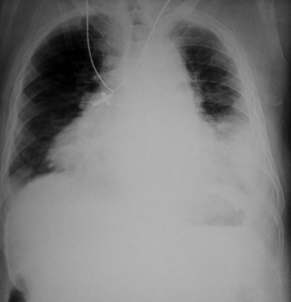
On admission the patient was pale, with dilated jugular veins, abdominal respiration with tachypnea (30 breaths/min), tachycardia (150 beats/min), and pulsus paradoxus (20 mmHg), while his blood pressure was with in normal range (100/60 mmHg). At auscultation, heart sounds were reduced and gallop rhythm was present; no murmurs or pericardial rub were audible. The chest examination revealed bilateral expiratory wheezing, that was attributed to the coexisting COPD. The abdomen was distended and the liver was palpable approximately 1 cm subcostally. The admission ECG showed sinus tachycardia with reduction of the amplitude of the QRS complexes and ST-segment depression in leads V4 to V6 (<1 mm). Chest X-ray demonstrated an enlarged cardiac silhouette, a small left lung lesion accompanied by a moderate pleural effusion on the left hemithorax and a minor one on the right, and elevation of the right dome of the diaphragm due to a calcified hepatic lesion (Figure 1). Arterial blood gas measurement revealed acute on chronic respiratory acidosis. Echocardiography revealed a significant pericardial effusion with compression of the right heart chambers. Due to his critical condition, an emergency pericardiocentesis was performed at the time of the admission. A quantity of 1100 cc of exudative bilious stained fluid was drained and his condition improved rapidly. Pericardial fluid showed total protein to be 2.2 g/dL, total bilirubin 4.7 mg/dL, indirect bilirubin 4.5 mg/dL and LDH 209 U/L, with absence of hemoglobin.
Figure 1.
The patient's admission chest x-ray demonstrating an enlarged cardiac silhouette, evidence of bilateral pleural effusions, more prominent on the left hemithorax, and a left lung lesion. An elevation of the right dome of the diaphragm due to the presence of a calcified subdiaphragmatic lesion can also be seen.
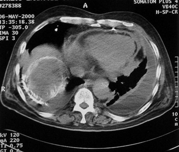
Chest CT-scan revealed a central mass of the left lung that was not prominent on the chest X-ray, a second smaller mass located in the left interlobar fissure, segmental atelectasis of the left upper lobe, a moderate pleural effusion on the left hemithorax and a small one on the right, a residual pericardial effusion, and a typical 9-cm hydatid cyst with calcified wall, located sub-diaphragmatically on the right lobe of the liver, in close contact with the diaphragm and the pericardial cavity (Figure 2).
Figure 2.

Chest CT-scan following pericardial drainage demonstrating a calcified hydatid cyst on the dome of the liver that was in close contact with the pericardial cavity. Additional findings included a bilateral pleural effusion (more prominent on the left) and the residual pericardial effusion.
A chest tube was inserted in the left hemithorax and 500 cc of sanguineous exudative fluid were consequently drained. In the complete blood count, white blood cells were 26,800/mm3 with marked neutrophilia (91.8%), whereas serum biochemistry values included glucose 234 mg/dL (normal range 70–110 mg/dL), total bilirubin 2.3 mg/dL (0–1 mg/dL), direct bilirubin 1.3 mg/dL, LDH 189 U/L (range 89–221 U/L), ALT 84 U/L (range 5–49 U/L), γ-GT 96 U/L (range 0–53 U/L), CK 311 U/L (range 26–174 U/L) and CK-MB 33 U/L (range 0–24 U/L). The titer of anti-echinococcal antibodies was negative (Elisa, bioMerieux®, titer of negative control <1:100). Culture of fluid from the left pleural and the pericardial effusion for common pathogens and Mycobacterium tuberculosis were negative. However, the cytology of the pericardial effusion was positive for non-small cell lung cancer (NSCLC), whereas the left pleural effusion was suspected for malignancy. Total bilirubin levels of the pericardial fluid were within normal range (0.5 mg/dL) after the third post-drainage day. Furthermore, no bilirubin was detected in the pleural fluid drained.
The chest tube was removed six days later after a successful slurry talk pleurodesis. A proper percutaneous subxiphoid pericardiotomy under local anaesthesia was performed one day later. Numerous local infiltrations on the pericardium and the myocardium were observed. Initially, drainage of 500–600 cc of fluid per day was observed, but with a diminishing trend. Pericardiodesis with bleomycin was performed and the pericardial drain was removed on the 10th post-pericardiotomy day. Histological examination of biopsies obtained from the resected pericardium revealed a metastatic poorly differentiated lung adenocarcinoma.
The patient was subsequently submitted to chemotherapy, but died 6 weeks later. During the follow-up no recurrent effusion was detected, clinically or radiologically. Unfortunately, the severe clinical condition of our patient did not allow us to investigate further the mechanism of pericardio-biliary communication. Postmortem examination was also not carried out.
Discussion
Cardiac tamponade may be the initial and potentially life-threatening manifestation of metastatic cancer. Malignant infiltrations of the myocardium are a rare consequence. Adenocarcinoma of the breast and lung are the most frequent causes of metastatic pericarditis [3,4]. Hepatic hydatid cyst is the most frequent presentation of hydatidosis. The diagnosis is mainly established by a CT-scan, due to its typical three-layered structure and the ring like, polycyclic calcification [5]. Immuno-antigenic anti-echinococcal titres may be low when the cysts contain clear liquid, or when they are calcified, as was the case in our patient [6].
Thoracic complications associated with a hepatic hydatid cyst include erosion and migration through the diaphragm into the thoracic cavity, leading to pleural hydatidosis, broncho-biliary fistula, pleural empyema, and pulmonary abscess [7]. Broncho-biliary fistula is an uncommon manifestation, occurring approximately in 2% of the cases of hepatic echinococcosis [8]. The mechanism of formation of a thoraco-biliary fistula probably involves the erosion of the diaphragm and the disruption of the biliary tree, due to the pericystic inflammation and the infiltration of the pleura. When the cyst crosses the diaphragm, the possibility of rupture into the great vessels or the pericardium increases [9].
In our case, the interesting finding was the presence of bile only in the large pericardial effusion, and its absence from the synchronous pleural effusion. Although the exact route of communication between the pericardium and the biliary tract in our patient has not been clarified, it is most likely that a mechanism similar to that described above may have resulted to the creation of a pericardio-biliary fistula, as it has previously been described by Song et al, [10]. The alternative of an esophago-pericardial communication was clinically and radiologically excluded from the absence of an air-fluid level in the pericardial cavity, both in the chest X-rays and the CT scans.
In summary, we report a case of non-small cell lung cancer with a coexisting hepatic hydatid cyst, presenting as a bilious pericardial effusion. The probable mechanism involves the formation of a pericardio-biliary fistula. This rare initial presentation of lung carcinoma is, to the best of our knowledge, the first to be reported in the literature.
Contributor Information
Christophoros S Kotoulas, Email: chrkotoulas@hol.gr.
Christophoros Foroulis, Email: foroulis@internet.gr.
Konstantinos Letsas, Email: k.letsas@mail.gr.
Konstantinos Kostikas, Email: ktk@otenet.gr.
Marios Konstantinou, Email: markonstantinou@yahoo.gr.
References
- Frazer RS, Viloria JB, Wang N. Cardiac tamponade as a presentation of extracardiac malignancy. Cancer. 1980;45:1697–1704. doi: 10.1002/1097-0142(19800401)45:7<1697::aid-cncr2820450730>3.0.co;2-j. [DOI] [PubMed] [Google Scholar]
- Johnson MM, Chin R, Jr, Haponik EF. Thoracobiliary fistula. South Med J. 1996;89:335–339. doi: 10.1097/00007611-199603000-00016. [DOI] [PubMed] [Google Scholar]
- Haskell RJ, French WJ. Cardiac tamponade as the initial presentation of malignancy. Chest. 1985;88:70–73. doi: 10.1378/chest.88.1.70. [DOI] [PubMed] [Google Scholar]
- DeLoach JF, Haynes JW. Secondary tumors of the heart and pericardium; review of the subject and report of one hundred thirty-seven cases. Arch Intern Med. 1953;91:224–249. doi: 10.1001/archinte.1953.00240140084007. [DOI] [PubMed] [Google Scholar]
- Wegener OH. The liver. In: Wegener OH, editor. In Whole Body Computed Tomography. 2. Boston: Blackwell SP; 1992. p. 271. [Google Scholar]
- Kardaras F, Kardara D, Tselikos D, Tsoukas A, Exadactylos N, Anagnostopoulou M, Lolas C, Anthopoulos L. Fifteen-year surveillance of echinococcal heart disease from a referral hospital in Greece. Eur Heart J. 1996;17:1265–1270. doi: 10.1093/oxfordjournals.eurheartj.a015045. [DOI] [PubMed] [Google Scholar]
- Kabiri EH, Maslout AE, Benosman A. Thoracic rupture of hepatic hydatidosis (123 cases) Ann Thorac Surg. 2001;72:1883–1886. doi: 10.1016/S0003-4975(01)03204-0. [DOI] [PubMed] [Google Scholar]
- Mazziotti S, Gaeta M, Blandino A, Barone M, Salamone I. Hepatobronchial fistula due to transphrenic migration of hepatic echinococcosis: MR demonstration. Abdom Imaging. 2000;25:497–499. doi: 10.1007/s002610000080. [DOI] [PubMed] [Google Scholar]
- Moumen M, El Farres F. Les fistules bilio-bronchiques d'origine hydatique: a propos de 8 cas. J Chir. 1991;128:188–192. [PubMed] [Google Scholar]
- Song ZL. Cholangiothoracic fistulae. Zhonghua Wai Ke Za Zhi. 1989;27:269–71. [PubMed] [Google Scholar]


