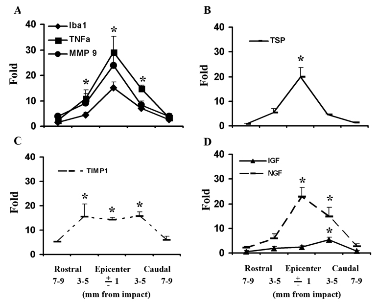
Figure 10.
Patterns of gene expression in 2 mm thick coronal tissue segments from a 2 cm length of cord centered on the injury epicenter at 3 days after SCI. (A) There was significantly higher expression of Iba1, TNFα, MMP9 at the epicenter as well as at 3–5 mm rostral and caudal to it, compared with the uninjured tissue. (B) TSP was only significantly higher than control at the epicenter. (C) TIMP1 was more broadly up-regulated with similar high expression at the epicenter and 3–5 mm rostral and caudal to it. (D) The expression of IGF was only significantly altered at 3–5 mm caudal to epicenter, while NGF mRNA was found significantly greater at the epicenter as well as 3–5 mm caudal to it. N = 5. *Significantly different from the uninjured spinal cord.

