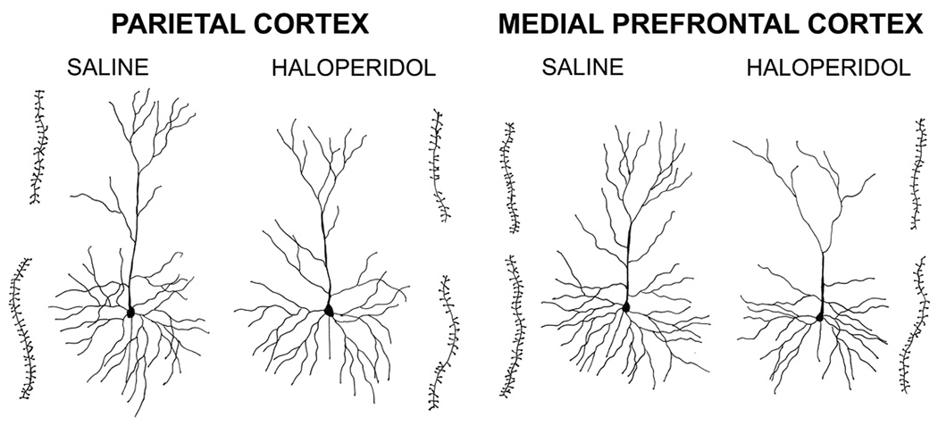
Fig. 1.
Camera lucida drawings of typical pyramidal cells in the parietal and medial prefrontal cortices of mice treated with saline or haloperidol on P3-10. The insets immediately adjacent to each cell are drawings of terminal apical (upper) or third order basal (lower) dendritic segments that were used to calculate spine densities. The cells shown were selected because they were close to the group averages for our 3 measures of dendritic form.

