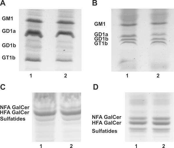
Figure 2. Deletion of Ugcg in oligodendrocytes does not affect the ganglioside profile of PND 18 Ugcgflox/flox;Cnp/Cre brains.
(A) The predominant brain gangliosides, GM1, GD1a, GD1b, and GT1b, were detected in the total brain lipid extracts of wild-type (lane1) and Ugcgflox/flox;Cnp/Cre mice (lane 2). (B) The same pattern of gangliosides was observed in myelin lipid extracts of wild-type (lane 1) and mutant brains (lane 2). (C) The neutral lipid fraction was isolated from brains of wild-type (lane1) and Ugcgflox/flox;Cnp/Cre mice (lane 2). HFA-GalCer, NFA-containing GalCer, and sulfatides were detected in both mutant and wild-type mice. (D) The presence of HFA-GalCer, NFA-GalCer, and sulfatides was also observed in the myelin of wild-type (lane 1) and Ugcgflox/flox;Cnp/Cre mice (lane 2).

