Background
The most common type of nasopharyngeal tumor is nasopharyngeal carcinoma. The etiology is multifactorial with race, genetics, environment and Epstein-Barr virus (EBV) all playing a role. While rare in Caucasian populations, it is one of the most frequent nasopharyngeal cancers in Chinese, and has endemic clusters in Alaskan Eskimos, Indians, and Aleuts. Interestingly, as native-born Chinese migrate, the incidence diminishes in successive generations, although still higher than the native population.
EBV is nearly always present in NPC, indicating an oncogenic role. There are raised antibodies, higher titers of IgA in patients with bulky (large) tumors, EBERs (EBV encoded early RNAs) in nearly all tumor cells, and episomal clonal expansion (meaning the virus entered the tumor cell before clonal expansion). Consequently, the viral titer can be used to monitor therapy or possibly as a diagnostic tool in the evaluation of patients who present with a metastasis from an unknown primary.
The effect of environmental carcinogens, especially those which contain a high levels of volatile nitrosamines are also important in the etiology of NPC. Chinese eat salted fish, specifically Cantonese-style salted fish, and especially during early life. Perhaps early life (weaning period) exposure is important in the “two-hit” hypothesis of cancer development.
Smoking, cooking, and working under poor ventilation, the use of nasal oils and balms for nose and throat problems, and the use of herbal medicines have also been implicated but are in need of further verification. Likewise, chemical fumes, dusts, formaldehyde exposure, and radiation have all been implicated in this complicated disorder.
Various human leukocyte antigens (HLA) are also important etiologic or prognostic indicators in NPC. While histocompatibility profiles of HLA-A2, HLA-B17 and HLA-Bw46 show increased risk for developing NPC, there is variable expression depending on whether they occur alone or jointly, further conferring a variable prognosis (B17 is associated with a poor and A2B13 with a good prognosis, respectively).
Clinical Features
Nasopharyngeal carcinoma (NPC) is primarily a tumor of adults with a peak occurrence between 40 and 60 years, although the tumor can occur in children. Curiously, African children are more commonly affected than Chinese children. There are about 65,000 new cases each year, and about 38,000 deaths. There is a strong male to female ratio of about 3:1, irrespective of geographic location.
Most patients present with an asymptomatic cervical mass (typically in the apex of the posterior cervical triangle or in the superior jugular chain of nodes), serous otitis media, epistaxis, or nasal obstruction. A blood-tinged post-nasal drip is also seen. Endoscopic evaluation of the upper aerodigestive tract with gross lesion biopsy and random biopsies of the lateral, superior, and posterior walls of the nasopharynx for patients with a high suspicion of NPC is standard of care. Interestingly, 12% of patients with dermatomyositis have NPC although only 1% of patients with NPC have dermatomyositis—limited in general to endemic areas.
Magnetic resonance (MR) is superior to CT in demonstrating extent of disease and defining the borders of the process. Work-up for metastatic tumor is also suggested by a variety of radiographic and laboratory studies. As already suggested, about 90% of patients have EBV positive serologies, with IgA against the viral capsid antigen (VCA) and early antigens (EA) used most commonly. Other tests used in combination show promise but require further evaluation.
Pathology Features
The world health organization (WHO) has proposed a number of different classifications, the most recent published in 2005 (Table 1). The definition is as follows: “A carcinoma arising in the nasopharyngeal mucosa that shows light microscopic or ultrastructural evidence of squamous differentiation.” It encompasses squamous cell carcinoma, non-keratinizing carcinoma (differentiated or undifferentiated) and basaloid squamous cell carcinoma. Adenocarcinoma and salivary gland-type carcinoma are excluded.
Table 1.
Classification of nasopharyngeal carcinoma
| World Health Organization (1978) | |
| 1. Squamous cell carcinoma | |
| 2. Non-keratinizing carcinoma | |
| 3. Undifferentiated carcinoma | |
| World Health Organization (1991) | |
| 1. Squamous cell carcinoma | |
| 2. Non-keratinizing carcinoma | |
| A. Differentiated non-keratinizing carcinoma | |
| B. Undifferentiated carcinoma | |
| World Health Organization (2005) | |
| 1. Keratinizing squamous cell carcinoma | 8071/3 |
| 2. Non-keratinizing carcinoma | 8072/3 |
| A. Differentiated type | |
| B. Undifferentiated type | |
| 3. Basaloid squamous cell carcinoma | 8083/3 |
It encompasses squamous cell carcinoma, non-keratinizing carcinoma (differentiated or undifferentiated) and basaloid squamous cell carcinoma. Adenocarcinoma and salivary gland-type carcinoma are excluded.
A host of synonyms have been applied in the past, including lymphoepithelioma, lymphoepithelial carcinoma, and anaplastic carcinoma, to name just a few. However, using the anatomic and histologic moniker of “Nasopharyngeal Carcinoma” with subclassification into squamous, non-keratinizing, and basaloid squamous cell will yield the most useful classification system to pathologists, surgeon’s and oncologists/radiologists alike.
Macroscopic
Most tumor arise on the lateral wall of the nasopharynx, especially common in the fossa of Rosenmüller. Most tumors are exophytic (about 75%), with a few described as ulcerated (about 10%). It is usually a smooth, discrete raised nodule below the mucosa. Cervical lymph node metastasis is also common.
Microscopic
Tumors are separated into three major categories with two variants. This is different from the original WHO 1, 2, and 3 (from 1978) and from the 1991 modification. The separation boundaries between the categories and subtypes/variants is not always clear, sampling error can be a significant problem with small biopsies, and there is considerable intra- and inter-observer reproducibility variability.
In-situ disease has been described, but is quite uncommon, suggesting a rapid development of invasive tumor or origin from deeper epithelium.
The term “squamous cell carcinoma” is applied to tumors showing obvious squamous differentiation at the light microscopic level (intercellular bridges, keratinization) in the majority of the tumor (Fig. 1). It can be graded as well, moderately, or poorly differentiated. The tumor grows in irregular islands separated by desmoplastic stroma and with variable numbers of lymphocytes, plasma cells, neutrophils, and eosinophils. The cell borders are distinct, separated by intercellular bridges. Areas of keratinization are seen. The nuclei are usually hyperchromatic. If the tumor is radiation induced there is no EBV association. However, if no radiation association, they tend to have lower copy numbers of EBV and the nuclear signal is usually only identified in the basaloid/less differentiated cells.
Fig. 1.
This squamous cell carcinoma shows prominent intercellular bridges, keratinization and an infiltrative growth pattern. The tumor is classified as keratinizing squamous cell carcinoma
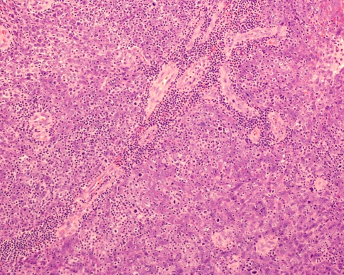
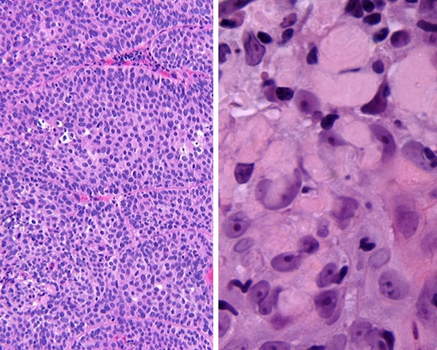
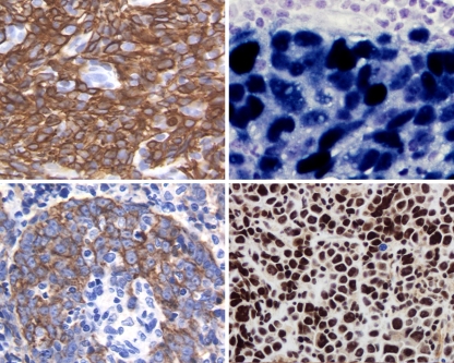
Non-keratinizing carcinoma is the classic NPC. There are two types: differentiated and undifferentiated. There are solid sheets of syncytial appearing, large tumor cells arranged in irregular islands and trabeculae of carcinoma, intimately associated and intermingled with inflammatory elements (Fig. 2). There is often cellular overlap. The nuclear chromatin is vesicular, accentuating the prominent nucleoli (Fig. 3). There is a high nuclear to cytoplasmic ratio with amphophilic cytoplasm. Sometimes the tumor may have areas reminiscent of transitional epithelium (bladder carcinoma-like; Fig. 4). A desmoplastic stroma is uncommon. When the lymphoid component is dominant the “lymphoepithelioma” concept is brought to mind. Well defined epithelial islands are termed Regaud pattern, while individual cells in ill-defined sheets are called “Schmincke pattern.” Granulomatous response to the tumor may be the dominant finding in a few cases, while a heavy eosinophilic infiltrate can simulate Hodgkin lymphoma. Occasionally, amyloid globules can be seen within the tumor (intracellularly) derived from keratin-type intermediate filaments (Fig. 4). Keratin is strongly and diffusely immunoreactive in this variant, specifically CK5/6 and 34βE12 (Fig. 5). CK7 & CK20 are negative. When the lymphoid population is dominant, the thin wisps of cytoplasm reacting with the keratin create a “meshwork” pattern. The lymphoid population is polytypic with B- and T-cell markers. S100 protein cells can form a background dendritic cell network.
Fig. 2.
Sheets of epithelial cells show a syncytial architecture with lymphocytes intimately associated with the neoplastic cells
Fig. 3.
This quartet of non-keratinizing carcinoma show a spectrum of cytologic features. The nuclei range from vesicular to lightly granular. Nucleoli are prominent. There is a high nuclear to cytoplasmic ratio. Inflammatory cells are identified within the syncytium of tumor cells
Fig. 4.
Left: Sheets of monotonous cells simulate a “transitional” epithelium seen in the “differentiated” non-keratinizing carcinoma. Right: Amyloid globules within the cytoplasm of neoplastic cells
Fig. 5.
Left: Keratin CK5/6 (upper) and 34βE12 (K903, lower) accentuated the neoplastic cells, showing wisps of cytoplasmic extensions. Right: EBER technique showing strong and diffuse reactivity in the nuclei of the tumor cells
Trying to separate this non-keratinizing category into differentiated and undifferentiated is arbitrary and difficult to achieve and of no clinical or prognostic significance at this time. There is much overlap both within a single tumor and in the same tumor over time that it is impractical to try and separate these lesions further.
Basaloid squamous cell carcinoma contains a basaloid neoplasm with focal areas of squamous differentiation, squamous cell carcinoma in situ or invasive squamous cell carcinoma. There are only a few reported cases, making a meaningful discussion of limited clinical value.
Special Studies
EBV is usually found in the non-keratinizing type rather than the keratinizing variant of NPC. In-situ hybridization or polymerase chain reaction (PCR) is generally needed to document the EBV, since the EBV-LMP is not sufficiently reliable to make this determination (positive in only about 40% of cases). The EBV encoded early RNA (EBER) is the most sensitive and specific analysis available at present (Fig. 5), although a number of other tests show promise.
Inactivation of the p16 tumor suppressor gene on 9p21 by homozygous deletion and methylation has been shown to be the most common molecular alteration in NPC tumorigenesis. There are a whole host of other genetic alterations, too incompletely studied to be clinical significant at this time.
FNA of an enlarged lymph node can help with initial diagnosis or staging. Usually there are irregular clusters of large cells with overlapping vesicular nuclei and large nucleoli. Others have used nasopharyngeal aspirates or brush samples, but they are not as sensitive as biopsy.
Differential Diagnosis
Crush artifacts are common, mandating careful evaluation of better preserved areas. Keratin staining will help in uncertain cases. Keratinizing squamous cell carcinoma is not difficult to separate from other lesions. However, what percent of the tumor is squamous versus “undifferentiated” before it is placed in a different category is still open to discussion. In radiation cases, the stromal atypia and cytologic atypia within the epithelium may make reactive versus neoplastic separation more difficult.
A non-keratinizing carcinoma has a much broader differential diagnosis and includes melanoma, rhabdomyosarcoma, lymphoma, olfactory neuroblastoma, Ewing sarcoma and primitive neuroectodermal tumors. In fact, even floridly reactive germinal centers can sometimes contain large vesicular nuclei and lack a well-defined mantle zone. Plump endothelial cells in reactive lymphoid aggregates can also have vesicular nuclei which can be confused with NPC. These cells usually do not contain nucleoli and are negative for keratin. Sinonasal undifferentiated carcinoma (SNUC) is a completely separate tumor based on location and pattern of growth (Table 2). The differential considerations can often be confirmed with a pertinent immunohistochemistry panel.
Table 2.
Non-keratinizing carcinoma versus sinonasal undifferentiated carcinoma
| Feature | Non-keratinizing carcinoma | Sinonasal undifferentiated carcinoma |
|---|---|---|
| Growth | Syncytial | Trabecular |
| Cytology | Large, vesicular nuclei with prominent nucleoli | Medium sized nuclei with inconspicuous nucleoli |
| Necrosis | Limited | Prominent |
| Mitoses | Uncommon | Frequent |
| Vascular invasion | Rare | Very common |
| Lymphocytes | Yes | No |
| EBV | Yes | No |
| Location | Nasopharynx | Sinonasal tract |
| Clinical | Small primary and lymph node metastases | Large primary with ± lymph node metastases |
| Radiographic | Little destruction/spread | Marked destruction and spread |
| CK 5/6 & CK13 | Positive | Negative |
Prognosis and Management
Because of the strategic location of the nasopharynx and the tendency for the tumor to invade surrounding tissues, the first line of therapy for NPC is irradiation. Surgery, if used at all, is reserved for radioresistant and/or locally recurrent tumors. These tumors are highly malignant with extensive and early lymphatic spread (due to a rich lymphatic plexus) and a high incidence of hematogenous spread. Direct extension into the base of the skull, paranasal sinuses, orbit and basal foramina is common. About 50% of patients will have lymph node metastasis at presentation. Specifically, the jugulo-digastric node and the posterior cervical chain is more frequently affected than by any other head and neck cancer. As the local and regional disease can be adequately managed by radiotherapy, the role of chemotherapy is usually reserved for disseminated disease. The most common sites are bone, lung, and liver. If metastasis is going to develop, it usually does so within 3 years of initial presentation.
If re-assessment of the nasopharynx is performed post-radiation therapy, at least a 10–12 week interval should have passed before obtaining as biopsy. Radiation changes can sometimes be difficult to separate from residual tumor. Usually, the radiation changes are limited to a few cells and there is a normal N:C ratio. These changes in the mucosa are usually gone within a year. If there is a positive EBER, it favors recurrent/residual tumor. The radiation changes in the fibroblasts can last for years and should not be over interpreted as spindled carcinoma.
The current TNM is a better staging system than past models, with an overall 5-year survival in the United States of about 40–80% (dependent on endemic versus sporadic tumor). Squamous cell carcinoma has a lower survival (20–40%) than undifferentiated (65%), although basaloid squamous has the lowest survival (<10%). A better prognosis is seen in young patients (<40 years) and women. Poor prognostic indicators include advanced clinical stage, cranial nerve involvement, keratinizing histology and an absence of EBV. EBV titers have been correlated with survival when EBV is present, with serum EBV DNA showing promise in monitoring disease status. Keratinizing squamous cell carcinoma has the greatest propensity for locally advanced tumor growth, but a lower chance of metastasis than other types.
Bibliography
- 1.Akao I, Sato Y, Mukai K, et al. Detection of Epstein-Barr virus DNA in formalin-fixed paraffin-embedded tissue of nasopharyngeal carcinoma using polymerase chain reaction and in-situ hybridization. Laryngoscope. 1991;101:279–83. doi: 10.1288/00005537-199103000-00010. [DOI] [PubMed] [Google Scholar]
- 2.Buell P. The effect of migration on the risk of nasopharyngeal cancer among Chinese. Cancer Res. 1974;34:1189–91. [PubMed] [Google Scholar]
- 3.Burt RD, Vauhn TL, McKnight B, et al. Associations between human leukocyte antigen type and nasopharyngeal carcinoma in Caucasians in the United States. Cancer Epidemiol Prevent. 1995;5:879–87. [PubMed] [Google Scholar]
- 4.Carbone A, Micheau C. Pitfalls in microscopic diagnosis of undifferentiated carcinoma of nasopharyngeal type (lymphoepithelioma) Cancer. 1982;50:1344–51. doi: 10.1002/1097-0142(19821001)50:7<1344::AID-CNCR2820500721>3.0.CO;2-O. [DOI] [PubMed] [Google Scholar]
- 5.Chan ATC, Teo PML, Leung TWT, et al. The role of chemotherapy in the management of nasopharyngeal carcinoma. Cancer. 1998;82:1003–12. doi: 10.1002/(SICI)1097-0142(19980315)82:6<1003::AID-CNCR1>3.0.CO;2-F. [DOI] [PubMed] [Google Scholar]
- 6.Chan JKC, Bray F, McCarron P, Foo W, Lee AWM, Yip T, Kuo TT, Pilch BZ, Wenig BM, Huang D, Lo KW, Zeng YX, Jia WH. Nasopharyngeal carcinoma. In: Barnes EL, Eveson JW, Reichart P, Sidransky D, editors. Pathology and genetics of head and neck tumours. Kleihues P, Sobin LH, series editors. World Health Organization Classification of Tumours. Lyon, France: IARC Press, 2005:85–97.
- 7.Chua DTT, Sham JST, Kwong DLW, et al. Treatment outcome after radiotherapy alone for patients with stage I–II nasopharyngeal carcinoma. Cancer. 2003;98:74–80. doi: 10.1002/cncr.11485. [DOI] [PubMed] [Google Scholar]
- 8.Easton JM, Levine PH, Hyams VJ. Nasopharyngeal carcinoma in the United States. A pathologic study of 177 US and 30 foreign cases. Arch Otolaryngol. 1980;106:88–91. doi: 10.1001/archotol.1980.00790260020007. [DOI] [PubMed] [Google Scholar]
- 9.Franchi A, Moroni M, Mossi D, et al. Sinonasal undifferentiated carcinoma, nasopharyngeal-type undifferentiated carcinoma, and keratinizing and non-keratinizing squamous cell carcinoma express different cytokeratin patterns. Am J Surg Pathol. 2002;26:1597–604. doi: 10.1097/00000478-200212000-00007. [DOI] [PubMed] [Google Scholar]
- 10.Goldsmith DB, West TM, Morton R. HLA associations with nasopharyngeal carcinoma in Southern Chinese: a meta-analysis. Clin Otolaryngol. 2002;27:61–7. doi: 10.1046/j.0307-7772.2001.00529.x. [DOI] [PubMed] [Google Scholar]
- 11.Heng DMK, Wee J, Fong K-W, et al. Prognostic factors in 677 patients in Singapore with nondisseminated nasopharyngeal carcinoma. Cancer. 1999;86:1912–20. doi: 10.1002/(SICI)1097-0142(19991115)86:10<1912::AID-CNCR6>3.0.CO;2-S. [DOI] [PubMed] [Google Scholar]
- 12.Jeannel D, Hubert A, Vathaire F, et al. Diet, living conditions and nasopharyngeal carcinoma in Tunisia—a case-control study. Inst J Cancer. 1990;46:421–5. doi: 10.1002/ijc.2910460316. [DOI] [PubMed] [Google Scholar]
- 13.Jeng Y-M, Sung M-T, Fang C-L, et al. Sinonasal undifferentiated carcinoma and nasopharyngeal-type undifferentiated carcinoma. Two clinically, biologically, and histopathologically distinct entities. Am J Surg Pathol. 2002;26:371–376. doi: 10.1097/00000478-200203000-00012. [DOI] [PubMed] [Google Scholar]
- 14.Jenkin RDT, Anderson JR, Jereb B, et al. Nasopharyngeal carcinoma - a retrospective review of patients less than thirty years of age: A report from Childrens Cancer Study Group. Cancer. 1981;47:360–366. doi: 10.1002/1097-0142(19810115)47:2<360::AID-CNCR2820470225>3.0.CO;2-3. [DOI] [PubMed] [Google Scholar]
- 15.Lee AWM, Poon YF, Foo W, et al. Retrospective analysis of 5037 patients with nasopharyngeal carcinoma treated during 1976–1985. Overall survival and patterns of failure. Int J Radiat Oncol Biol Phys. 1992;23:261–70. doi: 10.1016/0360-3016(92)90740-9. [DOI] [PubMed] [Google Scholar]
- 16.Marks JE, Phillips JL, Menek HR. The National Cancer Data Base Report on the relationship of race and national origin to the histology of nasopharyngeal carcinoma. Cancer. 1998;83:582–8. doi: 10.1002/(SICI)1097-0142(19980801)83:3<582::AID-CNCR29>3.0.CO;2-R. [DOI] [PubMed] [Google Scholar]
- 17.Pak W, To KF, Lo YMD, et al. Nasopharyngeal carcinoma in-situ (NPCIS)—pathologic and clinical perspectives. Head Neck. 2002;24:989–95. doi: 10.1002/hed.10161. [DOI] [PubMed] [Google Scholar]
- 18.Pathmanathan R, Prasad U, Sadler R, et al. Clonal proliferation of cells infected with Epstein-Barr virus in preinvasive lesions related to nasopharyngeal carcinoma. N Engl J Med. 1995;333:693–8. doi: 10.1056/NEJM199509143331103. [DOI] [PubMed] [Google Scholar]
- 19.Reddy SP, Raslan WF, Gooneratne S, et al. Prognostic significance of keratinization in nasopharyngeal carcinoma. Am J Otolaryngol. 1995;16:103–8. doi: 10.1016/0196-0709(95)90040-3. [DOI] [PubMed] [Google Scholar]
- 20.Sanmugaratnam K, Chan SH, de-The G, et al. Histopathology of nasopharyngeal carcinoma. Correlations with epidemiology, survival rates, and other biological characteristics. Cancer. 1979;44:1029–44. doi: 10.1002/1097-0142(197909)44:3<1029::AID-CNCR2820440335>3.0.CO;2-5. [DOI] [PubMed] [Google Scholar]
- 21.Tai S-T, Jin Y-T, Su I-J. Expression of EBER1 in primary and metastatic nasopharyngeal carcinoma tissues uing in situ hybridization. A correlation with WHO subtypes. Cancer. 1996;77:231–6. doi: 10.1002/(SICI)1097-0142(19960115)77:2<231::AID-CNCR2>3.0.CO;2-P. [DOI] [PubMed] [Google Scholar]
- 22.Thompson LDR. Malignant neoplasms of the nasal cavity, paranasal sinuses and nasopharynx. In: Thompson LDR, editor. Head and neck pathology. Goldblum JR, series editor. Foundations in Diagnostic Pathology. Vol 3. Philadelphia: Churchill Livingstone Elsevier, 2006:155–213.
- 23.Vaughan TL, Shapiro JA, Burt RD, et al. Nasopharyngeal cancer in a low risk population: Defining risk factors by histological type. Cancer Epidemiol. 1996;5:587–93. [PubMed] [Google Scholar]
- 24.Wei WI. Nasopharyngeal cancer: current status of management. Arch Otolaryngol Head Neck Surg. 2001;127:766–9. [PubMed] [Google Scholar]
- 25.Yu MC, Ho JH, Lai S-H, et al. Cantonese-style salted fish as a cause of nasopharyngeal carcinoma. Report of a case-control study in Hong Kong. Cancer Research. 1986;46:956–61. [PubMed] [Google Scholar]


