Abstract
The follicular variant of the adenomatoid odontogenic tumor (AOT) is thought to originate from the reduced enamel epithelium of the dental follicle. The origin of the extra-follicular variant however, remains less clear. This paper presents a case of an extra-follicular AOT, which we believe originated from the epithelial lining of a unicystic ameloblastoma, and reviews the literature. The available evidence seems to indicate that some extra-follicular AOTs might arise as secondary phenomena within pre-existing odontogenic cysts or cystic tumors.
Keywords: Adenomatoid odontogenic tumor, Extra-follicular, Unicystic ameloblastoma
Most adenomatoid odontogenic tumors (AOTs) occur intra-osseously. They surround the crowns and are attached to the necks of unerupted teeth in a true follicular relationship. Some however have no association with unerupted teeth and a few occur on the gingiva in an extra-osseous location [1, 2]. For the follicular variant there is histologic and immunohistochemical evidence that they originate from the reduced enamel epithelium (REE) of the dental follicle [3].
Origin of the extra-follicular variant is less clear. Philipsen et al. [2] argue that the fact that all AOT variants show identical histology, points towards a common origin and implicate the dental lamina or its remains.
The aim of this paper is to present a case of an extra-follicular AOT, which we believe originated in the wall of a unicystic ameloblastoma, to review the literature and to propose that some extra-follicular AOTs might arise as a secondary phenomenon within pre-existing odontogenic cysts or cystic tumors.
Case Report
The patient was a 40-year old male with a 7-month history of a bony hard swelling of the anterior mandible. There was no history of trauma. The swelling extended from the second bicuspid on the right to the first molar on the left with eggshell crackling noted anteriorly. There was both buccal and lingual expansion.
Radiographic examination showed a well-defined radiolucent lesion extending from tooth #19 to tooth #28 with root resorption of the incisor teeth.
The surgical specimen comprised a cystic lesion measuring 35 × 20 × 8 mm with a wall thickness of 3–4 mm.
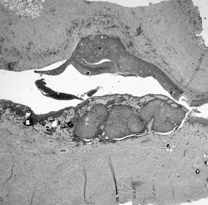
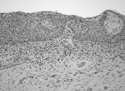
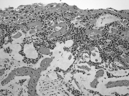
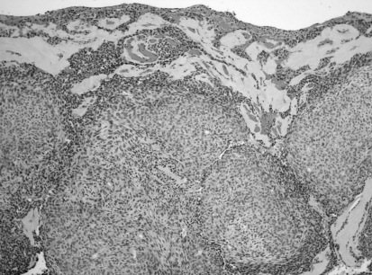
Sections showed the presence of a cystic lesion consisting of a single locule that was lined for the most part by hyperplastic, non-keratinising stratified squamous epithelium (Figs. 1, 2). In areas the squamous epithelial lining gave origin to clusters and strands of smaller cuboidal cells arranged in a lace-like pattern with associated hyaline material and surrounded by a vascular stroma (Fig. 3). This epithelium was continuous with nodules of fusiform odontogenic epithelial cells showing rosette formation, small pseudo-glandular structures and eosinophilic hyaline globules (Fig. 4). The wall of the cyst contained many odontogenic cell rests and foci of dystrophic calcification.
Fig. 1.
The lesion consisted of a unilocular epithelial lined cyst with a mural nodule characteristic of AOT (H & E × 64)
Fig. 2.
The cyst was lined by a hyperplastic non-keratinising stratified squamous epithelium (H & E × 248)
Fig. 3.
Strands of odontogenic epithelium originating from the cyst lining and extending into the underlying vascular stroma (H & E × 248)
Fig. 4.
Nodules of fusiform cells with rosette formation, pseudo-glandular structures and hyaline globules characteristic of AOT (H & E × 124)
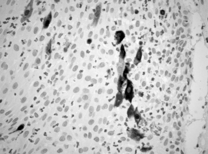
Calretinin immunohistochemistry showed positive nuclear and cytoplasmic staining of both single and clusters of epithelial cells in the inflamed cyst lining characteristic of ameloblastoma (Fig. 5).
Fig. 5.
Immunohistochemistry showing focal positive staining of epithelial lining cells with Calretinin anti-serum (Calretinin × 248)
The sign out diagnosis was that of an adenomatoid odontogenic tumor originating within a unicystic ameloblastoma.
Discussion
The hypothesis that follicular AOTs arise from the REE which lines the follicles of unerupted teeth is fairly conclusive and is supported by evidence that is both morphological and immunocytochemical in nature [3]. They surround the crowns and are attached to the necks of unerupted teeth in a true follicular relationship. Many present as cystic lesions with only mural nodules of AOT lesional tissue and in some instances origin of the lesional tissue from the REE can be demonstrated histologically. Immunohistochemically, it has been shown that the immuno-phenotypic profile of REE and AOT epithelium is virtually identical, further supporting a common derivative [3].
Whether origin of the follicular variant occurs before or after cystic expansion has taken place is open to conjecture. If it occurs after cystic expansion then this effectively means origin from a dentigerous cyst and several such case reports have been published [4–6].
If it occurs before cystic expansion then the tumor tissue will fill the follicular space and the AOT will present as a solid tumor. It is reasonable to assume that given enough time even those originating from a cyst may grow and fill the lumen completely.
When considering the question of the possible origin of AOTs from an ameloblastoma, cognisance must be taken of the fact that some lesions may have features of both tumors and pathologists can be divided as to the most likely diagnosis. This is the case in the so-called ‘adenoid ameloblastoma with dentinoid’. Matsumoto et al. [7] reported such a case and added another 11 cases from the literature. In six cases the sign out diagnosis was AOT while in the remaining six cases it was ameloblastoma. There is little doubt that the clinico-pathological features of these lesions are those of ameloblastoma.
Two cases of unicystic ameloblastoma associated with AOTs have been published by Raubenheimer et al. [8]. These cases were not described in detail but formed part of a study, which demonstrated the varying differentiation potential of epithelium in the ameloblastoma. They both occurred in the anterior mandible.
A report of an AOT originating in the wall of a calcifying odontogenic cyst (COC) has been published by Zeitoun et al. [9]. They showed transformation of the epithelial lining of a COC to form an AOT. The lesion presented as a large cyst extending from the left to the right bicuspid regions in the mandible. It had caused resorption of the associated teeth and radiographically had a scalloped margin.
Several cases of so-called ‘combined AOT and calcifying epithelial odontogenic tumor’ have been published [10] but it is now widely agreed that these are not true hybrid lesions but represent a histologic variation within an AOT [11].
Several of the cases previously reported as being extra-follicular AOTs which were large and sometimes showed aggressive behavior and which occurred in the mandibular second or third molar region are now regarded as a specific odontogenic tumor. Some of these, Vargas and colleagues [12] have termed adenomatoid odontogenic hamartomas. This includes the four cases described by Allen et al. [13] as adenomatoid dentinomas as well as the lesions reported by Dunlap and Fritzlen [14], Orlowski et al. [15] and Tajima et al. [16].
A report of an unusually large AOT occurring in the anterior mandible was published by Geist and Mallon [17]. The lesion had caused expansion and resorption of the lower border of the mandible and displacement of the anterior teeth and extended across the midline. The canine was contained within the lesion. Although conclusive evidence is not provided the clinical and radiographic presentation of this lesion could be consistent with a unicystic ameloblastoma.
A further very unusual case, which could represent an AOT developing in an ameloblastoma, was reported by Nomura et al. [18]. This lesion occurred in a 64 year old Japanese man who exhibited a large destructive lesion in the mandible, which had proliferated intra-orally into the overlying gingivae. Although the histology showed features of an AOT, in the periphery of the epithelial sheets the tumor cells differentiated into columnar cells similar to ameloblasts. An extensive amount of osteodentine or cementum-like material was present at the periphery. The lesion showed aggressive clinical and radiographic behavior, uncharacteristic of AOT.
Similarly an aggressive AOT which recurred four times and which extended to the base of the skull was published by Takigami et al. [19]. Again the diagnosis of AOT must be questioned.
In the case we are reporting the clinical and radiographic features of a large uni-locular lesion, which had caused both buccal and lingual expansion and root resorption of the associated teeth, were characteristic of unicystic ameloblastoma. The histology although not diagnostic, was consistent with this diagnosis. Confirmation of the diagnosis of ameloblastoma was provided by the calretinin immunohistochemistry, which showed a characteristic pattern of positive staining of the cyst lining [20, 21]. Previous unpublished work in our department has shown that AOTs do not stain with calretinin neither in the solid areas nor in the cyst linings [22].
The available evidence seems to point towards the existence of a subset of extra-follicular AOTs which present in the symphyseal region or body of the mandible and have no association with an unerupted tooth, are aggressive in nature and cause root resorption and expansion. Histologically they focally show features of an AOT but the clinical presentation and behavior is inconsistent with such a diagnosis. We suggest that such lesions represent unicystic ameloblastomas with secondary AOT proliferation.
Acknowledgment
This study was supported by NHLS Research Trust No 93919.
References
- 1.Philipsen HP, Reichart PA, Zhang KH, Nikai H, Yu QX. Adenomatoid odontogenic tumor: biologic profile based on 499 cases. J Oral Pathol Med. 1991;20:149–158. doi: 10.1111/j.1600-0714.1991.tb00912.x. [DOI] [PubMed] [Google Scholar]
- 2.Philipsen HP, Samman N, Ormiston IW, Wu PC, Reichart PA. Variants of the adenomatoid odontogenic tumor with a note on tumor origin. J Oral Pathol Med. 1992;21:348–352. doi: 10.1111/j.1600-0714.1992.tb01363.x. [DOI] [PubMed] [Google Scholar]
- 3.Crivelini MM, Araújo VC, Sousa SOM, Araújo NS. Cytokeratins in epithelia of odontogenic neoplasms. Oral Dis. 2003;9:1–6. doi: 10.1034/j.1601-0825.2003.00861.x. [DOI] [PubMed] [Google Scholar]
- 4.Tajima Y, Sakamoto E, Yamamoto Y. Odontogenic cyst giving rise to an adenomatoid odontogenic tumor: report of a case with peculiar features. J Oral Maxillofac Surg. 1992;50:190–193. doi: 10.1016/0278-2391(92)90370-F. [DOI] [PubMed] [Google Scholar]
- 5.Garcia-Pola Vallejo M, Gonzalez Garcia M, Lopez-Arranz JS, Herrero Zapatero A. Adenomatoid odontogenic tumor arising in a dental cyst: report of unusual case. J Clin Pediatr Dent. 1998;23:55–58. [PubMed] [Google Scholar]
- 6.Takahashi H, Fujita S, Shibata Y, Yamaguchi A. Adenomatoid odontogenic tumour: immunohistochemical demonstration of transferrin, ferritin and alpha-one-antitrypsin. J Oral Pathol Med. 2001;30:237–244. doi: 10.1034/j.1600-0714.2001.300408.x. [DOI] [PubMed] [Google Scholar]
- 7.Matsumoto Y, Mizoue K, Seto K. Atypical plexiform ameloblastoma with dentinoid: adenoid ameloblastoma with dentinoid. J Oral Pathol Med. 2001;30:251–254. doi: 10.1034/j.1600-0714.2001.300410.x. [DOI] [PubMed] [Google Scholar]
- 8.Raubenheimer EJ, Heerden WF, Noffke CE. Infrequent clinicopathological findings in 108 ameloblastomas. J Oral Pathol Med. 1995;24:227–232. doi: 10.1111/j.1600-0714.1995.tb01172.x. [DOI] [PubMed] [Google Scholar]
- 9.Zeitoun IM, Dhanrajani PJ, Mosadomi HA. Adenomatoid odontogenic tumor arising in a calcifying odontogenic cyst. J Oral Maxillofac Surg. 1996;54:634–637. doi: 10.1016/S0278-2391(96)90650-3. [DOI] [PubMed] [Google Scholar]
- 10.Esquiche-Leon J, Martinez-Mata G, Rodriguez-Fregnani E, et al. Clinicopathological and immunohistochemical study of 39 cases of adenomatoid odontogenic tumour: a multicentric study. Oral Oncol. 2005;41:835–842. doi: 10.1016/j.oraloncology.2005.04.008. [DOI] [PubMed] [Google Scholar]
- 11.Mosqueda-Taylor A, Carlos-Bregni R, Ledesma-Montes C, Fillipi RZ, Almeida OP, Vargas PA. Calcifying epithelial odontogenic tumor-like areas are common findings in adenomatoid odontogenic tumors and not a specific entity. Oral Oncol. 2005;41:214–215. doi: 10.1016/j.oraloncology.2004.08.003. [DOI] [PubMed] [Google Scholar]
- 12.Vargas PA, Carlos-Bregni R, Mosqueda-Taylor A, Cuairan-Ruidiaz V, Lopes MA, Almeida OP. Adenomatoid dentinoma or adenomatoid odontogenic hamartoma: what is the better term to denominate this uncommon odontogenic lesion. Oral Dis. 2006;12:200–203. doi: 10.1111/j.1601-0825.2005.01163.x. [DOI] [PubMed] [Google Scholar]
- 13.Allen CM, Neville BW, Hammond HL. Adenomatoid dentinoma. Report of four cases of an unusual odontogenic lesion. Oral Surg Oral Med Oral Pathol Oral Radiol Endod. 1998;86:313–317. doi: 10.1016/S1079-2104(98)90178-0. [DOI] [PubMed] [Google Scholar]
- 14.Dunlap CL, Fritzlen TJ. Cystic odontoma with concomitant adenoameloblastoma (adenoameloblastic odontoma) Oral Surg Oral Med Oral Pathol. 1972;34:450–456. doi: 10.1016/0030-4220(72)90324-6. [DOI] [PubMed] [Google Scholar]
- 15.Orlowski WA, Doyle JL, Salb R. Unique odontogenic tumor with dentinogenesis and features of plexiform ameloblastoma. Oral Surg Oral Med Oral Pathol. 1991;72:91–94. doi: 10.1016/0030-4220(91)90196-J. [DOI] [PubMed] [Google Scholar]
- 16.Tajima Y, Sakamoto E, Yamamoto Y. Odontogenic cyst giving rise to an adenomatoid odontogenic tumor: report of a case with peculiar features. J Oral Maxillofac Surg. 1992;50:190–193. doi: 10.1016/0278-2391(92)90370-f. [DOI] [PubMed] [Google Scholar]
- 17.Geist SM, Mallon HL. Adenomatoid odontogenic tumor: report of an unusually large lesion in the mandible. J Oral Maxillofac Surg. 1995;53:714–717. doi: 10.1016/0278-2391(95)90180-9. [DOI] [PubMed] [Google Scholar]
- 18.Nomura M, Tanimoto K, Takata T, Shimosato T. Mandibular adenomatoid odontogenic tumor with unusual clinicopathologic features. J Oral Maxillofac Surg. 1992;50:282–285. doi: 10.1016/0278-2391(92)90327-v. [DOI] [PubMed] [Google Scholar]
- 19.Takigami M, Ueda T, Imaizumi T et al. A case of adenomatoid odontogenic tumour with intra-cranial extension. No Shinkei Geka 16:775–779; 1988. Cited by Toida M, Hyodo I, Okuda T, Tatematsu N. Adenomatoid odontogenic tumours. Report of two cases and survey of 126 cases in Japan. J Oral Maxillofac Surg 1990;48:407–408.
- 20.Altini M, Coleman H, Doglioni C, Favia G, Maiorano E. Calretinin expression in ameloblastomas. Histopathol. 2000;37:27–32. doi: 10.1046/j.1365-2559.2000.00940.x. [DOI] [PubMed] [Google Scholar]
- 21.Coleman H, Altini M, Ali H, Doglioni C, Favia G, Maiorano E. Use of calretinin in the differential diagnosis of unicystic ameloblastoma. Histopathol. 2001;38:312–317. doi: 10.1046/j.1365-2559.2001.01100.x. [DOI] [PubMed] [Google Scholar]
- 22.Altini M, Coleman HD, Ali H. Calretinin – a specific marker for ameloblastomas? In: Proceedings of the 40th annual congress of the federation of South African Societies of Pathology, Aventura Spa, Warmbaths, South Africa, 2–5 July 2000. (Abstract AP3).


